HAL Id: inserm-00821397
https://www.hal.inserm.fr/inserm-00821397
Submitted on 9 May 2013
HAL is a multi-disciplinary open access
archive for the deposit and dissemination of
sci-entific research documents, whether they are
pub-lished or not. The documents may come from
teaching and research institutions in France or
abroad, or from public or private research centers.
L’archive ouverte pluridisciplinaire HAL, est
destinée au dépôt et à la diffusion de documents
scientifiques de niveau recherche, publiés ou non,
émanant des établissements d’enseignement et de
recherche français ou étrangers, des laboratoires
publics ou privés.
Negative regulation of NF-κB signaling in T
lymphocytes by the ubiquitin-specific protease USP34.
Konstantinos Poalas, Emeline Hatchi, Nelia Cordeiro, Sonia Dubois, Héloïse
Leclair, Claire Leveau, Catherine Alexia, Julie Gavard, Aimé Vazquez, Nicolas
Bidère
To cite this version:
Konstantinos Poalas, Emeline Hatchi, Nelia Cordeiro, Sonia Dubois, Héloïse Leclair, et al.. Negative
regulation of NF-κB signaling in T lymphocytes by the ubiquitin-specific protease USP34.. Cell
Communication and Signaling, BioMed Central, 2013, 11 (1), pp.25. �10.1186/1478-811X-11-25�.
�inserm-00821397�
S H O R T R E P O R T
Open Access
Negative regulation of NF-κB signaling in T
lymphocytes by the ubiquitin-specific protease
USP34
Konstantinos Poalas
1,2,3, Emeline M Hatchi
1,2,3, Nelia Cordeiro
1,2,3, Sonia M Dubois
1,2,3, Héloïse M Leclair
4,5,6,
Claire Leveau
1,2,3, Catherine Alexia
1,2,3, Julie Gavard
4,5,6, Aimé Vazquez
1,2,3and Nicolas Bidère
1,2,3*Abstract
Background: NF-κB is a master gene regulator involved in plethora of biological processes, including lymphocyte
activation and proliferation. Reversible ubiquitinylation of key adaptors is required to convey the optimal activation
of NF-κB. However the deubiquitinylases (DUBs), which catalyze the removal of these post-translational
modifications and participate to reset the system to basal level following T-Cell receptor (TCR) engagement
continue to be elucidated.
Findings: Here, we performed an unbiased siRNA library screen targeting the DUBs encoded by the human
genome to uncover new regulators of TCR-mediated NF-κB activation. We present evidence that knockdown of
Ubiquitin-Specific Protease 34 (USP34) selectively enhanced NF-κB activation driven by TCR engagement, similarly
to siRNA against the well-characterized DUB cylindromatosis (CYLD). From a molecular standpoint, USP34 silencing
spared upstream signaling but led to a more pronounced degradation of the NF-κB inhibitor IκBα, and culminated
with an increased DNA binding activity of the transcription factor.
Conclusions: Collectively, our data unveils USP34 as a new player involved in the fine-tuning of NF-κB upon TCR
stimulation.
Keywords: DUBs, NF-κB, Ubiquitinylation, T-Cell receptor
Findings
Nuclear factor-κB (NF-κB) transcription factors initiate
transcription of genes essential for mounting an adequate
immune response [1]. Ubiquitously expressed NF-κB
heterodimers of Rel family proteins are normally
seques-tered in the cytosol of the cells by Inhibitors of NF-κB
(IκBs) proteins [2]. In lymphocytes, the ligation of
anti-gen receptors assembles the so-called CBM complex that
consists of the scaffold CARMA1 and the heterodimer
BCL10/MALT1 [3]. The CBM microenvironment drives
oligomerized BCL10 and MALT1 to undergo K63-linked
non-degradative ubiquitinylation [4-7]. This authorizes
the recruitment and activation of the IκB kinase (IKK)
complex that comprises two catalytic subunits (IKKα and
IKKβ) and a regulatory subunit (NEMO, also called
IKKγ) [8]. IKK phosphorylation of IκBs precipitates their
K48-linked ubiquitinylation and proteasomal elimination,
and thereby allows NF-κB to translocate to the nucleus
where it binds DNA and initiates transcription [8].
NF-κB-dependent neosynthesis of IκBs subsequently
drives NF-κB to shuttle back to the cytosol [1]. Although
reversible ubiquitinylation processes are central for
T-cell receptor-(TCR)-mediated NF-κB activation, the
deubiquitinylases (DUBs) in charge of trimming these
poly-ubiquitin chains to ensure optimal signaling, as
well as to reset the system to basal levels remain poorly
defined [9]. Thus far, two DUBs, namely cylindromatosis
(CYLD) and A20 (also known as TNFAIP3), were
demon-strated to negatively regulate antigen receptor signaling
[9,10]. Herein, we provide evidence that Ubiquitin-Specific
Protease 34 (USP34) also contributes to the fine-tuning of
NF-κB upon TCR engagement.
* Correspondence:nicolas.bidere@inserm.fr
1INSERM UMR_S 1014, Hôpital Paul Brousse, Villejuif 94800, France 2Université Paris-Sud P11, Orsay 91400, France
Full list of author information is available at the end of the article
© 2013 Poalas et al.; licensee BioMed Central Ltd. This is an Open Access article distributed under the terms of the Creative Commons Attribution License (http://creativecommons.org/licenses/by/2.0), which permits unrestricted use, distribution, and reproduction in any medium, provided the original work is properly cited.
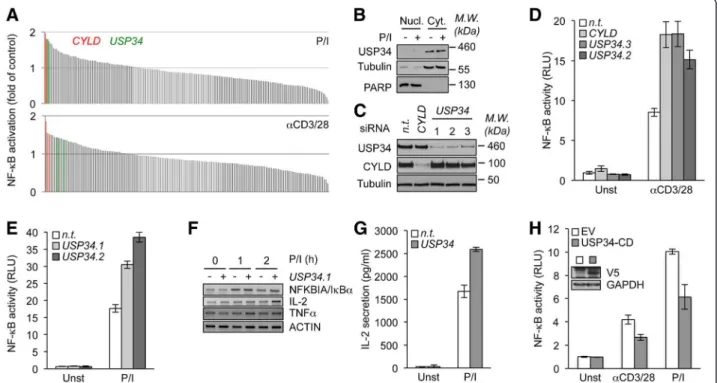
To identify additional negative regulators of
TCR-mediated NF-κB activation, we conducted a siRNA library
screen against 98 DUBs through a gene reporter luciferase
assay in Jurkat T cells stimulated with either anti-CD3 and
anti-CD28 antibodies or PMA plus ionomycin to mimic
TCR engagement (Figure 1A and Additional files 1 and 2).
As expected, CYLD silencing led to an enhanced NF-κB
activity upon TCR stimulation (Figure 1A). Furthermore,
this screening also uncovered siRNA sequences specific
for USP34 that potentiated NF-κB activation with a
similar magnitude to CYLD siRNA (Figure 1A). USP34
encompasses a 404 kDa protein with a central catalytic
domain [11]. However, little is known about this DUB,
albeit it was previously linked to the Wnt developmental
signaling pathway [12]. Subcellular fractionation
experi-ments showed that USP34 was essentially distributed in
the cytosol of cells regardless of TCR stimulation, and
was notably absent from the nucleus and organelles
(Figure 1B and Additional file 3A). We next verified by
immunoblot that CYLD and USP34 endogenous levels
were efficiently decreased by their respective siRNA
sequences (Figure 1C). Of note, an additional siRNA
duplex specific for USP34 was also included to reinforce
our initial findings (named sequence 3). Consistent with
the primary screening, NF-κB reporter activity was similarly
boosted upon TCR stimulation in USP34- and
CYLD-silenced Jurkat when compared to control non-targeting
siRNA transfected cells (Figure 1D and E). As a
conse-quence, the levels of the NF-κB targets NFKBIA (IκBα),
interleukin-2 (IL-2) and TNFα, as measured by RT-PCR
were increased in USP34-knocked down cells (Figure 1F).
Accordingly, downstream IL-2 secretion was enhanced in
supernatants of USP34-silenced cells (Figure 1G). Finally,
ectopic expression of a plasmid encoding for the catalytic
domain of USP34 (USP34-CD [13]) markedly dampened
TCR-mediated NF-κB activity (Figure 1H). Because
USP34-CD is a large segment (383 amino acids), it is
possible that in addition to the catalytic domain, it also
comprises a domain required for the binding to its
partners to regulate NF-κB in lymphocytes. Collectively,
Figure 1 Identification of USP34 as a negative regulator of TCR-mediated NF-κB activation. (A) NF-κB reporter luciferase assay screen of a siRNA library targeting 98 DUBs (2 siRNA/target) in Jurkat T lymphocytes stimulated with 20 ng.ml-1PMA plus 300 ng.ml-1ionomycin
(P/I, top panel), or with 0.5 μg.ml-1anti-CD3 and anti-CD28 (bottom panel). Fold activation compared to non-targeting (n.t.) siRNA-treated cells is
shown. Green and red histograms indicate siRNA against USP34 and CYLD, respectively. (B) Nuclear (Nucl.) and cytosolic (Cyt.) fractions from Jurkat T cells stimulated with P/I as in (A) for 0 and 15 min were analyzed by immunoblot. PARP and tubulin served as loading and purity controls for nucleus and cytosol, respectively. Molecular weight markers (M.W.) are indicated. (C) Lysates from Jurkat cells transfected for four days with siRNA against CYLD, USP34 (three individual sequences), or with control n.t. siRNA were analyzed by immunoblot as indicated. (D and E) NF-κB reporter luciferase assay (mean ± S.D. of triplicate experiments) of n.t.-, CYLD- or USP34-silenced Jurkat cells stimulated as in (A). RLU, Relative Light Units; Unst, unstimulated cells. (F) Cells as in (C) were stimulated with 20 ng.ml-1PMA plus 300 ng.ml-1ionomycin (P/I) for 1 and
2 hours. mRNA levels of NFKBIA (IκBα), IL-2, TNFα, and ACTIN were measured by RT-PCR. (G) Enzyme-Linked ImmunoSorbent Assay (ELISA) of IL-2 secreted in the supernatant of Jurkat treated as in (C) and stimulated with P/I. (H) NF-κB reporter luciferase assay of Jurkat cells transfected with the catalytic domain of USP34 (V5-tagged USP34-CD) or with a control empty vector (EV) and stimulated as indicated. Histograms represent mean ± S.D. of triplicate experiments. Inset blot shows USP34-CD expression when overexpressed in HEK293T cells.
Poalas et al. Cell Communication and Signaling 2013, 11:25 Page 2 of 5 http://www.biosignaling.com/content/11/1/25
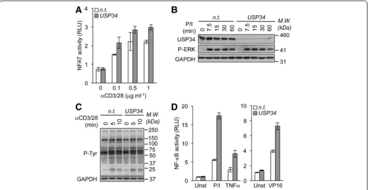
Figure 2 Knockdown of USP34 selectively potentiates NF-κB activation. (A) NFAT reporter luciferase assay (mean ± S.D. of triplicate experiments) of Jurkat cells transfected with control non-targeting (n.t.) or USP34 siRNA and stimulated with 0, 0.1, 0.5, and 1 μg.ml-1anti-CD3
and anti-CD28 antibodies (αCD3/28). RLU, Relative Light Units. (B) n.t.- and USP34-silenced Jurkat cells were stimulated with 20 ng.ml-1PMA plus
300 ng.ml-1ionomycin (P/I) for 0, 7.5, 15, 30 and 60 min. Cell extracts were prepared and analyzed by immunoblot as indicated. Molecular weight
markers (M.W.) are shown. (C) Cells as in (B) were stimulated with 1 μg.ml-1anti-CD3 and anti-CD28 antibodies. Cell extracts were prepared and
general tyrosine phosphorylation pattern (P-Tyr) was evaluated by immunoblot. GAPDH served as a loading control. (D) NF-κB reporter luciferase assay of Jurkat cells transfected as in (A) and stimulated with 10 ng.ml-1PMA plus 300 ng.ml-1ionomycin (P/I), with 10 ng.ml-1TNFα, or with
40 μM etoposide (VP16). Shown is the mean ± S.D. of triplicate experiments. Unst, unstimulated.
Figure 3 Silencing of USP34 enhances IκBα degradation and NF-κB binding to DNA. Jurkat cells were transfected with non-targeting- (n.t.) or USP34-targeting siRNA for 72-96 h. (A) Lysates from cells stimulated with 40 ng.ml-1PMA plus 300 ng.ml-1ionomycin (P/I) for 0, 10, 30 and
60 min were analyzed by immunoblot as indicated. M.W., molecular weight markers. (B) NF-κB binding to DNA was assessed on nuclear extracts from cells stimulated as in (A) for the indicated time by non-radioactive electrophoretic mobility shift assays (EMSA) using biotin-labeled κB element DNA sequence. Note that DNA:p65 complexes were chased away with a cold probe, and were shifted when a p65 antibody was added. (C) Nuclear and cytoplasmic extracts from cells stimulated as in (A) were analyzed by immunoblot as indicated. GAPDH and PARP served as loading controls. (D) Lysates from cells stimulated as in (A) were assessed by immunoblot as indicated.
our data suggest that USP34 is a cytosolic protein,
which functions as a negative regulator of NF-κB upon
TCR engagement.
In addition to NF-κB, TCR ligation kindles various
signal-ing pathways includsignal-ing Nuclear factor of activated T-cells
(NFAT) and the Mitogen-activated protein kinase (MAPK)
Extracellular signal-regulated kinases (ERK) [14]. Gene
reporter assays showed only modest increase in NFAT
activation in USP34-silenced when compared to control
cells (Figure 2A). Furthermore, ERK phosphorylation
occurred normally without USP34 (Figure 2B). Keeping
with this, no overt change in the general pattern of tyrosine
phosphorylation was observed upon TCR stimulation,
further arguing against a general impairment of TCR
signaling in the absence of USP34 (Figure 2C). We next
investigated whether USP34 also curtailed NF-κB activity
emanating from TCR-autonomous signaling triggers. To
this end, USP34-silenced Jurkat cells were stimulated
with the cytokine TNFα or with the genotoxic stress
agent etoposide that functions via an unconventional
ATM/PIASy/Sumoylated-NEMO axis [15]. Paralleling
the situation with TCR, knocking down USP34 markedly
increased NF-κB in cells treated with TNFα or etoposide
(Figure 2D). Supporting previous studies with
CYLD-deficient cells [10,16], CYLD silencing in Jurkat cells also
increased TNFα- and etoposide-mediated NF-κB activation
(Additional file 4). Combined, these results indicate that
USP34 shares some functional similarities with CYLD and
selectively targets the NF-κB signaling pathway.
To gain insights on the signaling basis for the exacerbated
NF-κB activity in USP34-depleted cells, we first examined
BCL10 and MALT1 ubiquitinylation status since it governs
the strength of TCR-mediated NF-κB activation [4-6].
BCL10 ubiquitinylation, which can be assessed in fractions
enriched with heavy membranes [17], remained unchanged
without USP34 (Additional file 3A). Moreover,
pull-down of CK1α to precipitate the CBM complex [7,17],
showed similar amounts of ubiquitinylated MALT1 bound
in both control- and USP34-siRNA transfected cells
(Additional file 3B). Keeping with this, BCL10
associ-ation to CARMA1 occurred normally without USP34
(Additional file 3C). We finally evaluated the impact of
USP34 on the phosphorylation of IKK, which reflects
its activation [8]. IKK phosphorylation was not exacerbated
in those cells, and rather appeared slightly decreased
(Figure 3A, and Additional file 3D). Although puzzling,
this might result from a feedback loop triggered by
enhanced NF-κB. We next assessed directly NF-κB DNA
binding activity. Consistent with the gene reporter assays,
more active NF-κB-DNA complexes were detected in
nuclear extracts from TCR stimulated cells when USP34
was silenced (Figure 3B). Although no obvious difference
in NF-κB subunit p65 translocation into the nucleus was
detected, the degradation of the primary NF-κB inhibitor
IκBα was more pronounced in cytosolic fractions from
USP34-silenced cells when compared to control cells
(Figure 3C). Accordingly, IκBα degradation was also
prolonged or more dramatic in lysates from cells transfected
with USP34 siRNA even on longer time points up to 3 hours
(Figure 3D). Hence, our results suggest that USP34 likely
functions downstream of the CBM-IKK nexus to enhance
NF-κB activation.
Almost 100 DUBs were identified in the human genome
and yet, only a few have been ascribed a function [11]. As
for TCR signaling, the well studied A20 and CYLD thwart
NF-κB at different levels [10]. CYLD targets and inhibits
the ubiquitin-dependent IKKβ kinase TAK1 and therefore
prevents aberrant lymphocyte activation [18,19], while A20
dampens NF-κB activity by trimming K63-ubiquitin chains
attached to MALT1 [20,21]. Our study now unveils USP34
as an additional negative regulator of NF-κB in
lympho-cytes. How USP34 tempers NF-κB activity remains unclear.
In contrast to CYLD and A20, which target apical signaling
[18,20], USP34 rather seems to function downstream of the
IKK complex. Given that USP34 does not bind to the
NF-κB core components (our unpublished results), we
favor a model in which USP34 impacts on the activity of
a cytosolic co-activator to ensure IκBα fine-tuning
[22,23]. Alternatively, USP34 might also intervene in
other checkpoints to control NF-κB signal outcome and
intensity such as post-translational modifications, nuclear
shift, or DNA three-dimensional structure [22,24,25].
Nevertheless, our data illustrates how various layers of
control cooperate to ensure the fine-tuning of NF-κB
following engagement of the TCR.
Additional files
Additional file 1: Methods description.
Additional file 2: Design of the siRNA library screen. Additional file 3: BCL10 and MALT1 ubiquitinylation, CBM assembly, and IKK phosphorylation in USP34-silenced cells. Additional file 4: CYLD and USP34 silencing enhanced etoposide-and TNFα-mediated NF-κB activation.
Abbreviations
CYLD:Cylindromatosis; DUBs: Deubiquitinylases; ERK: Extracellular signal-regulated kinases; IκBs: Inhibitors of NF-κB; IKK: IκBs kinase; MAPK: Mitogen-activated protein kinase; NFAT: Nuclear factor of activated T-cells; NF-κB: Nuclear factor-κB; PMA: phorbol 12-myristate 13-acetate; TCR: T-Cell receptor.
Competing interests
The authors declare that they have no competing interests. Authors’ contributions
KP designed and conducted most experiments, analyzed the data and wrote the manuscript. EMH, NC, SMD, HML, CL, and CA designed and conducted experiments, and analyzed the data. EMH and JG helped drafting the manuscript. JG and AV analyzed the data. NB conceived the project, analyzed the data and wrote the manuscript. All authors read and approved the final version of the manuscript.
Poalas et al. Cell Communication and Signaling 2013, 11:25 Page 4 of 5 http://www.biosignaling.com/content/11/1/25
Acknowledgements
We thank V. Quesada and C. Lopez-Otín (Universidad de Oviedo, Spain) for providing USP34-CD plasmid, and D. Arnoult (INSERM U1014) for helpful discussions. This work was supported by grants from ANR JCJC, Fondation ARC, Fondation de France, Ligue Nationale contre le Cancer, and by Fondation pour la Recherche Médicale. KP and SMD are supported by fellowships from Université Paris Sud, and EMH by la Ligue contre le Cancer. Author details
1INSERM UMR_S 1014, Hôpital Paul Brousse, Villejuif 94800, France. 2Université Paris-Sud P11, Orsay 91400, France.3Equipe Labellisée Ligue
contre le Cancer, Villejuif, France.4Université Paris Descartes, Sorbonne Paris Cite, 6 rue de l’Ecole de Medecine, Paris 75006, France.5INSERM, U1016, 22
rue Méchain, Paris 75014, France.6CNRS, UMR8104, 22 rue Méchain, Paris
75014, France.
Received: 8 February 2013 Accepted: 3 April 2013 Published: 16 April 2013
References
1. Hayden MS, Ghosh S: Shared principles in NF-kappaB signaling. Cell 2008, 132(3):344–362.
2. Hinz M, Arslan SC, Scheidereit C: It takes two to tango: IkappaBs, the multifunctional partners of NF-kappaB. Immunol Rev 2012, 246(1):59–76. 3. Thome M, Charton JE, Pelzer C, Hailfinger S: Antigen receptor signaling to
NF-kappaB via CARMA1, BCL10, and MALT1. Cold Spring Harb Persp Biol 2010, 2(9):a003004.
4. Oeckinghaus A, Wegener E, Welteke V, Ferch U, Arslan SC, Ruland J, Scheidereit C, Krappmann D: Malt1 ubiquitination triggers NF-kappaB signaling upon T-cell activation. EMBO J 2007, 26(22):4634–4645. 5. Sun L, Deng L, Ea CK, Xia ZP, Chen ZJ: The TRAF6 ubiquitin ligase and
TAK1 kinase mediate IKK activation by BCL10 and MALT1 in T lymphocytes. Mol Cell 2004, 14(3):289–301.
6. Wu CJ, Ashwell JD: NEMO recognition of ubiquitinated Bcl10 is required for T cell receptor-mediated NF-kappaB activation. Proc Natl Acad Sci U S A2008, 105(8):3023–3028.
7. Bidere N, Ngo VN, Lee J, Collins C, Zheng L, Wan F, Davis RE, Lenz G, Anderson DE, Arnoult D, et al: Casein kinase 1alpha governs antigen-receptor-induced NF-kappaB activation and human lymphoma cell survival. Nature 2009, 458(7234):92–96.
8. Israel A: The IKK complex, a central regulator of NF-kappaB activation. Cold Spring Harb Perspect Biol2010, 2(3):a000158.
9. Skaug B, Jiang X, Chen ZJ: The role of ubiquitin in NF-kappaB regulatory pathways. Annu Rev Biochem 2009, 78:769–796.
10. Harhaj EW, Dixit VM: Regulation of NF-kappaB by deubiquitinases. Immunol Rev2012, 246(1):107–124.
11. Nijman SM, Luna-Vargas MP, Velds A, Brummelkamp TR, Dirac AM, Sixma TK, Bernards R: A genomic and functional inventory of deubiquitinating enzymes. Cell 2005, 123(5):773–786.
12. Lui TT, Lacroix C, Ahmed SM, Goldenberg SJ, Leach CA, Daulat AM, Angers S: The ubiquitin-specific protease USP34 regulates axin stability and Wnt/beta-catenin signaling. Mol Cell Biol 2011, 31(10):2053–2065. 13. Quesada V, Diaz-Perales A, Gutierrez-Fernandez A, Garabaya C, Cal S,
Lopez-Otin C: Cloning and enzymatic analysis of 22 novel human ubiquitin-specific proteases. Biochem Biophys Res Commun 2004, 314(1):54–62. 14. Samelson LE: Immunoreceptor signaling. Cold Spring Harb Perspect Biol
2011, 3(12):a011510.
15. McCool KW, Miyamoto S: DNA damage-dependent NF-kappaB activation: NEMO turns nuclear signaling inside out. Immunol Rev 2012, 246(1):311–326.
16. Niu J, Shi Y, Iwai K, Wu ZH: LUBAC regulates NF-kappaB activation upon genotoxic stress by promoting linear ubiquitination of NEMO. EMBO J2011, 30(18):3741–3753.
17. Carvalho G, Le Guelte A, Demian C, Vazquez A, Gavard J, Bidere N: Interplay between BCL10, MALT1 and IkappaBalpha during T-cell-receptor-mediated NFkappaB activation. J cell sci 2010, 123(Pt 14):2375–2380.
18. Reiley WW, Jin W, Lee AJ, Wright A, Wu X, Tewalt EF, Leonard TO, Norbury CC, Fitzpatrick L, Zhang M, et al: Deubiquitinating enzyme CYLD negatively regulates the ubiquitin-dependent kinase Tak1 and prevents abnormal T cell responses. J Exp Med 2007, 204(6):1475–1485.
19. Staal J, Driege Y, Bekaert T, Demeyer A, Muyllaert D, Van Damme P, Gevaert K, Beyaert R: T-cell receptor-induced JNK activation requires proteolytic inactivation of CYLD by MALT1. EMBO J 2011, 30(9):1742–1752. 20. Duwel M, Welteke V, Oeckinghaus A, Baens M, Kloo B, Ferch U, Darnay BG,
Ruland J, Marynen P, Krappmann D: A20 negatively regulates T cell receptor signaling to NF-kappaB by cleaving Malt1 ubiquitin chains. J Immunol2009, 182(12):7718–7728.
21. Stilo R, Varricchio E, Liguoro D, Leonardi A, Vito P: A20 is a negative regulator of BCL10- and CARMA3-mediated activation of NF-kappaB. J Cell Sci2008, 121(Pt 8):1165–1171.
22. Perkins ND: The diverse and complex roles of NF-kappaB subunits in cancer. Nat Rev Cancer 2012, 12(2):121–132.
23. Wan F, Lenardo MJ: The nuclear signaling of NF-kappaB: current knowledge, new insights, and future perspectives. Cell Res 2010, 20(1):24–33.
24. Chen LF, Greene WC: Shaping the nuclear action of NF-kappaB. Nat Rev Mol Cell Biol2004, 5(5):392–401.
25. Smale ST: Hierarchies of NF-kappaB target-gene regulation. Nat Immunol 2011, 12(8):689–694.
doi:10.1186/1478-811X-11-25
Cite this article as: Poalas et al.: Negative regulation of NF-κB signaling in T lymphocytes by the ubiquitin-specific protease USP34. Cell Communication and Signaling2013 11:25.
Submit your next manuscript to BioMed Central
and take full advantage of:
• Convenient online submission
• Thorough peer review
• No space constraints or color figure charges
• Immediate publication on acceptance
• Inclusion in PubMed, CAS, Scopus and Google Scholar
• Research which is freely available for redistribution
Submit your manuscript at www.biomedcentral.com/submit