HAL Id: hal-02291949
https://hal.sorbonne-universite.fr/hal-02291949
Submitted on 19 Sep 2019
HAL is a multi-disciplinary open access
archive for the deposit and dissemination of
sci-entific research documents, whether they are
pub-lished or not. The documents may come from
teaching and research institutions in France or
abroad, or from public or private research centers.
L’archive ouverte pluridisciplinaire HAL, est
destinée au dépôt et à la diffusion de documents
scientifiques de niveau recherche, publiés ou non,
émanant des établissements d’enseignement et de
recherche français ou étrangers, des laboratoires
publics ou privés.
influenza vaccination in COPD
K. Staples, N. P. Williams, O. Bonduelle, A.J. Hutton, D. Cellura, A.
Marriott, B. Combadière, T. Wilkinson
To cite this version:
K. Staples, N. P. Williams, O. Bonduelle, A.J. Hutton, D. Cellura, et al.. Acquired immune responses
to the seasonal trivalent influenza vaccination in COPD. Clinical and Experimental Immunology,
Wiley, 2019, 198 (1), pp.71-82. �10.1111/cei.13336�. �hal-02291949�
© 2019 The Authors. Clinical & Experimental Immunology published by John Wiley & Sons Ltd on behalf of British Society 71
Acquired immune responses to the seasonal trivalent influenza
vaccination in COPD
K. J. Staples ,*† N. P. Williams,*‡ O. Bonduelle,§¶ A. J Hutton,§ D. Cellura,* A. C. Marriott,** B. Combadière§¶ and T. M. A. Wilkinson*†‡* Clinical and Experimental
Sciences, University of Southampton Faculty of Medicine, Sir Henry Wellcome Laboratories, Southampton General Hospital, Tremona Road, † Wessex Investigational Sciences
Hub, University of Southampton Faculty of Medicine, Southampton General Hospital, Tremona Road, ‡ Southampton NIHR
Respiratory Biomedical Research Unit, Southampton General Hospital, Tremona Road, Southampton, UK, § Sorbonne
Universités, UPMC Univ Paris 06, Unité Mixte de Recherche de Santé (UMR S) CR7, Centre d’Immunologie et des Maladies Infectieuses –Paris (Cimi-Paris), ¶ Institut
National de Santé et de Recherche Médicale (INSERM) U1135, Cimi-Paris, Paris, France, and ** National Infection Service, Public Health
England, Porton Down, UK
Accepted for publication 29 May 2019 Correspondence: Dr K. J. Staples, Clinical and Experimental Sciences, University of Southampton Faculty of Medicine, Sir Henry Wellcome Laboratories, Mailpoint 810, Southampton General Hospital, Tremona Road, Southampton SO16 6YD, UK. E-mail: k.staples@southampton.ac.uk
Summary
Epidemiological data suggest that influenza vaccination protects against all-cause mortality in chronic obstructive pulmonary disease (COPD) pa-tients. However, recent work has suggested there is a defect in the ability of some COPD patients to mount an adequate humoral response to in-fluenza vaccination. The aim of our study was to investigate humoral and cell-mediated vaccine responses to the seasonal trivalent influenza vaccina-tion (TIV) in COPD subjects and healthy controls. Forty-seven subjects were enrolled into the study; 23 COPD patients, 13 age-matched healthy controls (HC ≥ 50) and 11 young healthy control subjects (YC ≤ 40). Serum and peripheral blood mononuclear cells (PBMC) were isolated pre-TIV vaccination and at days 7 and 28 and 6 months post-vaccine for haemagglutinin inhibition (HAI) titre, antigen-specific T cell and antibody-secreting cell analysis. The kinetics of the vaccine response were similar between YC, HC and COPD patients and there was no significant differ-ence in antibody titres between these groups at 28 days post-vaccine. As we observed no disease-dependent differences in either humoral or cellular responses, we investigated if there was any association of these measures with age. H1N1 (r = −0·4253, P = 0·0036) and influenza B (r = −0·344, P = 0·0192) antibody titre at 28 days negatively correlated with age, as did H1N1-specific CD4+ T helper cells (r = −0·4276, P = 0·0034). These results suggest that age is the primary determinant of response to trivalent vaccine and that COPD is not a driver of deficient responses per se. These data support the continued use of the yearly trivalent vaccine as an adjunct to COPD disease management.
Keywords: COPD, influenza, vaccination
Introduction
Chronic obstructive pulmonary disease (COPD) is char-acterized by airway inflammation, resulting from the inhalation of tobacco smoke or other irritants [1]. The course of the disease is one of progressive, irreversible airflow limitation, functional impairment and periods of acute symptom deterioration called exacerbations [2]. Acute exacerbations are important events in the natural history of COPD associated with impaired quality of life
and accelerated decline in lung function, placing a con-siderable burden on health-care resources [3–5]. Respiratory infection is a key determinant, with viral infections implicated in between 40 and 60% of COPD exacerbations [6,7]. Consequently, preventative strategies aimed at reducing the impact of respiratory viral infection are key interventions in reducing the morbidity and health-care burden associated with COPD exacerbations.
Influenza A and B are single-stranded RNA viruses of the Orthomyxoviridae family that circulate in the human
population, causing acute infection of both the upper and lower respiratory tract. In patients with COPD, influenza is associated with seasonal exacerbations, with the virus detected in up to 28% of exacerbating COPD patients [8,9]. The seasonal influenza vaccination is currently the best method for reducing the incidence of influenza infection and the associated complications such as pneumonia, and is recommended for elderly people and in those with chronic conditions such as COPD [10]. Therapies to treat influenza infection, once present in COPD, are currently very limited, both in terms of the window of opportunity for treatment and therapeutic efficacy. Thus, vaccination is a critical component in exacerbation prevention. In the United Kingdom, the most commonly used formulation is the trivalent influenza vaccine (TIV) which currently contains inactive viral haemagglutinin (HA) from H1N1, H3N2 and influenza B strains. A recognized correlate of vaccine protection is the production of sufficient concen-trations of antibodies to HA, as assessed by haemagglutinin inhibition (HAI) assays [11]. The response to TIV can be demonstrated by either seroprotection (HAI antibody titre ≥1 : 40) or seroconversion (minimum fourfold rise in HAI antibody titre) [12]. While the use and efficacy of TIV in COPD is supported by epidemiological studies [13,14], recent laboratory-based work has begun to question the effectiveness of these vaccines in disease [15–17].
While trials and epidemiological data point to a benefit of TIV in COPD patients [18], a small study from Australia demonstrated that COPD patients had lower titres of H1N1-specific antibodies 28 days post-vaccination and lower serum interleukin (IL)-21 levels, which is thought to play a role in B cell antibody synthesis [16]. In addi-tion, the same group reported a reduced proportion of COPD patients who seroconverted 28 days after
vaccination compared to healthy controls [15]. More recently, Parpaleix et al. demonstrated decreased geometric mean HAI antibody titres in COPD patients at 30 days post-vaccine compared to healthy controls, but no sig-nificant difference in the proportion of volunteers who experienced seroconversion between health and disease [17]. Furthermore, this decrease in geometric mean HAI antibody titres was associated with a decrease in vaccine-specific CD4+ T cell responses prior to vaccination.
The aim of our study was to conduct a detailed assess-ment of the HAI antibody, T cell and B cell responses to TIV of COPD patients compared to age-matched healthy and young, healthy controls, enabling the identification of any differences in vaccine responses between the groups. Methods
Volunteer recruitment
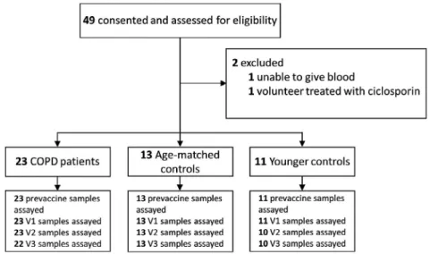
Twenty-three COPD patients, 13 age-matched aged ≥50 years) healthy control subjects and 11 younger (aged <40 years) healthy control subjects were studied (Fig. 1). Power calculations were based on the proportion of responders achieving at least a fourfold increase in the H1N1 HAI antibody titre in each group. A minimum sample size of 22 subjects in the COPD group and 11 subjects in the control groups was determined as suf-ficient for a two-sided, type I error rate of 0·05 and a power of 80%. A diagnosis of COPD was confirmed by post-bronchodilator spirometry, with a forced expiratory volume in 1 s (FEV1)/forced vital capacity (FVC) ratio
of <0·7 required for enrolment. Spirometry was con-ducted in accordance with ATS standards [19]. COPD subjects had a smoking history of at least 10 pack-years.
Fig. 1. Consolidated Standards of Reporting Trials (CONSORT) flow diagram detailing subject enrolment and subgroupings in the study. Where samples are missing, this is due to a visit being missed and a sample not collected.
All subjects had received the TIV in the year prior to the study (2014). The vaccine strains for the 2014/2015 northern hemisphere vaccine were A/California/7/2009 (H1N1) pdm09-like virus, A/Texas/50/2012 (H3N2)-like virus and B/Massachusetts/2/2012-like virus. Exclusion criteria included a history of other pulmonary disease, long-term use of immunosuppressant medications (including oral corticosteroids) and an exacerbation within the month prior to recruitment. All subjects gave written informed consent and the study was approved by the National Research Ethics Service (NRES) South Central – Oxford C Committee (15/SC/0528).
Study procedures
As this was a purely observational study, the intramuscular influenza vaccine was administered by the volunteer’s usual health-care provider as part of usual care between October 2015 and December 2015. All the young healthy group were health-care professionals. The vaccine strains for the 2015/2016 northern hemisphere were A/California/7/2009 (H1N1) pdm09-like virus, A/Switzerland/9715293/2013 (H3N2)-like virus and B/Phuket/3073/2013-like virus. Phlebotomy was performed at a prevaccine visit and then at 7−10 days (visit 1), 28 ± 3 days (visit 2) and 180 ± 14 days (visit 3) post-vaccination.
Serum HAI antibody titres
The HAI assays were performed by the Public Health England (PHE) laboratories, Porton Down, Salisbury, UK. Briefly, serial twofold dilutions for each set of sera were incubated with standardized concentrations (4 HA units) of influenza virus representing either the H1N1, H3N2 and influenza B 2015/16 viral strains. Chicken red blood cells were then added and allowed to settle. After 30 min, the strain-specific HAI antibody titres at each time-point for each individual were calculated as the highest dilution of sera that inhibited haemagglu-tination. Seroprotection rates (i.e. numbers of individuals with HAI antibody titres ≥40) and seroconversion rates (i.e. numbers of individuals with HAI antibody titres <10 at day 0 and HAI antibody titres ≥40 after vac-cination or with HAI antibody titres ≥10 at day 0 and a fourfold or greater increase in HAI antibody titres after vaccination) are also indicated.
Peripheral blood mononuclear cells (PBMC) isolation and storage
PBMCs from volunteers were isolated from heparinized blood by means of centrifugation on Ficoll-Paque (GE Healthcare, Little Chalfont, UK). Purified PBMC were frozen in heat-inactivated fetal bovine serum containing 10% (v/v) dimethylsulphoxide (DMSO) (Sigma, Poole, UK) and stored in liquid nitrogen until analysis.
Flow cytometry analysis
Flow cytometry was performed as previously described [20]. To analyze influenza-specific T cells, PBMCs were stimulated overnight with inactivated A/H3N2 Wisconsin/67/2005 and A/H1N1/California/04/2009 in the presence of monensin/brefeldin A mix (Sigma). Cells were first stained for viability and surface markers: live/dead (Molecular Probes, Eugene, OR, USA), CD4 (OKT4 clone), CD3 (SK7 clone), CD8 (RPA-T8 clone), CD49d (9F10 clone) and CD49a (SR84 clone; BD Biosciences, Oxford, UK). Cells were then resuspended in Cytofix/Cytoperm (BD Biosciences) before staining for intracellular markers: IL-2 (N7.48 A clone; Miltenyi Biotech, Woking, UK), interferon (IFN)-β (B27 clone), IL-17a (N49-653 clone), granzyme B (GzmB) (GB11 clone) and tumour necrosis factor (TNF)-α (MAb11 clone; BD Biosciences). Flow cytometric analysis was performed on a fluorescence acti-vated cell sorter (FACS) Fortessa using FACSDiva software version 5.0.3 (BD Biosciences). At least 1 × 10⁶ live events,
according to forward- and side-scatter parameters, were accumulated and analyzed for Boolean combination gating with FlowJo software (Tree Star Inc., Ashland, OR, USA). The percentage of influenza A virus-specific CD4+ or CD8+
T cells expresses the sum of the 15 different cytokine Boolean combinations (IL-2, IFN-β IL-17a and/or TNF-α). Background cytokine responses detected in negative con-trols were subtracted from those detected in stimulated samples for every specific combination.
Antibody-secreting cell (ASC) detection
A/California/07/2009 (H1N1) influenza HA-specific IgG-secreting B cells were performed as previously described [21]. Briefly, PBMC were stimulated with a mixture of pokeweed mitogen (Sigma), Staphylococcus aureus protein A (Sigma) and CpG ODNs (Invivogen, San Diego, CA, USA) for 6 days before enzyme-linked immunospot (ELISPOT) assay. H1N1 HA protein (Protein Sciences, Meriden, CT, USA) was used to coat the plate (Millipore, Watford, UK), and immunoglobulin (Ig)G- or IgA-paired antibodies (Mabtech/Oxford lmmunotec Limited, Abingdon, UK) were used to reveal. ELISPOT readouts were expressed as the number of HA-specific IgG or IgA ASC/106 PBMC.
Statistics and analysis
Statistical analyses were performed using either a Wilcoxon’s matched-pairs signed-rank test, Mann– Whitney U-test, Kruskal–Wallis or Friedman’s test with Dunn’s multiple comparison testing as appropriate (GraphPad Prism version 7.0; GraphPad Software Inc., San Diego, CA, USA). For the paired analysis, if data were missing for a given visit, all data from that volunteer
were excluded and there was no data imputation. Data are expressed as medians. Results were considered sig-nificant if P < 0·05. The radar charts were designed with
r, a free software environment for statistical computing
and graphics (http://www.r-proje ct.org/). Results
Subject demographics
Forty-seven subjects were successfully enrolled into the study, 23 COPD patients, 13 age-matched healthy controls and 11 young control subjects (Fig. 1). The COPD and healthy control subjects were well-matched for age and gender. As expected, there were significant differences in the proportion of current smokers and FEV1 measures.
To ensure that recent vaccination history was known, our inclusion criteria required subjects to have received the prior year’s (2014/15) influenza vaccination. The descriptive characteristics are shown in Table 1.
Humoral responses to TIV
To ascertain the effectiveness of the vaccine to induce a humoral response, we first analyzed the antibody titres to all three vaccine components via individual haemag-glutinin inhibition (HAI) assays in the whole cohort (Fig. 2a–c). The median [interquartile range (IQR)] level of prevaccine HAI titres were 40 (10−40) for A/H1N1, 20 (10−40) for A/H3N2 and 17·5 (5−40) for B/Phuket strains. These data suggest that half the cohort were already seroprotected against A/H1N1 virus but not the A/H3N2 or B/Phuket strains before vaccination. There was a significant increase in antibody titres 7 days (visit 1) and 28 days (visit 2) following vaccination for all vaccine components, although this was not sustained
to 6 months (visit 3) for B/Phuket strain. In addition, the HAI antibody increases in FluB were not as strong and remained consistently lower than the titres induced by the FluA components.
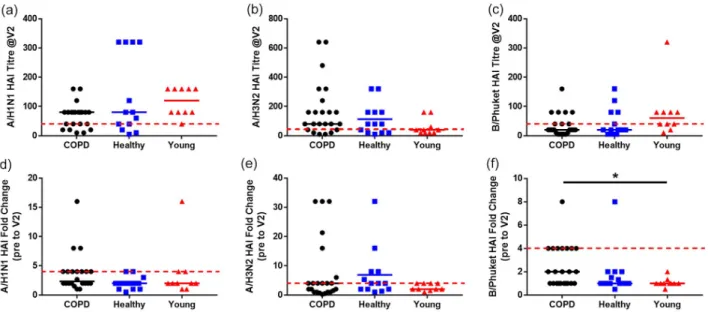
In line with other studies [17], we next compared the HAI titres to all three vaccine components between groups at 28 days post-vaccine, when all groups dem-onstrated a significant increase in titres against the FluA components (Fig. 3). Only the COPD group demonstrated a significant increase in HAI antibody titres to B/Phuket at 28 days post-vaccination (P = 0·0127, Fig. 3c). However, there was no significant difference in the HAI antibody titres between the groups for any of the vaccine com-ponents at day 28 (Fig. 4a–c), nor was there a significant difference in the fold change in HAI antibody titres between groups at visit 2 compared to the prevaccina-tion visit, except for B/Phuket between COPD patients and young controls (Fig. 4d–f). While there appeared to be differences in the proportion of volunteers sero-protected (titre ≥40) between groups for each vaccine component, the proportion was again only significantly different for B/Phuket, where 80% of the young controls were seroprotected compared to the 62·5% of healthy controls and 53% of COPD patients (P = 0·0469, χ2).
As it is also recommended that seroconversion be taken into account, as well as seroprotection [12],we analyzed the number of patients who did not seroconvert at visit 2 (Fig. 4d–f). The only significant difference in serocon-version we observed was a decrease in the response of young controls to B/Phuket compared to COPD patients (Fig. 4f). In order to assess any differences in the response to vaccine using either definition, we also assessed the proportions of volunteers who were not seroprotected at baseline and also did not seroconvert by day 28 (Table 2). While there was evidence of differential responses to different vaccine components, there were no significant differences in the proportions of the groups that were not protected against the individual vaccine components. Cellular responses to TIV
In addition to antibodies, T cell responses are also key parameters of influenza protection [20,22–26]. We there-fore analyzed the polyfunctional CD4+ and CD8+ T cell
responses to inactivated whole virus (A/H1N1 and A/H3N2) in PBMC from our volunteer cohort at each time-point (Supporting information, Fig. S1). We did not observe any significant difference in the quality of poly-functional CD4+ and CD8+ T cell responses between
groups (Supporting information, Fig. S2). We observed a significant increase (P = 0·0035) in the proportion of virus-specific CD4+ T cells 28 days post-vaccination
(Fig. 5a) compared to prevaccination in the whole cohort. In contrast, there was no increase in the proportion of Table 1. Demographics and clinical characteristics
COPD n = 23 Healthy control n = 13 P-value Young control n = 11 Age* 69·0 (55–83) 65·1 (56–72) 0·112 33·2 (22–40) Gender, male, n, % 13 (56·5) 5 (38·5) 0·489 5 (45·5) Current smoker, n, % 8 (34·8) 1 (7·7) 0·001 1 (9·1) Pack years 38 (22) 4 (20) <0·001 0 (2) FEV1 (l) 1·46 (1·0) 2·95 (1·3) <0·001 3·53 (0·85) FEV1% 55 (25) 109 (18) <0·001 98 (20)
Data represented as median (interquartile range) or frequency (%). *Represented as mean (range). Statistical significance was determined using a Mann–Whitney U-test and is indicated in bold.
COPD, chronic obstructive pulmonary disease; FEV, forced expira-tory volume in 1 s.
virus-specific CD8+ T cells (Fig. 5b). However, there was
a significant decrease in the proportion of virus-specific GzmB+ CD8+ T cells at all time-points following
vaccina-tion (Fig. 5c). We also compared the proporvaccina-tions of
virus-specific T cells between groups at 28 days post-vaccine (Fig. 5d–f). Similarly to the HAI data, we again observed no significant differences in CD4+ or CD8+ T
cell proportions between the groups.
Fig. 2. Cohort haemagglutinin inhibition (HAI) antibody responses to trivalent influenza vaccine (TIV). HAI antibody titres of trivalent influenza vaccine constituents [(a) A/H1N1, (b) A/H3N2 and (c) B/Phuket] in serum from the whole cohort of volunteers at each individual visit [prevaccine, 7 days (visit 1), 28 days (visit 2) and 6 months (visit 3) following vaccination]. For all graphs, y-axes are presented as log2 scale and median values are indicated by line, while + indicates the mean. The dotted red line indicates seroprotection (titre of ≥40). Statistical significance was determined by Friedman’s analysis of variance (ANOVA) with a Dunn’s post-hoc test. P-values from Dunn’s test are shown; *P < 0·05, ***P < 0·001, ****P < 0·0001.
Fig. 3. Haemagglutinin inhibition (HAI) antibody responses of the cohort subgroups to different vaccine components. HAI antibody titre to (a) A/H1N1, (b) A/H3N2 and (c) B/Phuket constituents of the trivalent influenza vaccine in serum from chronic obstructive pulmonary disease (COPD) patients, age-matched healthy controls and young controls at each individual visit [prevaccine, 7 days (visit 1), 28 days (visit 2) and 6 months (visit 3) following vaccination]. For all graphs, median values are indicated by line. Statistical significance was determined by Friedman’s analysis of variance (ANOVA) with a Dunn’s post-hoc test. P-values from Dunn’s test are shown; *P < 0·05, ***P < 0·001, ****P < 0·0001.
At the same time we also analyzed the proportions of both A/H1N1 HA-specific IgG-secreting B cells in these samples using ELISPOT (Fig. 6a–d). However, we observed no significant change in the proportion of A/H1N1 HA-specific IgG-secreting B cells either in response to vaccination or between the groups at 28 days post-vaccine. A/H1N1 HA-specific IgA-secreting B cells are very low (data not shown).
In order to present a complete picture of our work, we have also analyzed all the variables reported above at 7 days and 6 months post-vaccination and present these data as radar plots (Fig. 7b–d). Furthermore, we evaluated the capacity of CD4 and CD8 T cells to acquire the cell migration capacity to mucosal with the expres-sion of integrins CD49a and CD49d. There were no statistically significant differences in any of the
measurements between groups at any time-point post-vaccination. Thus, overall there appear to be no differ-ences in vaccine responses across the groups. This is despite a statistically significant difference in B/Phuket HAI antibody titres between groups at baseline, which appears to be due more to age rather than COPD (Fig. 7a). Furthermore, there are also differences in the proportions of influenza-specific cytokine-secreting CD4+
T cells at baseline which, surprisingly, are significantly lower in healthy age-matched controls compared to both young controls.
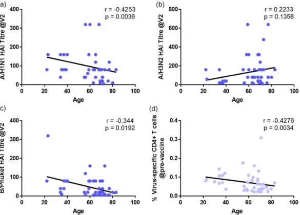
Age correlates with antibody titres
To further investigate the effects of age on responses to vaccination, we correlated the age of the volunteers with the antibody titres of the separate vaccine components at 28 days (Fig. 8a–c). There were weak but significant negative correlations between age and A/H1N1 but not A/H3N2 titres.
As we have previously shown that pre-existing CD4+
T helper cells are essential for adequate responses to influ-enza [24], we next analyzed the correlation between age and pre-existing virus-specific CD4+ T cells (Fig. 8d).
Similarly to the HAI data, we observed a weak, but sig-nificant negative correlation between age and the proportion of virus-specific CD4+ cells at the pre-vaccination visit.
Fig. 4. Haemagglutinin inhibition (HAI) antibody responses of the cohort subgroups to the different vaccine components 28 days post-vaccination. HAI antibody titre to (a) A/H1N1, (b) A/H3N2 and (c) B/Phuket constituents of the trivalent influenza vaccine in serum from chronic obstructive pulmonary disease (COPD) volunteers (filled circles), age-matched healthy controls (filled squares) and young healthy controls (filled triangles) at visit 2 (day 28 post-vaccination). Fold change in HAI antibody titre to (d) A/H1N1, (e) A/H3N2 and (f) B/Phuket constituents of the trivalent influenza vaccine in serum from COPD volunteers (filled circles), age-matched healthy controls (filled squares) and young healthy controls (filled triangles) at visit 2 (day 28 post-vaccination) compared to the prevaccination visit. For all graphs, median values are shown. The dotted red line indicates (a–c) seroprotection (titre of ≥40) or (d–f) seroconversion (fourfold increase in titre). Statistical significance was determined by Kruskal– Wallis analysis of variance (ANOVA) with a Dunn’s post-hoc test. P-values from Dunn’s test are shown; *P < 0·05.
Table 2. Proportions of volunteers who did not seroconvert (less than a fourfold increase in HAI titre) by visit 2 and were not seroprotected (HAI titre <1 : 40) at baseline to each of the vaccine components
COPD Healthy control Young control
H1N1 26·1% 30·8% 9·1%
H3N2 17·4% 23·1% 36·4%
FluB 47·8% 53·8% 36·4%
Discussion
In this study we have shown that in the majority of vol-unteers among disease and age groups who were vaccinated with TIV, there was adequate seroprotection (titre ≥40) to at least one component of the vaccine at 28 days post-vaccination. Importantly, there were minimal differences in the vaccine responses of COPD patients, compared to age-matched healthy controls and young controls at any time-point. Although there were no significant age-related differences between groups in many of the immune meas-ures analyzed prior to vaccine administration, there was evidence of a relationship between age and vaccine responses. Further analysis demonstrated a negative correlation between age and HAI antibody titres for two out of three vaccine components. Taken together, these data suggest that age-related immune senescence may have a stronger dampening effect on vaccine responses than COPD per se.
In the western world, COPD is a disease associated with lifelong smoking, and due to the pernicious nature
of symptom onset is not usually diagnosed until the late 40s/early 50s. Thus, COPD itself is a disease already asso-ciated with ageing, and therefore it is essential that the age of any control group is well matched with the COPD group. In the previous studies from the Australian group demonstrating a disease effect on vaccine responses, there were significant differences between the ages of the control group and the COPD group [15,16]. Indeed, Burel et al. demonstrated that age was associated with post-vaccination A/H1N1 antibody titres by univariate, although not mul-tivariate, analysis [16].
The use of HAI antibody titres as a correlate of pro-tection is further complicated by the use of different methods to assess vaccine responsiveness. The Committee for Proprietary Medicinal Product (CPMP) criteria state that post-vaccination serum is considered seroprotected if the HAI antibody titre is ≥40 [12]. However, the CPMP have two definitions of seroconversion, with the sero-protected level of ≥40 only being considered a serocon-version if the prevaccine levels were negative. If the serum Fig. 5. T cell responses to influenza virus pre and post vaccination. H3N2 and H1N1 inactivated virus-specific CD4 and CD8 T cells were measured by presence of intracellular cytokines [Boolean gates for interferon (IFN)-β, interleukin (IL)-2, IL-17 and/or tumour necrosis factor (TNF)-α] or granzyme B (GzmB) in each subject. (a–c) Proportions of T cells responding to whole H1N1 and H3N2 viral stimulation from the whole cohort of volunteers after each individual visit [prevaccine, 7 days (visit 1), 28 days (visit 2) and 6 months (visit 3) following vaccination]. For all graphs, median values are indicated by line, while + indicates the mean. Statistical significance was determined by Friedman’s analysis of variance (ANOVA) with a Dunn’s post-hoc test. (d–f) Proportion of T cells responding to whole virus from chronic obstructive pulmonary disease (COPD) volunteers (n = 23; filled circles), age-matched healthy controls (n = 13; filled squares) and young healthy controls (n = 10; filled triangles) at visit 2 (day 28 post-vaccination). For all graphs, median values are shown. Statistical significance was determined by Kruskal–Wallis ANOVA with a Dunn’s
Fig. 6. B cell responses to H1N1 HA pre- and post-vaccination. (a) Number of H1N1-specific B cells releasing immunoglobulin (Ig)G per million peripheral blood mononuclear cells (PBMC) from the whole cohort of volunteers after each individual visit [prevaccine, 7 days (visit 1), 28 days (visit 2) and 6 months (visit 3) following vaccination]. Median values are indicated by line, while + indicates the mean. Statistical significance was determined by Friedman’s analysis of variance (ANOVA) with a Dunn’s post-hoc test. (b) Number of B cells releasing IgG from chronic obstructive pulmonary disease (COPD) volunteers (filled circles), age-matched healthy controls (filled squares) and young healthy controls (filled triangles) at visit 2 (day 28 post-vaccination). Median values are shown and statistical significance was determined by Kruskal–Wallis ANOVA with a Dunn’s
post-hoc test.
Fig. 7. Radar plot of responses in each volunteer group pre- and post-vaccination. Radar charts comparing flu virus-specific immune compartments of chronic obstructive pulmonary disease (COPD) (black), healthy control (blue) and young healthy control (red) groups (a) prevaccination, and fold change (visit/prevaccination) of influenza specific-immune response at visit 1 (b), visit 2 (c) and visit 3 (d). Radar charts show A/H3N2, A/H1N1 and B/Phuket strain haemagglutinin inhibition (HAI) antibody titres, A/H1N1 HA-specific immunoglobulin ( Ig)G and IgA-secreting B cells, virus-specific CD4 and CD8 T cells, granzyme B (GzmB)+ CD8 T cells and CD49a+CD49d+ homing markers evaluated on interferon (IFN)-β+ CD4 and
CD8 T cells. The values on the axis represent the mean of each parameter derived from the upper and lower 95% confidence intervals of the mean of each assay for all tested subjects. Statistical significance was determined by Kruskal–Wallis analysis of variance (ANOVA) with a Bonferroni correction; **P < 0·01.
sample already has a positive HAI titre, then a minimum fourfold increase in HAI antibody titre is required to be considered a seroconversion [12]. However, it is unclear what the relevance of the definition of seroconversion is if the baseline titre is already 40 or greater, and the volunteer is thus already considered to be seroprotected. For example, Nath et al. demonstrate an average 120-fold increase in the HAI antibody titre in controls and only an average twofold increase in COPD patients [15]. Nevertheless, the majority of COPD patients were already seroprotected with a median prevaccine titre of 320, which was significantly higher than the prevaccine titre of con-trols (median titre of 60) [15]. Parpaleix et al. present geometric mean titres, but when we analyze the geometric means of our own HAI data (not shown), there are no impacts on the conclusions we reached using the mean titres. However, while the titres are reported as being lower in COPD compared to controls, there were no significant differences in the proportions of these groups that underwent seroconversion to any of the vaccine components [17].
The data presented here and by Parpaleix et al. highlight that there are differential responses to the different com-ponents of the TIV [17]. We demonstrate that the mag-nitude of the response to B/Phuket in the whole cohort is approximately twofold lower than the two FluA
components, and is reflected in both the seroprotection and seroconversion rates. This observation may be explained by the decreased sensitivity of the HAI assay for influenza B whole viral antigens [27]. While the high level of pre-existing seroprotection to A/H1N1 in all groups is probably a result of the inclusion of the same com-ponents in both the 2014 and 2015 TIV, the other dif-ferences between the groups are harder to reconcile. For example, the young controls appeared to respond less well to the A/H3N2 components than either the COPD or age-matched controls. The reason for this is unclear, but may result from the original antigenic sin hypothesis, where exposure to different circulating viral strains that prime the immune system during childhood which may then go on to determine which response is able to be boosted by the vaccine [28]. Thus, the A/H3N2 may have been more dominant during the maturation of the immune response in the older volunteers, whereas A/H1N1 may have been more dominant at the time of immune matu-ration in the young volunteers. This is supposition, and further work will be required to confirm or refute this notion. This difference in response to A/H3N2 strain certainly impacts on the correlation with age, reversing the direction of the association compared to the other components even though the association is not statistically significant.
Fig. 8. Correlations of viral titres and pre-exisiting CD4+ T cells with age. Associations between (a–c) viral titres at visit 2 (28 days post-vaccine) to
The impact of ageing on the antibody responses to the influenza vaccination has long been recognized, with the clinical vaccine efficacy being reduced from 70 to 90% in the young to 17−53% in those subjects who were aged ≥65 years [29]. When this older age group were further categorized into those above or below 75 it was the over-75s who had significantly lower vaccine responses, sug-gesting that it is only those aged >75 years who have a diminished HAI antibody response [29]. The mean age of our COPD and age-matched healthy volunteers was 69 and 65 years, respectively, and only five COPD patients were aged >75 years at the time of sampling, which may explain the weak negative correlation with age in our study. It has been proposed that the manifestation of COPD is a result of accelerated ageing, but current evi-dence suggests that this phenomenon is confined to the lung rather than a manifestation of systemic disease, as would be required for an impact on vaccine response [30]. In order to overcome the effects of ageing on the vaccine response, recent studies have demonstrated that increasing the dose of the vaccine components leads to better vaccine responses in elderly people [31,32]. If the administration of high-dose vaccine is taken into practice, our data would suggest that COPD patients may be just as likely to respond as other elderly patients. However, clinical trials of the high-dose vaccine in COPD would be needed to confirm this.
A limitation of our study, that we share with others, is the differences in the number of current and ex-smokers between the COPD groups and the controls [15–17]. This is an important issue, as smoking is associated with increased susceptibility to influenza infection [33]. However, given the toxic effects of smoking there is some debate about whether there is such a thing as healthy smoker. Despite this, Burel et al. demonstrate that smok-ing was not statistically associated with vaccine respon-siveness in a univariate analysis [16]. A further limitation of our study was that there was no age continuum in either our healthy or COPD cohort, but rather we recruited to discrete age groups. This was deliberate on our part to try to ensure that if there was an effect of age it would be clearly visible. Furthermore, our ability to only demonstrate associations with age may be a function of the study being powered to detect differences due to disease rather than age differences. The biggest limitation to this and all other studies using HAI antibody titre as an outcome is that this is not a functional measure of protection from influenza vaccination, but is only a surrogate marker of protection. Thus, larger-scale rand-omized controlled trials with clinical end-points and confirmed influenza diagnosis are required to fully inves-tigate the impact of both age and COPD on vaccine responses.
In summary, our data suggest that there is no primary defect in the responsiveness of COPD patients to the TIV. However, there was substantial heterogeneity in the responses to the three vaccine components among the different age groups, suggesting that age is the primary driver to reduced vaccine responsiveness for at least two of the vaccine components. These data would support continuing the yearly influenza vaccine schedule as an adjunct to COPD disease management in an effort to reduce the burden of influenza in this patient group. Acknowledgements
We extend our gratitude to all the volunteers who par-ticipated in this study. We would like to thank Farzaneh Sanei for their assistance in volunteer recruitment, as well as the staff of the Southampton NIHR Biomedical Research Centre and NIHR Wellcome Trust Clinical Research Facility, in particular Sarah Bawden and Pedro Rodrigues. The authors also gratefully acknowledge the support of the Southampton AAIR charity. This study was funded by the University of Southampton, in part through an award from the Global Partnerships Fund.
Author contributions
Conception and design: K. J. S., N. P. W. and T. M. A.W.; data acquisition, analysis and interpretation: K. J. S., N. P. W., O. B., A. J. H., D. C., A. C. M. and D. B. C.; drafting of the manuscript for important intellectual content: K. J.S., N. P. W., O. B., B. C. and T. M. A. W. Disclosures
KJS and TMAW have received grants from GlaxoSmithKline and AstraZeneca outside the current work. The rest of the authors have no relevant conflicts of interest.
References
1 Decramer M, Janssens W, Miravitlles M. Chronic obstructive pulmonary disease. Lancet 2012; 379:1341–51.
2 Rabe KF, Hurd S, Anzueto A et al. Global strategy for the diagnosis, management, and prevention of chronic obstructive pulmonary disease: GOLD executive summary. Am J Respir Crit Care Med 2007; 176:532–55.
3 Miravitlles M, Ferrer M, Pont A et al. Effect of exacerbations on quality of life in patients with chronic obstructive pulmonary disease: a 2 year follow up study. Thorax 2004; 59:387–95. 4 Donaldson GC, Seemungal TA, Bhowmik A, Wedzicha JA.
Relationship between exacerbation frequency and lung function decline in chronic obstructive pulmonary disease. Thorax 2002; 57:847–52.
5 Anzueto A. Impact of exacerbations on COPD. Eur Respir Rev 2010;19: 113–8.
6 Wilkinson TMA, Aris E, Bourne S et al. A prospective, observational cohort study of the seasonal dynamics of airway pathogens in the aetiology of exacerbations in COPD. Thorax 2017; 72:919–27.
7 Rohde G, Wiethege A, Borg I et al. Respiratory viruses in exacerbations of chronic obstructive pulmonary disease requiring hospitalisation: a case–control study. Thorax 2003; 58:37–42. 8 Papi A, Bellettato CM, Braccioni F et al. Infections and airway
inflammation in chronic obstructive pulmonary disease severe exacerbations. Am J Respir Crit Care Med 2006; 173:1114–21.
9 Sapey E, Stockley RA. COPD exacerbations. 2: aetiology. Thorax 2006; 61:250–8.
10 Excellence NIfHaC. Immunizations – seasonal influenza. Clinical Knowledge Summary. Available at: https ://cks.nice.org.uk/ immun izati ons-seaso nal-influ enza#!scenario: National Insititutes for Health and Care Excellence (accessed May 2018). 11 Sanei F, Wilkinson T. Influenza vaccination for patients with
chronic obstructive pulmonary disease: understanding immunogenicity, efficacy and effectiveness. Ther Adv Respir Dis 2016; 10:349–67.
12 Trombetta CM, Perini D, Mather S, Temperton N, Montomoli E. Overview of serological techniques for influenza vaccine evaluation: past, present and future. Vaccines (Basel) 2014; 2:707–34.
13 Poole PJ, Chacko E. Wood-Baker RW, Cates CJ. Influenza vaccine for patients with chronic obstructive pulmonary disease. Cochrane Database Syst Rev 2006;1:CD002733.
14 Schembri S, Morant S, Winter JH, MacDonald TM. Influenza but not pneumococcal vaccination protects against all-cause mortality in patients with COPD. Thorax 2009; 64:567–72.
15 Nath KD, Burel JG, Shankar V et al. Clinical factors associated with the humoral immune response to influenza vaccination in chronic obstructive pulmonary disease. Int J Chron Obstruct Pulmon Dis 2014; 9:51–6.
16 Burel JGNK, Pritchard AL, White OJ, Davies JM, Towers M, Looke D, Upham JW. Evaluation of immune responses to influenza vaccination in chronic obstructive pulmonary disease. J Vaccines Vaccin 2012;S4:001.
17 Parpaleix A, Boyer L, Wiedemann A et al. Impaired humoral and cellular immune responses to influenza vaccination in chronic obstructive pulmonary disease patients. J Allergy Clin Immunol 2017; 140:1754–7 e6.
18 Bekkat-Berkani R, Wilkinson T, Buchy P et al. Seasonal influenza vaccination in patients with COPD: a systematic literature review. BMC Pulm Med 2017; 17:79.
19 Culver BH, Graham BL, Coates AL et al. Recommendations for a standardized pulmonary function report. An official American Thoracic Society technical statement. Am J Respir Crit Care Med 2017; 196:1463–72.
20 Bonduelle O, Carrat F, Luyt CE et al. Characterization of pandemic influenza immune memory signature after vaccination or infection. J Clin Invest 2014; 124:3129–36.
21 Bonduelle O, Yahia N, Siberil S et al. Longitudinal and integrative biomodeling of effector and memory immune compartments after inactivated influenza vaccination. J Immunol 2013; 191:623–31.
22 Rimmelzwaan GF, Fouchier RA, Osterhaus AD. Influenza virus-specific cytotoxic T lymphocytes: a correlate of protection and a basis for vaccine development. Curr Opin Biotechnol 2007; 18:529–36.
23 McMichael AJ, Gotch FM, Noble GR, Beare PA. Cytotoxic T-cell immunity to influenza. N Engl J Med 1983; 309:13–7. 24 Wilkinson TM, Li CK, Chui CS et al. Preexisting influenza-specific CD4+ T cells correlate with disease protection against influenza challenge in humans. Nat Med 2012; 18:274–80. 25 Huang KY, Li CK, Clutterbuck E et al. Virus-specific antibody
secreting cell, memory B-cell, and sero-antibody responses in the human influenza challenge model. J Infect Dis 2014; 209:1354–61.
26 Pleguezuelos O, Robinson S, Fernandez A et al. A Synthetic influenza virus vaccine induces a cellular immune response that correlates with reduction in symptomatology and virus shedding in a randomized Phase Ib live-virus challenge in humans. Clin Vaccine Immunol 2015; 22:828–35.
27 Kendal AP, Cate TR. Increased sensitivity and reduced specificity of hemagglutination inhibition tests with ether-treated influenza B/Singapore/222/79. J Clin Microbiol 1983; 18:930–4. 28 Monto AS, Malosh RE, Petrie JG, Martin ET. The doctrine of
original antigenic sin: separating good from evil. J Infect Dis 2017; 215:1782–8.
29 Goodwin K, Viboud C, Simonsen L. Antibody response to influenza vaccination in the elderly: a quantitative review. Vaccine 2006; 24:1159–69.
30 Mercado N, Ito K, Barnes PJ. Accelerated ageing of the lung in COPD: new concepts. Thorax 2015; 70:482–9.
31 DiazGranados CA, Dunning AJ, Kimmel M et al. Efficacy of high-dose versus standard-dose influenza vaccine in older adults. N Engl J Med 2014; 371:635–45.
32 Izurieta HS, Thadani N, Shay DK et al. Comparative effectiveness of high-dose versus standard-dose influenza vaccines in US residents aged 65 years and older from 2012 to 2013 using Medicare data: a retrospective cohort analysis. Lancet Infect Dis 2015; 15:293–300.
33 Cruijff M, Thijs C, Govaert T, Aretz K, Dinant GJ, Knottnerus A. The effect of smoking on influenza, influenza vaccination efficacy and on the antibody response to influenza vaccination. Vaccine 1999; 17:426–32.
Supporting Information
Additional supporting information may be found in the online version of this article at the publisher’s web site:
Fig. S1. Representative gating strategy for identification of IFN-γ, IL-2, IL-17, TNF-α and Granzyme B CD4 and CD8 T cells. Gate represents percentage of mother gate.
Fig. S2. No difference of polyfunctional influenza-specific T-cell responses between COPD, Healthy controls and Younger
controls at V2 (28d post-vaccine). Detail analyses of qua-druple (blue), triple (red), double (green) and single (yellow) virus-specific CD4 T-cell response (a) and CD8 T-cell response (b) are shown on the x-axis for COPD (left graph), Healthy controls (middle graph) and Younger controls (right graph).