Decoding Observational Learning: A Circuit Level Analysis of
the Social Brain
By
Stephen Azariah Alisop B.S. in Biology
North Carolina Central University (2010)
MASSACHUSETTS INSTITUTE OF
TECHNOLOGY-JUL 2
0
2010
LIBRARIES
ARCHIVES
Submitted to the Department of Brain and Cognitive Sciences in Partial Fulfillment ofthe requirements for the degree of
DOCTOR OF PHILOSOPHY IN NEUROSCIENCE at the
MASSACHUSETTS INSTITUTE OF TECHNOLOGY June 2016
@ 2016 Massachusetts Institute of Technology. All rights reserved
Signature of Author:
Signature redacted
Department of Brain and Cognitive Sciences April 2 5th 2015
Certified by:
Signature
redactedm-Lw
Accepted :S
ig
Kay M. Tye Assistant Professor of Neuroscience
Theis Su pennrviscr
nature redacted
_ C- Matthew A. Wilson
MITLibraries
77 Massachusetts Avenue
Cambridge, MA 02139 http://Iibraries.mit.edu/ask
DISCLAIMER NOTICE
Due to the condition of the original material, there are unavoidable flaws in this reproduction. We have made every effort possible to provide you with the best copy available.
Thank you.
The images contained in this document are of the
Decoding Observational Learning: A Circuit Level Analysis of
the Social Brain
By
Stephen Azariah Allsop
Submitted to the Department of Brain and Cognitive Sciences on April 2 5th, 2016 in
partial fulfillment of the requirements for the degree of DOCTOR OF PHILOSOPHY IN NEUROSCIENCE
Abstract
The ability to engage in appropriate social interaction is a critical component of daily life that requires integration of multiple neural processes and can be perturbed in numerous psychiatric diseases (Adolphs et al. 2003; Frith et al. 2008). One approach to begin understanding how the brain supports a complex array of social behaviors is to study innate, evolutionarily conserved social behaviors. Observational fear learning is one such social behavior that offers a distinct advantage for survival and is thus highly conserved across various species including rodents (Heyes et al. 1990; Kavaliers et al. 2001), monkeys (Mineka et al. 1984), and humans (Olsson et al. 2007). The data presented in this thesis combines in vivo electrophysiology, optogenetics, and rodent behavior in order to answer a number of questions about the role of the anterior cingulate cortex (ACC) and the basolateral amygdala (BLA) in observational fear learning. We show that both the ACC and the BLA contain neurons that show conditioned responses to the cue and are therefore neural correlates of observational fear learning. We photo-identify neurons within the ACC-BLA network and show that the ACC-BLA network has an enhanced representation of cue information when compared to out of network neurons. In addition, we show that ACC neurons that project to the BLA encode cue information. Next, we inhibit ACC input to the BLA during the cue and show that this impairs observational learning but not classical fear conditioning. Further, inhibition of ACC input to the BLA changes the cue response of a subset of BLA neurons. Lastly, we show that ACC input to the BLA is necessary for normal social interaction. Together, this data provides the first circuit level analysis of observational fear learning. It establishes that the transfer of cue information from the ACC to the BLA plays a causal role in enabling observational learning and that this same input is needed for general social behavior.
Thesis Supervisor: Kay M. Tye
Table of Contents
A b s tra c t... 2
Acknowledgem ents ... 4
Chapter 1- Introduction... 5
Understanding social cognition ... 5
A translational m otivation for understanding social behaviors ... 8
Observational fear learning: an innate behavior ... 14
Fear conditioning: how the brain learns from direct experience ... 21
The anterior cingulate cortex and amygdala in fear conditioning ... 23
The anterior cingulate cortex and am ygdala in social behavior... 26
Towards understanding observational learning at the circuit level ... 28
Chapter 2-Neural dynamics of the anterior cingulate cortex and amygdala during Observational fear learning ... 33
A b stra c t ... 3 4 Background ... 34
R e s u lts ... 3 6 Discussion ... 59
M ethods ... 61
Chapter 3-A cortico-amygdala circuit encodes observational fear learning... 71
Abstract ... 72
Background ... 72
R e s u lts ... 7 4 Discussion ... 94
M ethods ... 96
Chapter 4-Conclusions and a way forward... 112
Sum m ary ... 112
Observational versus classical fear conditioning ... 115
Understanding deficits in social behavior in autistic m ouse m odels ... 119
Acknowledgements
There are so many people who have had a positive impact on my life. While I cannot thank them all here, this dissertation is a product of their love, hard work, commitment, and belief in me.
I would like to thank my Mom and Dad, Cynthia and Inskip Allsop for their unfailing love and support. You were my first teachers and instilled in me every good thing that has allowed me to come this far. None of this would be possible without you. I would like to thank my sisters Keziah and Sarah, for always believing in their big brother and being a constant source of love, fun, and laughter. To my grandparents, aunts, uncles, and cousins, and friends, I thank you for constant encouragement and adulation. Knowing I have so much love and support is invaluable.
I would like to thank Antonio Baines, PhD for allowing me to enter his lab as a freshman at NCCU. Without his early career mentorship and guidance, who knows where I would be now. I would like to thank Emery Brown MD, PhD, Guoping Feng, PhD, Li-Huei Tsai, PhD and Ziv Williams, MD for serving on my thesis committee. Your support, enthusiasm, and thoughtful criticism helped mold this thesis into something I am proud of.
I would like to thank Kay Tye, PhD for being the most amazing graduate advisor anyone could ask for. She has been extremely supportive, understanding, caring, fun, and critical (always at the right time). Your imprint on my approach to science will remain obvious for the rest of my career.
I would like to thank the Tye Lab for being remarkable colleagues in and out of the lab. I could have never imagined that graduate school would have been such an amazing time in my life. You all made every day enjoyable.
I would like to thank Tynesha Allsop for her love and friendship. Thank you for the sacrifices you made so that I could finish graduate school, pursue my passion for music, and have a beautiful family. To my children, Malachi and Hadassah Fahrai, this thesis is dedicated to you. You both remain a constant source of motivation and joy.
Chapter 1
Introduction
Chapter adapted from Allsop et al, 2014, Frontiers in Behavioral Neuroscience
Understanding social cognition
Survival in the world is predicated on an animal's ability to appropriately respond to stimuli in the environment. Thus animals have various mechanisms that allow them to avoid dangerous or aversive stimuli and approach rewarding stimuli or stimuli that increase their ability to survive and pass their genes on to their offspring. Towards this end, one of the most important abilities animals can have is the ability to learn which environmental stimuli are dangerous and which are rewarding, in order to proactively interact with the environment in flexible, beneficial ways.
Many animals also have the ability to assess and appropriately respond to a wide range of social stimuli (Weitekamp and Hofmann 2014). This ability to engage in social behavior also has a survival advantage. Here, we define social behavior as any behavior that is dependent on the processing of social stimuli from a conspecific. Using this definition, social behaviors are seen in a wide range of animals from nematodes to humans (Insel and Young 2000; Adolphs 2009; Sokolowski 2010). Indeed animals with simple nervous systems such as flies, ants, fish, birds, and bees engage in social behaviors seen in more complex animals such as courtship, mating, parenting, and aggression (Sokolowski 2010; Weitekamp and Hofmann 2014). Yet it is clear that with
such as alliance formation, cooperative hunting, empathy, and altruism (de Waal 1986; Soares et al. 2010; de Waal and Suchak 2010).
Whether simple or complex, it is thought that social behaviors are ubiquitous because they offer distinct evolutionary advantages such as decreased susceptibility to predation, increased success of foraging, efficient parenting, and enhanced reproductive selection and fitness (Alexander 1974; SchOlke et al. 2010). However, as advantageous as the ability to engage in social behavior is, the addition of interactions with conspecifics to an animal's environment adds to the unpredictability of that environment. An animal that is solely concerned with its "non-social abiotic or biotic environment" has an often-fixed set of challenges to overcome (Taborsky and Oliveira 2012). This would include problems such as finding appropriate resources for food and shelter or avoiding predators. The addition of a social domain to this environment drastically increases the complexity of the challenges that an animal has to overcome (Taborsky and Oliveira 2012). Not only is a conspecific's own behavior variable and less predictable than other abiotic or biotic components of the environment, but an animal must also take into consideration that conspecific's interaction with the environment as well as other animals in order to engage in adaptive behaviors. Thus, it is not surprising that brain size is correlated with an animal's ability to engage in complex social behaviors (Dunbar and Shultz 2007). In fact, many have posited the idea of the social brain: a set of neural circuits, networks, and signaling molecules uniquely designed to sense and interpret social information and respond appropriately (Newman 1999; Insel and Young 2000; Brothers 2002; Adolphs 2009; Soares et al. 2010; O'Connell and Hofmann 2012; Rushworth, Mars, and Sallet 2013). Much work has been done in order
to try to define the brain regions responsible for various social behaviors (Adolphs 2001; Adolphs 2009; Bicks et al. 2015) (Table 1). Still it has become clear that many regions and signaling molecules involved in social behaviors also play roles in other non-social behaviors (Frith and Frith 2012; Allsop et al. 2014). Thus it is important to understand exactly what role various circuits and molecules play in generating social behaviors.
Brain Regions Function Citation
Fusiform face area Recognition of faces Kanwisher et al. 1997 Medial prefrontal Cortex Theory of mind Gallagher et al. 2000
Amygdala Facial expressions/emotion Adolphs et al. 1994; Whalen et al.
recognition 1998
Anterior Insula/Anterior Empathy Singer et al. 2004; de Vignemont
Cingulate Cortex and Singer, 2006
Temporo-parietal junction Theory of Mind Saxe and Kanwisher, 2003 Left inferior frontal cortex/ Imitation lacoboni et al. 1999 right superior parietal lobe
Left medial Orbitofrontal Cooperation Decety et al. 2004 cortex
Hippocampus Character Judgement Croft et al. 2010
One approach to begin understanding how the brain supports a complex array of social behaviors is to study innate, evolutionarily conserved social behaviors and the circuits that underlie them, in order to understand how these circuits are used to build more complex social and non-social behaviors. An understanding of the circuits involved in building social behaviors can provide a basic understanding of processes such as empathy (Eisenberg and Miller 1987; Preston and de Waal 2002), altruism, (Trivers 1971; Fehr and Fischbacher 2003) and other social behaviors that play important roles in the generation of a functional society.
A translational motivation for understanding social behaviors
Aside from the basic science motivations, there is also a translational appeal for understanding social behaviors. This appeal is that social deficits have emerged as one of the major symptoms observed in many psychiatric diseases including schizophrenia, depression, anxiety, obsessive compulsive disorder, and Fragile X (Kennedy and Adolphs 2012; American Psychiatric Association, 2013; Derntl and Habel 2013). For example, patients with schizophrenia show deficits in facial expression identification and emotion matching when compared to controls (Mueser et al. 1996; Penn et al. 1997). Likewise, patients with depression show social deficits that dramatically affect their quality of life (Segrin 2000; Steger and Kashdan 2009). Furthermore, in patients with general anxiety, social function is significantly affected and has been found to be an important cause for disability when comparing anxious patients to controls (Schonfeld et al. 1997; Kessler et al. 1999; Kroenke et al. 2007). In young adults with anxiety, these
deficits may be even more detrimental because they occur during a period vital for social development (Wittchen, Nelson, and Lachner 1998).
In addition to these diseases that aren't typically thought of as predominantly driven by social deficits, some diseases, such as autism and social anxiety disorder, are primarily characterized by deficits in the social domain (Stein and Stein 2008; Losh M et al. 2009; Kennedy and Adolphs 2012). Patients with social anxiety disorder suffer from significant distress or impairment during social interaction, which interferes with ordinary routine in social settings, at work or school, or during everyday activities (American Psychiatric Association 2013). Thus, individuals with social anxiety disorder avoid interpersonal interactions whenever possible. If they must endure one, it is with extreme emotional and physical discomfort (Schneier 2006; Stein and Stein 2008). Autism Spectrum Disorders are characterized by deficits in a myriad of social behaviors with dramatic impact on one's quality of life (Lord et al. 2000; American Psychiatric Association, 2013). Symptoms can be seen as early as infancy and include a lack of verbal and non-verbal communication, facial gaze, and emotion sharing (Lord et al. 2000). Studies estimate that there are currently over 3 million people in the United States suffering from autism (Fombonne 2003; Buescher et al. 2014) while social anxiety disorder is the most common anxiety disorder (Stein and Stein 2008). These disorders, coupled with other mental health disorders, represent a significant public health burden. In addition, the lack of specific pharmacological treatments for neuropsychiatric diseases such as social anxiety disorder and autism points to a need for a greater understanding of the neural mechanisms that mediate social behaviors.
Current pharmacological treatment approaches for social anxiety disorder and autism spectrum disorders utilize drugs which are used to treat other psychiatric disorders (e.g., anxiety and depression) (Gordon et al. 1993; Stein et al. 1998; Fedoroff and Taylor 2001; Malone et al. 2002; Rodebaugh, Holaway, and Heimberg 2004). In addition, treatments for autism are often ineffective at treating social pathologies (McDougle et al. 2005; but see Hollander et al. 2007; Andari et al. 2010). Thus, there remains a clinical need for a better understanding of the neural substrates underlying social behavior and how they become aberrant in psychiatric disorders.
Although research in humans has provided significant insights about brain regions involved in social behavior (Adolphs 2003; Lieberman 2007), there are considerable ethical and technological limitations to using humans as experimental subjects (Council for International Organizations of Medical Sciences 2002; Institute of Medicine (US) Forum on Neuroscience and Nervous System 2008). Establishing causal relationships between specific neuropsychiatric symptoms and precise brain mechanisms requires invasive techniques that are not suitable for human subjects. In addition, the expense of drug development for psychiatric disorders dictates that drug targets are validated in more economical systems prior to being tested in humans (Frantz 2004). Animal models are one important means to address the limitations of human neuroscience research (Cryan and Holmes 2005; Nestler and Hyman 2010). Animal models enable more invasive methodologies and the application of new technologies in order to provide information about the basic mechanisms involved in driving behavior (Nestler and Hyman 2010; Aston-Jones and Deisseroth 2013; Cruz et
al. 2013; Kim, Chung, and Deisseroth 2013). One such technology is optogenetics. Optogenetics involves the integration of light-sensitive proteins, called "opsins," into cell membranes allowing for millisecond temporal control of cellular activity by photostimulation (Boyden et al. 2005; Fenno, Yizhar, and Deisseroth 2011). The most commonly used light-sensitive opsins are channelrhodopsins (ChRs), halorhodopsins (NpHRs), and Archaerhodopsins (Archs) (Soliman and TrOper 1982; Mukohata et al. 1988; Nagel et al. 2002; Nagel et al. 2003; Zhang et al. 2007). ChRs are a class of cation channels that, when exposed to blue light, cause the depolarization of neuronal
membranes where opsins are expressed and results in neuronal excitability (Nagel et al. 2003; Boyden et al. 2005). In contrast, NpHRs are chloride pumps and Archs are proton pumps that, when exposed to yellow light, cause the hyperpolarization of neuronal membranes and results in subsequent inhibition (Zhang et al. 2007; Chow et al. 2010; Gradinaru et al. 2010). Through various targeting strategies, optogenetics allows a high level of spatial and temporal control of specific, molecularly defined neuronal circuits (Tye and Deisseroth 2012). Importantly, optogenetics has been successfully used to elucidate neuronal circuits involved in many complex behaviors relevant to rodent models of psychiatric disease (Nieh et al. 2013; Deisseroth 2014). However, whether it is possible to model psychiatric disease in animals is controversial. For instance, some diagnostic features of psychiatric diseases include terms such as sadness, guilt, delusions, and disorganized thinking (American Psychiatric Association, 2013). These symptoms are difficult, if not impossible, to ascertain in animal models. In addition, the variability in clinical presentation of psychiatric diseases makes modeling emotional disease states in animals a challenge. Nevertheless, scientists have been
able to successfully create models that recapitulate important features of various psychiatric diseases such as anxiety (Lister 1990; Lang, Davis, and Ohman 2000), depression (Willner 1984; Castagn6 et al. 2001), and autism (Lewis et al. 2007; Ting and Feng 2011).
Indeed, animal models have been a useful tool for scientific inquiry into the brain regions, connections, and signaling involved in social function (Cacioppo 2002b; Insel and Fernald 2004; Crawley 2007; Adolphs 2009; Silverman et al. 2010). Many animals are known to display a wide array of social behaviors that can be assayed in a laboratory setting (Hau, Schapiro, and Jr 2002). For example, Caenorhabditis elegans and Drosophila melanogaster have been successfully used to study the genetic basis of social behaviors such as aggregation, mating, and aggression (Antony and Jallon 1982; Liu and Sternberg 1995; Lee and Hall 2000; Srinivasan et al. 2008; Macosko et al. 2009). For a synopsis of insights provided by the rich genetic toolkits of these model organisms, refer to the review by Sokolowski (2010). Various studies have utilized the social behaviors found in rodents to find neural substrates of innate behaviors like aggression and mating (Choi et al. 2005; Lin et al. 2011; Anderson 2012). Others have made strides in understanding the basis of behaviors such as emotional contagion, empathic responses, and observational learning in rodents (Jeon et al. 2010; Atsak et al. 2011; Bartal, Decety, and Mason 2011). Social behavior has also been studied extensively in non-human primates (Brown and Schafer 1888; de Waal and Suchak 2010). Primates exhibit a very complex set of social behaviors including the formation of long-term alliances and "friendships" that lead to social interactions and hierarchies that
closely resemble human social structures (Cheney, Seyfarth, and Smuts 1986; Whiten et al. 1999; Adolphs 2009). Another important animal model for studying social behavior is the prairie vole. Prairie voles maintain long-term social attachments after mating, known as a pair bond (Getz, Carter, and Gavish 1981; Carter, Devries, and Getz 1995; Wang and Aragona 2004; Young and Wang 2004) and thus serve as an appropriate analog to the type of social bonds observed in humans (Cacioppo 2002a; Insel and Fernald 2004; Adolphs 2009). To date, anatomical and pharmacological techniques have been used in combination with behavioral assays of pair bonding in prairie voles to reveal the importance of molecules such as oxytocin, vasopressin, dopamine, and opioids in selective social attachment (Insel and Hulihan 1995; Cho et al. 1999; Aragona et al. 2003; Aragona et al. 2006; Resendez et al. 2012; Resendez et al. 2013).
Optogenetics offers a great opportunity to begin elucidating the circuits involved in social behavior. Various optogenetic manipulations have provided recent evidence about the neural basis for a number of different rodent social behaviors (Gunaydin et al. 2014; Anderson 2012; Yizhar 2012) and application of optogenetic approaches to models such as the prairie vole holds great promise for future insight into the neurobiology of social attachments and behavior. Optogenetic studies have already provided evidence that manipulation of a single population of synapses can effectively change social behavior in rodents (Gunaydin et al. 2014; Felix-Ortiz and Tye 2014), although social behavior is likely dependent on multiple circuits acting in concert (Baron-Cohen et al. 2000; File and Seth 2003; Bachevalier and Loveland 2006; Rushworth, Mars, and Sallet 2013). Thus clarifying which circuits governing social behaviors interact
with circuits governing other complex behaviors will likely provide insight about the mechanism by which social function is impaired in a wide array of psychiatric diseases. One way of approaching this problem is to study the neural circuits underlying innate', evolutionarily conserved social behaviors, how those circuits might be used to build the networks underlying more complex social behaviors, and how this process goes wrong in psychiatric disease.
Observational fear learning: an innate social behavior
There are various innate social behaviors that are critical to an animal's survival that have been conserved across various species. These include courtship behaviors, mating, maternal behaviors, aggression, and observational learning (Matarid 1997; Sokolowski and Corbin 2012). These simpler forms of social behavior provide a reductionist framework in which we can begin to understand the neural basis of social cognition. Observational learning can take many forms and enables animals to learn about action-outcome relationships, places to approach and avoid, and which objects or agents in the environment are aversive or positive (Frith and Frith 2012). For humans, observational learning represents a critical means by which we learn about the world (Meltzoff and Moore 1977; Baeyens et al. 1996; Hopper et al. 2008; Hopper et al. 2010). However, observational learning has also been validated experimentally in a number of
I Konrad Lorenz is credited with conceptualizing a distinction between innate and learned behaviors in the mid-20th century (Lorenz 1950; Lorenz 1965). In this view, innate behaviors are not dependent on an animal's experience and are based in set genetic programs, while learned behavior is variable and is dependent on the interaction of the animal with its environment. This dichotomous view has been met with criticism over the years (Lehrman 1953; Johnston 1987) as it has become apparent that the line of distinction is not always so clear and that most behaviors arise due to a combination of genetics as well as experiences. Here, I use this term "innate" to refer to behaviors that manifest without prior conditioning or trial and error processes.
different animal species as well as paradigms. One early seminal study showed that, through observation, cats learned to jump over a hurdle in order to avoid receiving footshocks and that after observation they performed better on an operant reward-conditioning paradigm (John et al. 1968). A similar ability to perform better on an operant reward task after observation was also seen in dogs (Adler and Adler 1977). Octopi learned to select a specific object after observing conditioned octopi perform the task (Fiorito and Scotto 1992). Even the "non-social", red-footed tortoise was able to learn to navigate around an obstacle after observing a demonstrator tortoise (Wilkinson et al. 2010). Not surprisingly, non-human primates have demonstrated the ability to learn various operant and reward tasks, including tool use, through observation (Darby and Riopelle 1959; Tomasello et al. 1987; Hopper et al. 2008; Whiten, Horner, and de Waal 2005). Lastly, multiple studies have detailed the ability of rats and mice to learn to attain rewards, avoid aversive outcomes, and solve puzzles through observation (Russo 1971; Lore, Blanc, and Suedfeld 1971; Heyes and Dawson 1990; Jurado-Parras, Gruart, and Delgado-Garcia 2012). Still, these more complex forms of observational learning are not the focus of my dissertation.
This thesis focuses on observational fear learning, the process by which an animal learns about which stimuli in the environment are predictive of aversive or dangerous consequences through observation2 of the experience of another animal. It
2 Observation in this case does not imply sole reliance on visual information. Although various
studies have shown visual information to be an important driver of observational learning in rodents, monkeys, and humans (Mineka et al. 1984; Kavaliers et al. 2001; Olsson et al. 2007; Jeon et al. 2010), here the term "observe" refers to a multi-modal perception of the other animal's behavior. Indeed, it has been shown that olfactory and auditory social cues can also
is dependent on the detection, processing, and integration of a social signal in order to adaptively change behavior. It is a behavior fundamental to survival as many naturally aversive outcomes such as predation or consuming poisonous food would be life threatening to learn about through direct experience (Neilsen et al 2014; Griffin 2004; Frith and Frith 2012). It has been theorized that at the core of observational fear learning is another process known as emotional contagion (Hatfield, Cacioppo, and Rapson 1994; Panksepp and Lahvis 2011). Emotional contagion is the process by which an emotional state in one animal is able to elicit that same emotion in another animal (Preston and de Waal 2002). Although it isn't possible to directly ascertain specific emotional states from animals, there are behaviors that give us insight into the emotional state of an animal. For instance, in rodents, defensive behaviors such as freezing suggest that animals are in a "fear" state (Davis et al. 1993; Davis et al. 2009). Thus, we can use animal models to study the process by which an aversive "fear" state in one animal elicits the same state in another animal that is not in direct danger.
The idea that animals' behavior can be influenced by the aversive experience of other conspecifics has been experimentally tested in a number of different ways since the 1950s (Church 1959; Rice and Gainer 1962; Greene 1969; Rottman and Snowdown 1972; Langford et al. 2006; Chen, Panksepp, and Lahvis 2009a; Kim et al. 2010; Atsak et al. 2011; Pereira et al. 2012). For instance, an early seminal study showed that rats will decrease responses for a food reward when the response was paired with a footshock delivered to another rat (Church 1959). Yet another study showed that rats observing distressed rats hung up in a harness were motivated to press a bar to lower
them (Rice and Gainer 1962). Additionally, it was found that rats would release trapped cagemates in distress (Bartal, Decety, and Mason 2011; Ben-Ami Bartal et al. 2014). Other recent studies have shown that pain sensitivity in mice can be modulated by observation of a cagemate in pain (Kavaliers, Choleris, and Colwell 2001; Langford et al. 2006) and that freezing behavior in conditioned rats is sufficient to drive freezing in rats that have not been conditioned (Atsak et al. 2011; Pereira et al. 2012). Thus there is a plethora of experimental evidence for not only rapid modulation of behaviors in animals who are observing the aversive experiences of other animals, but also shared defensive behaviors in animals when only one is undergoing an aversive experience. It has been thought that this highly conserved innate ability is the basis for more complex behaviors such as empathy and altruism (Trivers 1971; Preston and de Waal 2002; Bastiaansen, Thioux, and Keysers 2009; Panksepp and Lahvis 2011).
However, observational fear learning does not end with the notion that a fear state in one animal can lead to the same state in another animal. Critically, animals must be able to use this experience to learn new information about the environment. In fact, the idea that the aversive experiences of one animal could be used to drive learning during observation has been tested under experimental conditions in various animal species (Mineka et al. 1984; Jeon et al. 2010; Kim et al. 2010; Atsak et al. 2011; Pereira et al. 2012; Chen, Panksepp, and Lahvis 2009b; Langford et al. 2006). A landmark study showed that when rhesus monkeys watched videos of monkeys behaving fearfully to certain fear-relevant toys, they learned to fear those toys (Mineka et al. 1984). It has also been demonstrated that mice learn to avoid biting flies after
observing those flies biting conspecifics (Kavaliers, Choleris, and Colwell 2001). Additionally, it has been demonstrated that rodents can acquire contextual and cued fear conditioning through observation of other mice receiving aversive foot shocks or expressing fear responses to a conditioned stimulus (Chen, Panksepp, and Lahvis 2009; Jeon et al. 2010; Bruchey, Jones, and Monfils 2010; Kim et al. 2012; Jones et al. 2014) Lastly, human subjects have been shown to acquire conditioned fear responses after observation of another subject undergoing fear conditioning (Hygge and Ohman
1978; Vaughan and Lanzetta 1980; Olsson, Nearing, and Phelps 2007).
Despite the experimental evidence detailing the ability of rodents, monkeys, humans, and other animals to learn through observation, surprisingly little is known about its neural basis. However, some studies have sought to begin unraveling how the brain enables observational learning and have found a number of different regions involved. For instance, in rodents, the anterior cingulate cortex (ACC), amygdala, medial and intralaminar nuclei of the thalamus (MITN), prefrontal cortex, and nucleus accumbens have all been shown to have various roles in different observational learning paradigms (Knapska et al. 2006; Jeon et al. 2010; Kim et al. 2012; Jurado-Parras, Gruart, and Delgado-Garcia 2012). In humans, the ACC, amygdala, dorsolateral and ventromedial prefrontal cortex, ventral striatum, cuneus, and cerebellum have all been shown to play a role in various types of observational learning (Torriero et al. 2007; Shane et al. 2008; Burke et al. 2010; Cooper et al. 2011; Goubert et al. 2011).
In studies that have looked specifically at observational fear learning, the ACC and amygdala are repeatedly implicated. The amygdala is a structure within the medial temporal lobe comprised of approximately 13 nuclei, and their respective sub-nuclei (Brodal 1947; Krettek and Price 1978a; Pitkanen, Savander, and LeDoux 1997; Sah et al. 2003) Among these nuclei, the lateral (LA), basal (BA), basomedial (BM) and central amygdala (CeA) and the connections between them have been well characterized for their role in processing fear and reward (LeDoux et al. 1990; Pitkanen, Savander, and LeDoux 1997; Sah et al. 2003; Namburi et al. 2015; Janak and Tye 2015). The LA, BA, and BM are sometimes grouped together functionally and referred to as the basolateral complex (BLA). The amygdala has been though to be critical for valence assignment to stimuli and emotional processing (LeDoux 2000; Namburi et al. 2015; Janak and Tye 2015).
The ACC is a large region of cortex that lies just dorsal to the corpus callosum (Devinsky, Morrell, and Vogt 1995; Allman et al. 2001). It can be divided into dorsal, ventral, anterior and posterior components and these distinctions seem to underlie functional differences (Vogt, Finch, and Olson 1992; Devinsky, Morrell, and Vogt 1995; Bush, Luu, and Posner 2000; Beckmann, Johansen-Berg, and Rushworth 2009; Etkin, Egner, and Kalisch 2011). Thus, across various species the ACC has been implicated in a number of different functions including attention, autonomic functions, action selection, conflict monitoring, competitive effort, pain processing, learning and memory, social prediction, and empathy (Tow and Whitty 1953; Kennard 1955; Gabriel 1990; Devinsky, Morrell, and Vogt 1995; Bussey et al. 1996; Rainville et al. 1997; Cohen et al.
1999; Hutchison et al. 1999; Botvinick, Cohen, and Carter 2004; Johansen and Fields 2004; Singer et al. 2004; Williams et al. 2004a; Hillman and Bilkey 2012; Sheth et al. 2012; Haroush and Williams 2015). It is thought to have such a wide variety of functions due to its broad efferent and afferent connectivity with regions such as the amygdala, hippocampus, hypothalamus, peri-aqueductal grey, thalamus, sensory cortices, and motor cortex (Bush, Luu, and Posner 2000; Beckmann, Johansen-Berg, and Rushworth 2009; Etkin, Egner, and Kalisch 2011). Overall, a simplified model divides the ACC's function into two major categories: cognitive appraisal and emotional modulation (Vogt, Finch, and Olson 1992; Devinsky, Morrell, and Vogt 1995; Carter et al. 1998; Bush, Luu, and Posner 2000; Quirk and Beer 2006).
In rodents it was found through lesion studies that both the ACC and LA were necessary for observational fear learning (Jeon et al. 2010). Furthermore, that study also found that during observational fear learning there was increased theta frequency synchronization between the amygdala and ACC. A follow-up study showed that electrical stimulation of the ACC was sufficient to enhance observational fear learning (Kim et al. 2012). In another study, they found that observational fear learning increased c-fos in the dorsomedial prefrontal cortex (dmPFC), which includes the ACC, as well as the BLA (Ito, Erisir, and Morozov 2015). In addition, synaptic changes occurred at dmPFC inputs to the BLA as a result of observational fear learning (Ito, Erisir, and Morozov 2015). Yet another study in rodents showed that normal dopamine and serotonin signaling within the ACC was necessary for observational fear learning (Kim et al. 2014a). Lastly, in humans observing demonstrators undergoing fear conditioning,
there was increased BOLD activity in the ACC, anterior insula, and amygdala (Olsson, Nearing, and Phelps 2007). It is clear from all of these studies that the ACC and the amygdala play some important role in observational fear learning. Elucidating the exact nature of that role is the focus of the work presented in this dissertation.
Fear conditioning: how the brain learns from direct experience
In order to properly interpret data from an observational fear conditioning experiment, one must be familiar with the body of literature detailing the manner in which animals learn about what to fear through direct experience (Pavlov 1927; Davis 1992; Fendt and Fanselow 1999; LeDoux 2000; Maren and Quirk 2004; Pape and Pare 2010; Johansen et al. 2011; Janak and Tye 2015). Pavlovian fear conditioning has become well known as a learning paradigm in which a neutral stimulus (conditioned stimulus) such as a light or tone is paired to an innately aversive stimulus (unconditioned stimulus). The unconditioned stimulus (US) is able to elicit defensive behaviors such as freezing (Small 1899; Griffith 1920; Blanchard and Blanchard 1971; Bouton and Bolles 1980), changes in blood pressure and heart rate (Holdstock and Schwartzbaum 1965; Hoffman and Fitzgerald 1978), changes in pain sensitivity (Chance, Krynock, and Rosecrans 1978; Fanselow and Baackes 1982), and heightened reflexes such as the startle reflex (Lang, Bradley, and Cuthbert 1990; Davis et al. 1993). When the conditioned stimulus (CS) is paired in a predictive manner to the US, animals begin to show defensive behaviors to the CS (Rescorla 1988). These behaviors are referred to as conditioned responses (CR). These defensive behaviors have been seen as representative of a fear state in animal models such as rodents due to the fact that
the constellation of behavioral changes that occur during conditioning are similar to that seen in humans when they report being in a fear state (Darwin, Ekman, and Prodger 1998; Davis 1992; Fendt and Fanselow 1999; American Psychiatric Association 2013). However, it is useful to keep in mind that when in danger, animals show varying "innate species-specific defensive reactions", thus the defensive behaviors we see during fear conditioning experiments represent the constellation of behaviors animals likely use to respond to danger in the wild (Bolles 1970). For example, rodents display robust freezing during fear conditioning because in the wild, predators are more likely to attack moving prey than still prey (Fanselow and Lester 1988).
Pavlovian fear conditioning has been a powerful experimental paradigm in behavioral neuroscience for a few reasons. Firstly, the ability to learn about which stimuli in the environment represent a potential threat and respond with appropriate behaviors is critical to survival. Thus, this form of conditioning has been shown to work in a wide range of animals including flies, worms, fish, birds, rodents, cats, dogs, monkeys, and humans (Carew and Sahley 1986; LeDoux 2000). This highlights its fundamental importance to the evolutionary fitness of animals and the wide applicability of experimental findings that use this behavioral model. Secondly, fear conditioning is a very robust form of learning and memory (Squire 1987; Maren 2005). Thus it has served as an important model for detailing the neural and molecular mechanisms underlying associative learning and memory (Thompson 1986; Atkins et al. 1998; Rumpel et al. 2005; Fanselow and Poulos 2005). Thirdly, fear conditioning provides us with a tractable experimental paradigm to begin solving the complex problem of how the brain
enables emotion and how emotions influence behavior (Darwin, Ekman, and Prodger 1998; LeDoux 2003; Delgado, Olsson, and Phelps 2006). Lastly, the prevalence of psychiatric disorders such as general anxiety disorder, post-traumatic stress disorder, and specific phobias provide a translational motivation for understanding how fear is processed in the brain and how that process can become pathological (Lissek et al. 2005; Mahan and Ressler 2012; Calhoon and Tye 2015; American Psychiatric Association 2013).
The anterior cingulate cortex and amygdala and in fear conditioning
Decades of fear conditioning research have established the amygdala as a critical region necessary for fear learning and expression (Davis 1992; Fendt and Fanselow 1999; LeDoux 2000; Maren and Quirk 2004; Pape and Pare 2010; Johansen et al. 2011; Janak and Tye 2015). Early lesion studies in monkeys3 first provided
evidence that damage to the amygdala results in profound alterations in a wide range of emotional responses, most notably, fear (Brown and Schafer 1888; Kluver and Bucy 1939; Weiskrantz 1956). Later studies firmly established a role for the amygdala in fear learning and memory across a range of mammals including rodents, monkeys, and
3 In the landmark study by Brown and Schafer, they removed the temporal lobe (which includes
the amygdala) of monkeys and saw significant deficits in the fear and avoidance behavior of a subset of animals (Brown and Schafer 1888). Later, Kluver and Bucy would also show blunted fear responses in monkeys with temporal lobectomies (Kluver and Bucy, 1939). Finally, Weiskrantz would show that isolated amygdala lesions could reproduce some of the phenotypes seen by Kluver and Bucy and Brown and Schafer. For instance, monkeys with amygdala lesions "permitted petting and handling without visible excitement" and even "approached and reached for observers" while control animals "continued to display their fear of and hostility toward humans by running to the farthest corner of the cage, frequently urinating and defecating,
humans (Robinson 1963; Horvath 1963; Kellicutt and Schwartzbaum 1963; Fonberg 1965; Blanchard and Blanchard 1972; Adolphs et al. 1994; Bechara et al. 1995; LaBar et al. 1995; Morris et al. 1996; Anderson and Phelps 2001).
Numerous studies have lead to an understanding of how information during classical fear conditioning is processed in the amygdala. Sensory information about the CS is relayed to the BLA from the sensory thalamus as well as sensory cortex( LeDoux, Farb, and Ruggiero 1990; LeDoux et al. 1990; Clugnet and LeDoux 1990; McCabe et al. 1993; Romanski and LeDoux 1993; Campeau and Davis 1995; Quirk, Armony, and LeDoux 1997a; Rogan, Staubli, and LeDoux 1997; Kwon et al. 2014). Meanwhile, it is thought that information about the US also reaches the BLA from thalamic and cortical regions as well as regions such as the periaqueductal grey known to process nociceptive information (Romanski et al. 1993; Shi and Davis 1999; Lanuza, Moncho-Bogani, and LeDoux 2008; Johansen et al. 2010) while the CeA also receives US information from nociceptive areas (Bernard and Besson 1990; Burstein and Potrebic 1993). Thus the BLA is well suited to integrate CS and US information and neurons within the BLA have been shown to respond to CSs as well as USs and also show plasticity during or as a result of fear learning (Maren, Poremba, and Gabriel 1991; Uwano 1995; Quirk, Repa, and LeDoux 1995a; Quirk, Armony, and LeDoux 1997b; Rogan, Staubli, and LeDoux 1997; McKernan and Shinnick-Gallagher 1997; Johansen et al. 2010; Nabavi et al. 2014; Namburi et al. 2015). Once the CS-US association is integrated in the BLA it then relays this information to the CeA, which serves as the main behavioral output of the amygdala (LeDoux et al. 1988; Kapp et al. 1992; Killcross, Robbins, and Everitt 1997; Duvarci, Popa, and Par6 2011; Li et al. 2013). The CeA then
projects to a number of regions such as the peri-aqueductal grey, parabrachial nucleus, Bed nucleus of the stria terminalis, and the hypothalamus in order to generate the behavioral and autonomic changes seen during conditioning (Krettek and Price 1978b; LeDoux et al. 1988; Veening, Swanson, and Sawchenko 1984; Rizvi et al. 1991; Petrovich, Canteras, and Swanson 2001; Dong, Petrovich, and Swanson 2001). While this general idea of information flow through the amygdala has been a useful model and seems to be generally accurate, in fact, the picture is much more complicated than this model suggests (Cahill et al. 1999; Par6, Quirk, and Ledoux 2004; Wilensky et al. 2006; Gross and Canteras 2012; Herry and Johansen 2014). For example, recent studies have provided compelling evidence that expands the role of CeA microcircuitry and its direct thalamic input in fear acquisition and expression (Ciocchi et al. 2010; Haubensak et al. 2010; Penzo et al. 2015).
In addition to the amygdala, the prefrontal cortex (PFC), has also been shown to play a role in fear conditioning and expression (Fuster 2001; Herry and Johansen 2014; Etkin, Egner, and Kalisch 2011; Courtin et al. 2013; Courtin et al. 2014). The PFC is a term used to describe a number of separate but interconnected regions in the cortex. In humans and non-human primates, it is divided into orbital, lateral, and medial regions encompassing Brodman's areas 8-13, 24, 25, 32, 46, and 47 (Ongtr and Price 2000; Fuster 2001). In rodents, it encompasses the prelimbic (PL), infralimbic (IL), and ACC regions (Ongor and Price 2000; Quirk and Beer 2006). The PFC has been shown to play a role in fear conditioning and expression in humans as well as rodents (Gilmartin and McEchron 2005; Laviolette, Lipski, and Grace 2005; Milad et al. 2007; Corcoran
and Quirk 2007; Mechias, Etkin, and Kalisch 2010; Etkin, Egner, and Kalisch 2011; Burgos-Robles, Vidal-Gonzalez, and Quirk 2009; Julien Courtin et al. 2014). In a seemingly conflicting role, some regions of PFC have also been shown to be important for promoting and maintaining fear extinction (Morgan and LeDoux 1995; Milad and Quirk 2002; Phelps et al. 2004; Gottfried and Dolan 2004; Burgos-Robles et al. 2007; Maroun et al. 2012; Do-Monte et al. 2015). Within the PFC, the ACC has also been shown to have some role in fear conditioning (Frankland et al. 2004; Tang et al. 2005; Milad et al. 2007; Bissiere et al. 2008; Etkin, Egner, and Kalisch 2011). However, It remains unclear, to what extent the ACC's role in fear conditioning overlaps with its role in observational conditioning.
The anterior cingulate cortex and amygdala in social behavior
The ACC has been shown to have various roles in the social behavior of rodents, monkeys, and humans (Devinsky, Morrell, and Vogt 1995; Rudebeck et al. 2006; Chang, Gari6py, and Platt 2013; Rilling et al. 2002; Singer et al. 2004; de Vignemont and Singer 2006; Rushworth, Mars, and Sallet 2013; Haroush and Williams 2015). A combination of approaches across humans and animal models have implicated the ACC to be involved in social behaviors such as social interest (Rudebeck et al. 2006), maternal behavior (Slotnick 1967), vocalizations (Aitken 1981), cooperation (Rilling et al. 2002), and interaction (Hadland et al. 2003), as well as higher order social cognitive functions such as social prediction (Haroush and Williams 2015), social decision making (Chang, Gariepy, and Platt 2013), empathy (Singer et al. 2004; de Vignemont and Singer 2006), and theory of mind (Vogeley et al. 2001; Amodio and Frith 2006). Early
surgical interventions in patients revealed that Cingulate lesions lead to profound changes in personality and affect, most notably affecting their social lives (Jarvie 1954; Tow and Whitty 1953; Saver and Damasio 1991; Devinsky, Morrell, and Vogt 1995).
ACC lesions in monkeys have further validated the necessity of the ACC for normal social behavior (Hadland et al. 2003; Rudebeck et al. 2006). Lastly, ACC dysfunction has been implicated as part of the pathology for a number of psychiatric diseases including autism (Beasley et al. 2006; Thakkar et al. 2008; Oblak, Gibbs, and Blatt 2009; Dichter, Felder, and Bodfish 2009). However, as previously mentioned, the ACC has also been shown to be important in non-social domains of cognition and behavior (Vogt, Finch, and Olson 1992; Devinsky, Morrell, and Vogt 1995; Carter et al. 1998; Bush et al. 2002; Quirk and Beer 2006). Thus, it is still uncertain exactly what the primary role of the ACC is and whether or not its social functions are secondary to its other cognitive functions.
The amygdala's role in social behavior has also been well-established (Brown and Schafer 1888; Kluver and Bucy 1939; Jonason and Enloe 1971; Kling and Steklis
1976; Amaral 2003; Adolphs 2010). Diseases such as autism, Urbach-Wiethe disease, Kluver-Bucy syndrome, and Williams syndrome have provided clues regarding the involvement of the amygdala in social behavior as amygdala damage or dysfunction appears to precipitate aberrant sociality in these diseases (Baron-Cohen et al. 2000; Meyer-Lindenberg et al. 2005; Todd and Anderson 2009; Adolphs 2010; Haas et al. 2010) In support of the notion that the amygdala plays a role in social functioning, it has also been found that higher amygdala volume and stronger intrinsic connectivity is
correlated with having a larger, more complex social network (Bickart et al. 2011; Bickart et al. 2012). In animals, many lines of investigation has established a role for the amygdala in social behaviors such as aggression, sex, and social affiliation (Rosvold, Mirsky, and Pribram 1954; Emery et al. 2001; Amaral 2003; Machado and Bachevalier 2006; Machado et al. 2008; Adolphs 2010; Bliss-Moreau et al. 2013; Chang et al. 2015). However, it should be noted that the amygdala's role in anxiety and fear can also affect social behaviors, thus it can be sometimes difficult to dissociate whether social deficits after amygdala perturbation are specific or secondary to changes in anxiety and fear
( Felix-Ortiz et al. 2016; Siuda et al. 2016).
Towards understanding observational learning at the circuit level
In this chapter I have outlined the basic science and translational motivations for the pursuit of my thesis work. Social behaviors are a critical part of the behavioral
repertoire of humans and other animals. This is highlighted by the break down in quality of life that occurs when humans can no longer engage in appropriate social interaction due to psychiatric disease (Schonfeld et al. 1997; Segrin 2000; Steger and Kashdan 2009; Kennedy and Adolphs 2012). Furthermore, the generation of culture and the structure and function of society are dependent on social norms and behaviors such as empathy and altruism (Sherif 1936; Trivers 1971; Cialdini and Goldstein 2004). Thus, understanding the neural basis for the generation and maintenance of social behaviors and structures has the potential to allow us to understand fundamental aspects of humanity and society. In addition, the public health burden of social deficits seen in psychiatric diseases such as autism, anxiety, and schizophrenia provide motivation to
understand how abnormal neural function gives rise to aberrant social behavior. I made the case that a wide variety of animal models are useful in this pursuit as they allow for a level of detailed experimentation and analysis that isn't currently available in humans. However, because many social behaviors are complex and involve a variety of brain regions, I have suggested that one way to approach this problem is to focus on innate forms of social behaviors in order to delineate the neural circuits that are at the core of these conserved behaviors. These are likely to be the circuits that are involved in generating the more complex behaviors that are a part of an animal's social repertoire.
Overall, we make a case that studying observational fear learning provides an excellent basis to begin understanding how the social brain works on a fundamental level. Firstly, the idea that this is a robust, very early form of social cognition is evident from its widespread occurrence across the animal kingdom, even in solitary animals that don't normally engage in social behaviors (John et al. 1968; Adler and Adler 1977; Fiorito and Scotto 1992; Wilkinson et al. 2010). The fact that social isolation during development impairs an animals ability to learn through observation (Yusufishaq and Rosenkranz 2013) and that the type of social relationship between a demonstrator and observer modulates the efficiency of learning (Jeon et al. 2010a; Golkar, Castro, and
Olsson 2015) strongly suggests that observational learning is a social behavior and
cannot be thought of as simply another form of associative learning. Secondly, within observational fear learning, there are dissociable processes as discussed above that provide us with the opportunity to use tools such as optogenetics, electrophysiology, and imaging to interrogate the different circuit components necessary for specific
aspects of this behavior. For instance, we can ask at least three distinct questions during an observational learning paradigm:
1) How do animals detect and process social signals?
2) How are those signals incorporated into motivational systems to change behavior?
3) How does this processing enable learning?
We can then interrogate at multiple levels of analysis the circuits involved in each step of the process. Lastly, the rich literature detailing the mechanisms of classical fear conditioning allow us some context and insight for detangling the circuits that govern the social elements of observational learning, and thus are likely specialized for social cognition, as opposed to those circuits that are involved in general learning mechanisms.
It has become clear that the anterior cingulate cortex (ACC) and the amygdala play a role in observational learning. In mice, observational fear learning was impaired by injection of lidocaine into the ACC as well as the basolateral amygdala (BLA) (Jeon et al. 2010). Interestingly, when doing simultaneous recordings from the ACC and amygdala, there was increased theta frequency synchronization between the two regions during observational fear learning (Jeon et al. 2010). Lastly, in humans, it was shown that the ACC and the amygdala are recruited when subjects acquired a fear to a conditioned stimulus through observation with no direct experience of the aversive event (Olsson, Nearing, and Phelps 2007). The notion that these two regions may work
BLA have reciprocal connections with each other (Cassell and Wright 1986; Gabbott et al. 2005; Bissiere et al. 2008) and that optogenetic stimulation of ACC terminals in the BLA invoked LTP in BLA pyramidal neurons (Morozov, Sukato, and Ito 2011).
While there is evidence that the ACC and amygdala are necessary for observational fear learning, there are several outstanding, important questions and the combination of optogenetics and electrophysiology has not been utilized in order to provide a circuit level analysis of the temporal dynamics of neural responses in either region. Thus, how the ACC or amygdala encodes information necessary for observational learning is unknown. Furthermore, it is unknown if neurons in the ACC or BLA encode the learned meaning of a CS during observational learning and if so whether that information is directly transmitted between these two regions. In addition, the directionality of information flow during observational learning is also unknown. Along those lines, it has not been determined if neurons in the ACC that provide input to the amygdala encode specific information critical to observational learning. Additionally, whether ACC projections to the amygdala are necessary for observational learning has not been determined. Lastly, how neural activity in one region affects the neural encoding of information in the other region during observational learning has not been determined. Lastly, the concept that circuits involved in observational fear learning are also used for normal social interaction has not been directly tested.
Through combining optogenetics, electrophysiology, and rodent behavioral paradigms, we provide the first circuit level analysis of observational fear learning and in the following chapters introduce data that provides insight to all of the questions above.
This thesis work provides the basis for an increasingly advanced understanding of how neural circuits contribute to the generation of social behaviors.
Chapter 2
Neural dynamics of the Anterior Cingulate
Cortex and Amygdala during observational fear
learning
Stephen A Allsopl,2*, Ada C Felix-Ortiz1' 2
*, Romy Wichmann2, Alienor Vienne2, Edward
H. Nieh1,2, Demba Ba2, Anne C Smith2, Alexandriya Emonds', Amna Magzoubl, Emery
H Brown', Kay M. Tye1'2
Affiliations:
1 Picower Institute of Learning and Memory
2 Department of Brain and Cognitive Sciences at MIT
correspondence: [email protected] *authors contributed equally
Abstract
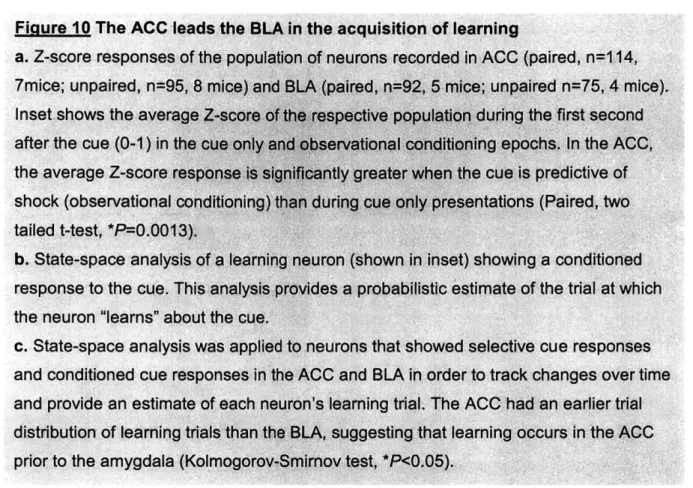
Observational fear learning is a powerful survival tool, allowing an individual to learn about environmental stimuli that predict specific threats without direct experience. This ability has been conserved from rodents to humans, and has been linked to the anterior cingulate cortex (ACC) and the basolateral amygdala (BLA). Here we record from the ACC as well as the BLA in an observer mouse during observational fear learning and show that both regions contain neurons that respond to the conditioned stimulus. In addition, we show that in an observer, both regions contain neurons that encode the learned predictive meaning of a cue during observational learning. Lastly, we provide evidence that on a population level, neurons in the ACC learn about the cue prior to those in the amygdala. Together this work demonstrates that the ACC and BLA contain neural correlates of observational learning at the neural level and that the ACC leads the BLA in acquiring the learned meaning of the cue.
Background
It is well-known that animals use direct sensory experiences to learn about aversive stimuli and their predictors (Pavlov 1927; Davis 1992; Fendt and Fanselow 1999; LeDoux 2000; Maren and Quirk 2004; Pape and Pare 2010; Johansen et al. 2011; Janak and Tye 2015). However, learning about aversive stimuli through direct experience often puts an animal in life-threatening situations (Griffin 2004; Nielsen et al. 2012). Thus, the ability to use the experiences of others to extract information about events that are predictive of aversive outcomes is critical to evolutionary fitness. This kind of observational learning is a fundamental social behavior that is highly conserved
across various species (Mineka et al. 1984; Heyes and Dawson 1990; Kavaliers, Choleris, and Colwell 2001; Olsson, Nearing, and Phelps 2007; Barber and Kimbrough 2015). For example, it has been shown that rodents display defensive behaviors when in the presence of conspecifics undergoing aversive or stressful experiences (Church 1959; Rice and Gainer 1962; Greene 1969; Chen, Panksepp, and Lahvis 2009b; Kim et al. 2010b; Atsak et al. 2011; Pereira et al. 2012). In addition, mice observing demonstrators undergo negative experiences showed increases in depression-like and anxiety-like behaviors (Warren et al. 2013) as well as fear learning (Guzman et al. 2009; Chen, Panksepp, and Lahvis 2009b; Bruchey, Jones, and Monfils 2010; Jeon et al. 2010; Kim et al. 2012; Yusufishaq and Rosenkranz 2013). However, despite the fundamental importance of observational learning to the survival of humans and other animal species, little is known about its neural basis.
In both rodents and humans, the ACC and amygdala have been implicated in observational fear learning and social cognition (Jeon et al. 2010; Olsson, Nearing, and Phelps 2007; Kim et al. 2012; Adolphs et al. 1994; Kim et al. 2014). In humans, both brain regions were recruited when subjects acquired fear responses to a conditioned stimulus through observation (Olsson, Nearing, and Phelps 2007). Likewise, in mice engaged in observational fear learning there was increased theta frequency synchronization between the ACC and the lateral amygdala (Jeon et al. 2010). Additionally, pharmacological lesions of either region inhibited learning (Jeon et al. 2010; Kim et al. 2012; Kim et al. 2014). The notion that these two regions may work together during observational learning is bolstered by studies showing that the ACC and
the basolateral complex of the amygdala (BLA), which includes the lateral amygdala (Janak and Tye 2015) have reciprocal connections with each other (Cassell and Wright 1986; Gabbott et al. 2005; Bissiere et al. 2008) and optogenetic stimulation of ACC terminals in the BLA invokes a persistent strengthening of synapses in putative pyramidal neurons (Morozov, Sukato, and Ito 2011). Despite this evidence, how these regions encode and transmit information necessary for observational learning remains unknown. Furthermore, the function, directionality, and necessity of the ACC-BLA circuit in observational learning have heretofore not been determined.
Results
In order to address the role of the ACC and BLA in observational fear learning, we designed a behavioral paradigm in which mice observe a demonstrator mouse undergo cued fear conditioning through a transparent divider with holes allowing for the transmission of auditory, visual, and olfactory information from one mouse to the other (Fig. 1a). In this study, observation refers to the multimodal perception of the behavior of the demonstrator. Mice termed "Experienced observers" (EO) first briefly explore the shock floor side of the chamber, receive a single shock, and are immediately placed on the plastic "safe" floor side of the chamber where they will no longer be shocked. A "demonstrator" familiar with the observer is then placed on the shock floor side of the chamber and 30 cue-shock pairings are delivered in which a cue is predictive of shock delivery to the demonstrator. 24 hrs later, observers are tested for cue learning by being placed back on the shock floor without a demonstrator and cues are played in the absence of shock (Fig. 1 b). EO mice demonstrated increased freezing to the cue during
test day as well as during observational conditioning (Fig. 1c, Fig. 2a) To verify that this learning was driven by the fear responses of the demonstrator we included experienced solo (ES) mice that received the initial shock experience but then observed cues and shocks delivered to an empty chamber (Fig. 1b). ES mice did not demonstrate conditioned responses to the cue during training or test day (Fig. 1c, Fig. 2a). To confirm that conditioned responses to the cue observed in the EO group were due to the fact that the cue predicted delivery of shock, we also had unpaired observers (UO).
r demonstrator mouse -. observer i mouse I Day 1: Training shock experience 1 trial observational conditioning 30 trials Es 20s Day 2: Test 5 trials 20s
2s
2s
1
d 24 hshock floor safe floor
b
Shock
experience
Paired cue and shock
Demonstrator present