HAL Id: inserm-02940444
https://www.hal.inserm.fr/inserm-02940444
Submitted on 16 Sep 2020
HAL is a multi-disciplinary open access
archive for the deposit and dissemination of sci-entific research documents, whether they are pub-lished or not. The documents may come from teaching and research institutions in France or abroad, or from public or private research centers.
L’archive ouverte pluridisciplinaire HAL, est destinée au dépôt et à la diffusion de documents scientifiques de niveau recherche, publiés ou non, émanant des établissements d’enseignement et de recherche français ou étrangers, des laboratoires publics ou privés.
European Association for Neuro-Oncology (EANO)
guideline on the diagnosis and treatment of adult
astrocytic and oligodendroglial gliomas
Michael Weller, Martin van den Bent, Jörg Tonn, Roger Stupp, Matthias
Preusser, Elizabeth Cohen-Jonathan-Moyal, Roger Henriksson, Emilie Le
Rhun, Carmen Balana, Olivier Chinot, et al.
To cite this version:
Michael Weller, Martin van den Bent, Jörg Tonn, Roger Stupp, Matthias Preusser, et al.. European Association for Neuro-Oncology (EANO) guideline on the diagnosis and treatment of adult astrocytic and oligodendroglial gliomas. Lancet Oncology, Elsevier, 2017, 18 (6), pp.e315-e329. �10.1016/S1470-2045(17)30194-8�. �inserm-02940444�
Zurich Open Repository and Archive University of Zurich Main Library Strickhofstrasse 39 CH-8057 Zurich www.zora.uzh.ch Year: 2017
European Association for Neuro-Oncology (EANO) guideline on the diagnosis and treatment of adult astrocytic and oligodendroglial gliomas
Weller, Michael ; van den Bent, Martin ; Tonn, Jörg C ; Stupp, Roger ; Preusser, Matthias ; Cohen-Jonathan-Moyal, Elizabeth ; Henriksson, Roger ; Le Rhun, Emilie ; Balana, Carmen ; Chinot, Olivier ; Bendszus, Martin ; Reijneveld, Jaap C ; Dhermain, Frederick ; French, Pim ; Marosi, Christine
; Watts, Colin ; Oberg, Ingela ; Pilkington, Geoffrey ; Baumert, Brigitta G ; Taphoorn, Martin J B ; Hegi, Monika ; Westphal, Manfred ; Reifenberger, Guido ; Soffietti, Riccardo ; Wick, Wolfgang ;
European Association for Neuro-Oncology (EANO)
DOI: https://doi.org/10.1016/S1470-2045(17)30194-8
Posted at the Zurich Open Repository and Archive, University of Zurich ZORA URL: https://doi.org/10.5167/uzh-141088
Journal Article Accepted Version
The following work is licensed under a Creative Commons: Attribution-NonCommercial-NoDerivatives 4.0 International (CC BY-NC-ND 4.0) License.
Originally published at:
Weller, Michael; van den Bent, Martin; Tonn, Jörg C; Stupp, Roger; Preusser, Matthias; Cohen-Jonathan-Moyal, Elizabeth; Henriksson, Roger; Le Rhun, Emilie; Balana, Carmen; Chinot, Olivier; Bendszus, Martin; Reijneveld, Jaap C; Dhermain, Frederick; French, Pim; Marosi, Christine; Watts, Colin; Oberg, Ingela; Pilkington, Geoffrey; Baumert, Brigitta G; Taphoorn, Martin J B; Hegi, Monika; Westphal, Manfred; Reifenberger, Guido; Soffietti, Riccardo; Wick, Wolfgang; European Association for Neuro-Oncology (EANO) (2017). European Association for Neuro-Neuro-Oncology (EANO) guideline on the diagnosis and treatment of adult astrocytic and oligodendroglial gliomas. Lancet Oncology, 18(6):e315-e329. DOI: https://doi.org/10.1016/S1470-2045(17)30194-8
1
EANO guideline on the diagnosis and treatment of adult astrocytic and oligodendroglial gliomas
Prof Michael Weller MD1, Prof Martin van den Bent MD2, Prof Jörg C. Tonn MD3, Prof
Roger Stupp MD4, Matthias Preusser MD5, Prof Elizabeth Cohen-Jonathan-Moyal
MD6, Prof Roger Henriksson MD7, Emilie Le Rhun MD8, Carmen Balana MD9, Prof
Olivier Chinot MD10, Prof Martin Bendszus MD11, Jacob C. Reijneveld MD12,
Frederick Dhermain MD13, Pim French PhD14, Christine Marosi MD5, Colin Watts
MD15, Ingela Oberg16, Geoffrey Pilkington PhD17, Brigitta G. Baumert MD18, Prof
Martin J.B. Taphoorn MD19, Monika Hegi PhD20, Prof Manfred Westphal MD21, Prof
Guido Reifenberger MD22, Prof Riccardo Soffietti MD23, Prof Wolfgang Wick MD24, for
the European Association for Neuro-Oncology (EANO) Task Force on Gliomas
1Department of Neurology & Brain Tumor Center, University Hospital and University
of Zurich, Zurich, Switzerland
2Neurooncology Unit, ErasmusMC Cancer Institute, Rotterdam, the Netherlands
3Department of Neurosurgery, University of Munich LMU, Munich, Germany
4Department of Oncology & Brain Tumor Center, University Hospital and University of
Zurich, Zurich, Switzerland
5Department of Medicine I and Comprehensive Cancer Center Vienna, Medical
University of Vienna, Vienna, Austria
6Département de Radiotherapie, Institut Claudius Regaud, IUCT-Oncopole,
Toulouse, France
7Regional Cancer Center Stockholm Gotland and Department of Radiation Sciences
& Oncology, Umeå University Hospital, Sweden
2
9Catalan Institute of Oncology (ICO), Hospital Germans Trias i Pujol, Carretera
Canyet sn, 08916 Badalona/Barcelona, Spain
10Aix-Marseille Université, APHM, CHU Timone, Department of Neuro-Oncology,
Marseilles, France
11Department of Neuroradiology, University Hospital Heidelberg, Germany
12Department of Neurology and Brain Tumor Center Amsterdam, VU University
Medical Center, Amsterdam, The Netherlands
13 Department of Radiotherapy, Gustave Roussy University Hospital, Villejuif, France
14 Department of Neurology, Erasmus MC, Rotterdam, The Netherlands
15 Department of Clinical Neurosciences, Division of Neurosurgery, University of
Cambridge, UK
16Division D – Neurosurgery, Addenbrooke’s Hospital CUHFT, Cambridge
Biomedical Campus, Hills Road, CB2 0QQ, United Kingdom
17Brain Tumor Research Centre, University of Portsmouth, Portsmouth, United
Kingdom
18Department of Radiation Oncology, MediClin Robert Janker Clinic & Clinical
Cooperation Unit Neurooncology, University of Bonn Medical Center, Bonn, Germany
19 Departments of Neurology, Leiden University Medical Center, and Medical Center
Haaglanden, The Hague, The Netherlands
20Department of Clinical Neurosciences, University Hospital Lausanne, Lausanne,
Switzerland
21Department of Neurosurgery, University Hospital Hamburg, Hamburg, Germany
22Department of Neuropathology, Heinrich Heine University Düsseldorf, and German
Cancer Consortium (DKTK), partner site Essen/Düsseldorf, Düsseldorf, Germany
23Department of Neuro-Oncology, University Hospital, Turin, Italy
3
Heidelberg, Germany and German Consortium of Translational Cancer Research
(DKTK), Clinical Cooperation Unit Neurooncology, German Cancer Research Center,
Heidelberg, Germany
Correspondence
Prof. Dr. Michael Weller, Department of Neurology & Brain Tumor Center, University
Hospital Zurich, Frauenklinikstrasse 26, CH-8091 Zurich, Switzerland, Tel. +41 44
255 5500, E-Mail: michael.weller@usz.ch
Funding
The preparation of this guideline was not funded. The members of the task force did
4
Summary
This guideline provides recommendations for the clinical care of patients with
astrocytic and oligodendroglial gliomas of adulthood. It is based on the 2016 WHO
classification of tumors of the nervous system and on scientific developments since
the 2014 version of the guideline. The recommendations focus on pathological and
radiological diagnostics as well as the major treatment modalities of surgery,
radiotherapy and pharmacotherapy. The results from contemporary
practice-changing clinical trials have been integrated. The guideline aims to provide guidance
for diagnostic and management decisions while limiting unnecessary treatment and
cost. It is a source of knowledge for professionals involved in the management of
glioma patients, for patients and caregivers, and for health care providers in Europe.
Implementing this guideline requires multidisciplinary and multiprofessional structures
of care and defined processes of diagnosis and treatment.
Key words
Astrocytoma, oligodendroglioma, glioblastoma, surgery, radiotherapy, temozolomide
Search strategy and selection criteria
This guideline was prepared by a task force nominated by the Executive Board of the
European Association for Neuro-Oncology (EANO) in cooperation with the Brain
Tumor Group of the European Organization for Research and Treatment of Cancer
(EORTC) in 2016. The task force represents the disciplines involved in the diagnosis
and care of glioma patients and reflects the multinational character of EANO.
References were retrieved onPubMed with the search terms “glioma”, “anaplastic”,
5
and “chemotherapy” from January 2001 to July 2016. Publications were identified
through searches of the authors` own files, too. Only papers in English were
reviewed. Data available only in Abstract form were only exceptionally included. The
definitive reference list was generated based on relevance to the broad scope of this
guideline.
Introduction
This guideline follows the revision of the fourth edition of the World Health
Organization (WHO) Classification of Tumors of the Central Nervous System1 and
builds on previous guidelines.2,3 It addresses astrocytic and oligodendroglial gliomas
of WHO grades II-IV of adulthood and their variants and covers prevention, early
diagnosis and screening, therapy, and follow-up. It does not address differential
diagnosis, adverse effects of treatment, or supportive or palliative care.
Diagnostics
Early diagnosis and screening
The annual incidence of gliomas is in the range of 6/100,000. Serum markers for
early detection have not been identified. Instead, brain magnetic resonance imaging
(MRI) has the highest sensitivity to detect small tumors. Gliomas can evolve rapidly
over several weeks or months, emphasizing the challenges for population-based
prevention or early intervention. Screening is therefore limited to persons at genetic
risk, for example, patients with neurofibromatosis type I, or Turcot and Li Fraumeni
syndromes. No repeat scanning is indicated in such individuals unless clinically
justified. A particular challenge is counseling and screening of relatives of patients
6
strategies are not available.
History and clinical examination
The evolution of neurological symptoms and signs allows to estimate the growth
dynamics of gliomas and may reveal familial risk or exogenous risk factors including
exposure to irradiation or other conditions associated with brain tumors. Relatives
may be required to obtain a reliable history. Characteristic modes of presentation are
new onset epilepsy, focal deficits including neurocognitive impairment, and indicators
of intracranial mass effect. The physical examination focuses on the detection of
systemic cancer and contraindications for neurosurgical procedures. Neurocognitive
assessment beyond documenting the Karnofsky performance score (KPS) or the
WHO performance status and performing a Mini Mental State Examination (MMSE)
or a Montreal Cognitive Assessment has become increasingly common.5,6
Ancillary studies
MRI before and after application of gadolinium is the standard method to detect a
glioma.7 In addition, cranial computed tomography (CT) demonstrates calcifications,
intra-arterial angiography may aid the surgical strategy, and amino acid positron
emission tomography (PET) helps define metabolic hotspots for biopsy.8
Standardized MRI sequences are also recommended to assess the efficacy of
therapeutic interventions.9 Cerebrospinal fluid studies play no major role in the
diagnostic work-up of gliomas and lumbar punctures carry the risk of neurological
deterioration in patients with large space-occupying tumors. Electroencephalography
helps for monitoring tumor-associated epilepsy and in determining causes of altered
7
Preoperative management
Management should follow written local standard operating procedures and
multidisciplinary discussion preferentially including dedicated neuroradiologists and
neuropathologists as well as neurosurgeons, radiation oncologists and
neurooncologists in a brain tumor board. Prior to surgery, unless there are
contraindications or the suspicion of primary cerebral lymphoma or inflammatory
lesions, corticosteroids may be administered to decrease tumor-associated edema.
Additional pharmacological measures such as osmotic agents are rarely necessary.
Glioma patients who have suffered epileptic seizures should receive anticonvulsant
drugs preoperatively. Primary prophylaxis is not indicated in patients without
seizures.10
Biopsy or resection
Treatment decisions in glioma patients are based on a tissue diagnosis and the
assessment of selected molecular markers. Surgery is thus commonly performed
with diagnostic and therapeutic objectives. The surgical management of glioma
patients should take place in high-volume specialist centers. A decision for palliative
care without histological diagnosis should be avoided unless the risk of the biopsy
procedure is considered too high or if the prognosis is likely to be very unfavourable,
e.g., in old patients with large tumors and rapid clinical decline. Stereotactic serial
biopsies along the trajectory under local anesthesia are associated with low morbidity
and a firm diagnosis aids counselling patients and relatives also when tumor-specific
therapy is not recommended. Serial sampling allows to avoid undergrading, the
procedure requires close cooperation between neuropathologist, neuroradiologist
8
Histological classification and molecular diagnostics
Intraoperative assessment of cytological specimens or frozen sections before the
surgical procedure is terminated assure that sufficient tissue is obtained to establish
a diagnosis. Tumor tissue is formalin-fixed and embedded in paraffin for conventional
histological staining, including routine hematoxylin-eosin staining and additional
immunohistochemical and molecular analyses. If possible, a part of the tissue should
be cryopreserved for future scientific molecular marker studies. The diagnostic
process follows the WHO classification and consists of histological tumor typing as
well as tumor grading using the four-tiered WHO grading scheme from WHO grade I
to IV, designed to provide clinicians with information on the tumor's biological
behavior and consequently the patient's prognosis and outcome (Fig. 1). The 2016
WHO classification recognizes the major diagnostic role of isocitrate dehydrogenase
(IDH) 1 codon 132 or IDH2 codon 172 missense mutations ("IDH mutation") and
defines diffuse and anaplastic astrocytic and oligodendroglial gliomas essentially as
IDH-mutant tumors.1 Oligodendroglial tumors additionally carry 1p/19q co-deletions.
IDH-wildtype diffuse and anaplastic astrocytomas are considered provisional entities.
“Not otherwise specified” (NOS) categories have been introduced for those gliomas
that cannot be tested for the diagnostically relevant markers or for which testing
remains inconclusive. Management recommendations for these NOS categories are
included in Table 1, but evidence is low. Oligoastrocytomas and gliomatosis cerebri
both lack distinctive genetic and epigenetic profiles11,12 and are thus no longer
considered as distinct glioma entities.1 Diffuse midline glioma, H3-K27M-mutant, has
been introduced as a novel entity characterized by midline tumor location and
presence of lysine to methionine mutation at codon 27 of histones 3.3 or 3.1.1
9
IDH mutation, 1p/19q co-deletion, H3-K27M mutation and O6-methylguanine DNA
methyltransferase (MGMT) promoter methylation. IDH mutation, 1p/19q co-deletion
and H3-K27M mutation are assigned a role in the revised WHO classification (Table
1) whereas MGMT promoter methylation status guides treatment decisions regarding
the use of chemotherapy.13 Immunohistochemical detection of loss of nuclear ATRX
expression is helpful to substantiate the diagnoses of IDH-mutant diffuse and
anaplastic astrocytomas, and if retained should prompt testing for 1p/19q co-deletion
in IDH-mutant gliomas, however, ATRX immunohistochemistry shall not substitute for
1p/19q codeletion testing. High-throughput assays will probably soon be introduced
instead of single marker assessments.
Therapy - General recommendations
Prognostic factors
Younger age and better performance status are important positive,
therapy-independent prognostic factors across glioma entities. Extent of resection is an
important therapy-dependent prognostic factor. Prognostically favorable molecular
markers such as IDH mutation and 1p/19q co-deletion are now at the core of the
WHO classification and define more homogeneous diagnostic and prognostic
entities.1,13
Surgical therapy
Beyond establishing a histological diagnosis, the goal of surgery is to remove as
much of the tumor as is safely possible with the goal of improving neurological
10
navigation systems housing functional MRI datasets, intraoperative MRI, ultrasound,
intraoperative functional monitoring and the fluorescent dye, 5-aminolevulinic acid
(ALA), to visualize tumor tissue14 help increase the extent of resection while keeping
the risk of new neurological deficits low. The use of evoked potentials,
electromyography or mapping in awake patients under local anesthesia to monitor
and preserve language and cognition should support resections in eloquent areas.
Preventing new permanent neurological deficits is more important than extent of
resection because gliomas are not cured by surgery: this is precluded by tumor cell
infiltration far beyond the lesion as delineated by neuroimaging and network-like
growth, hallmarks of diffuse gliomas.15 Postoperative deficits due to emerging
complications are a negative prognostic factor. Furthermore, quality of life is a high
priority to patients and carers. The result of surgery is assessed by early MRI – or CT
if MRI is not possible - without and with contrast and diffusion imaging within 24-72 h
of surgery.16
Radiotherapy (RT)
The goal of RT for patients with gliomas is to improve local control at a reasonable
risk benefit ratio. RT helps to preserve function and increases survival. Indications
for, timing, dosing and scheduling of RT are determined by diagnosis and prognostic
factors, including age, KPS and extent of resection.17 Focal RT is administered at
50-60 Gy in 1.8-2 Gy fractions, depending on prognosis defined by tumor type and
grade. Hypofractionated RT with higher fraction sizes and lower total dose e.g., to 15
x 2.67 Gy, is appropriate in older patients and those with poor prognostic factors, and
considered biologically equivalent to 30 x 2 Gy. The area of residual enhancement on
T1 imaging plus the surgical bed is defined as the gross tumor volume. A margin,
11
added to define the clinical target volume which is then modified in areas where
microscopic spread is unlikely, or to reduce the dose to critical structures. Finally
another margin, usually 0.3 to 0.5 cm, is added to allow for error setup and
movement during treatment, generating the planning target volume.18 PET is studied
for improving target delineation in clinical trials.8
The organs at higher risk of radiotherapy-associated toxicity including optic nerves,
optic chiasm, retinae, lenses, brainstem, pituitary, cochleas and hippocampus should
be delineated. Modern techniques of focused RT, e.g. stereotactic,
intensity-modulated or image-guided RT may improve the targeted delivery of RT to better
protect surrounding tissue. In children especially, but also in adults with deeply
localized tumors, interstitial brachytherapy and proton/heavy ions radiotherapy may
be alternatives. Randomized data comparing such novel approaches with standard
techniques are not available.
Pharmacotherapy
Cytotoxic chemotherapy is standard of care for most glioma patients (Table 2). It
requires regular hematology, hepatic and renal laboratory and exclusion of major
lung or heart disease and infection. Blood counts need to be monitored during
therapy. Temozolomide (TMZ), an oral DNA alkylating agent with good blood brain
barrier penetration, is widely used in glioma treatment and has a favourable safety
profile, with myelosuppression, notably thrombocytopenia as its main and
dose-limiting toxicity. Hepatic function should also be assessed regularly. In contrast to
TMZ, nitrosoureas such as lomustine (CCNU), carmustine (BCNU), nimustine
(ACNU) or fotemustine cause prolonged leukopenia and thrombocytopenia. This may
necessitate delays of further treatment at reduced dose or even discontinuation and
12
seen with carmustine. Nitrosoureas have become a second choice after TMZ for
glioma treatment in most European countries, although data from larger comparative
trials are missing and retrospective or subgroup analyses suggest a higher efficacy of
procarbazine, lomustine (CCNU) and vincristine (PCV) over TMZ in good prognosis
patients with anaplastic glioma.19,20 Locally delivered carmustine wafers (Gliadel®)
implanted into the surgical cavity have provided a moderate survival advantage to
patients with newly diagnosed grade WHO III or IV gliomas, or recurrent
glioblastoma,21 but are now a rarely considered option, and mostly in patients without
systemic treatment options. Their application requires careful patient selection and a
gross total resection. Among various candidate anti-angiogenic agents explored in
clinical trials in glioma patients, only bevacizumab, an antibody to vascular
endothelial growth factor, is approved for recurrent glioblastoma in the USA, Canada,
Switzerland and several other countries outside the European Union. Glioma patients
undergoing systemic therapy should carry a documentation of treatment including
laboratory results and information on complications and contraindications. Clinical
centers managing glioma patients should generate standard operating procedures
and instructions for handling side effects and complications from treatment.
Other therapeutic approaches
Other approaches to glioma therapy including various targeted and immunological
therapies, notably immune checkpoint inhibitors, are of unknown activity and should
be explored within clinical trials.
Monitoring and follow-up
In addition to clinical examination, MRI is the standard diagnostic measure for the
13
common practice initially for most patients, but longer intervals are appropriate in
case of durable disease control and less aggressive tumors. Vice versa, in case of
suspected disease progression short term control MRI may be reasonable to confirm
progression. Pseudoprogression and pseudoresponse are most likely to occur during
the first three months of treatment. Particular attention is needed when interpreting
scans during this period. The Response Assessment in Neuro-Oncology (RANO)
working group has recommended that assessment of non-contrast-enhancing tumor
components should be an additional key component of response criteria.9
Specific recommendations
Figures 2 and 3 and Table 1 provide an overview of therapeutic approaches. Table 3
provides the key recommendations.
Diffuse astrocytoma, IDH-mutant – WHO grade II
These are the most common WHO grade II astrocytomas. Gemistocytic
astrocytomas are a subtype of IDH-mutant diffuse astrocytoma.1 Maximum surgical
resection as feasible is increasingly considered the best initial therapeutic measure.
Watch-and-wait strategies without establishment of a diagnosis are less commonly
pursued. Asymptomatic younger patients with seizures only can be managed by
observation alone after gross total resection. Involved field RT (50 Gy) should be
considered for patients with incomplete resection and patients older than 40 years. It
prolongs progression-free survival (PFS), but not overall survival (OS).22
Chemotherapy alone as initial treatment should be considered investigational, but
14
than with RT.23 The RTOG 9802 trial reported a major prolongation of survival by
adding PCV polychemotherapy to RT (54 Gy) compared with RT alone from 7.8 to
13.3 years in patients with high-risk WHO grade II gliomas who were 18 to 39 years
of age and had undergone a subtotal resection or biopsy, or who were 40 years of
age or older. RT followed by PCV constitutes a new standard of care, given the lack
of other up-coming clinical trial results likely to challenge these data. Benefit was
reported across histological subgroups and, although data are limited, there was no
overt link between benefit from PCV and a particular molecular marker profile,
potentially due to limited power.24 Treatment at progression depends on first-line
therapy and may involve second surgery, radiotherapy in previously un-irradiated
patients or alkylating agent chemotherapy. TMZ is often preferred over PCV
polychemotherapy because of its favorable safety profile and ease of administration.
IDH-wildtype diffuse astrocytomas can take a rather aggressive course, resembling
glioblastoma, in particular in the elderly, but there are also less aggressive variants.25
Anaplastic astrocytoma, IDH-mutant – WHO grade III
These are the most common WHO grade III astrocytomas. Standard of care includes
maximal surgical removal or biopsy followed by RT at 60 Gy in 1.8–2 Gy fractions
(Table 1), largely based on trials where these tumors were pooled with
glioblastomas. The NOA-04 trial showed that PCV or TMZ alone were as active as
RT alone for PFS and OS.20,26 The EORTC 26053 trial (CATNON) explored whether
the addition to RT of concomitant or maintenance TMZ or both improved outcome
over RT alone in patients with newly diagnosed 1p/19q-non-codeleted anaplastic
gliomas in a 2 by 2 design. A first interim analysis showed that 12 cycles of
maintenance TMZ prolonged OS which should now be considered standard of care
15
Molecular marker studies in the CATNON trial are pending. A retrospective study of
pooled datasets indicated that specifically patients with IDH-wildtype tumors with
MGMT promoter methylation benefit from alkylating agent chemotherapy.28
First-line therapy informs on the choices of treatment in the recurrent setting. An
indication for second surgery should be explored. For patients relapsing after
radiotherapy re-irradiation is an option with a minimum in the range of 12 months
interval since the end of the first RT course. However, size and patterns of
recurrence limit the options of re-RT, and the overall efficacy remains uncertain;
randomized data are lacking. Alkylating agent chemotherapy should be considered
for chemo-naive patients who progress after RT, TMZ and nitrosoureas probably
being equally effective.29,30 Bevacizumab is used after failure of RT and
chemotherapy, with PFS rates at 6 months of 20-60%.31,32 Controlled data are
lacking, including for combining bevacizumab with chemotherapy.
Glioblastoma, IDH-wildtype - WHO grade IV
The majority of histological glioblastomas are IDH-wildtype, including the
morphological variants of giant cell glioblastoma, gliosarcoma and epitheloid
glioblastoma. There are to date no specific treatment recommendations for
glioblastoma variants. About 50% of the rare epitheloid glioblastomas carry a
druggable BRAF-V600E mutation, but the promising efficacy of BRAF inhibitors
remains to be evaluated systematically. The following applies to IDH-wildtype
glioblastoma; IDH-mutant glioblastomas are increasingly treated like IDH-mutant
anaplastic astrocytoma.
Surgery for glioblastoma should be gross total resection whenever feasible. A small
randomized trial in patients WHO grade III and IV tumors aged > 65 reported
16
sample size and KPS imbalances between groups. While some studies reported
gradually improved outcome with increasing extent of resection, only gross total
resection may be associated with improved outcome.34,35
RT has been standard of care for glioblastoma for decades, roughly doubling
survival.17,36 Standard dose is 60 Gy in 1.8-2 Gy fractions; 50 Gy in 1.8 Gy fractions
improved survival relative to best supportive care in patients 70 years or older with
good KPS.37 Patients with unfavorable prognostic factors defined by age or KPS are
treated with hypofractionated RT, e.g., 40 Gy in 15 fractions.38 In the elderly, this is
the standard of care for patients with tumors without MGMT promoter
methylation.39,40 Further hypofractionation to 5 x 5 Gy may be feasible without
compromising survival,41 but is unlikely to be well tolerated in terms of neurocognitive
side effects which will assume more relevance once other treatment options allow
long-term survival in elderly glioblastoma patients, too. Neither accelerated hyper- or
hypofractionated regimens nor brachytherapy, radiosurgery or a stereotactic RT
boost are superior to standard regimens for survival.
Concomitant and maintenance TMZ chemotherapy plus RT (TMZ/RT→TMZ) is the standard of care for newly diagnosed adult patients in good general and neurological
condition and aged up to 70 years.42-44 The benefit from TMZ is most prominent in
patients with MGMT promoter-methylated glioblastoma.45 Recent trials in MGMT
promoter unmethylated patients showing no detriment from omitting TMZ have raised
doubts whether TMZ should be used in every patient despite lack of MGMT promoter
methylation.46-48 There is no benefit from increasing the dose of TMZ in the newly
diagnosed setting49 and probably also not from extending the duration of
chemotherapy beyond 6 cycles.50,51
The NOA-08 and Nordic trials39,40 made MGMT promoter methylation testing
17
lacking MGMT promoter methylation should be treated with hypofractionated RT
alone. This is also the treatment of choice for elderly patients when the MGMT status
is unknown. Elderly patients with tumors with MGMT promoter methylation should
receive TMZ alone (5/28 until progression or for 12 months) or TMZ/RT→TMZ. In the NCIC CE.6/EORTC 26062 trial enrolling patients ≥ 65 years with newly diagnosed glioblastoma, the addition of concomitant and maintenance TMZ to 40 Gy/15
fractions radiotherapy significantly improved survival. MGMT promoter methylation
was not prognostic, but highly predictive for benefit from TMZ. Interpretation of the
benefit from TMZ in patients with MGMT-unmethylated tumors remains controversial.
There was overall no indication that the benefit from TMZ was reduced with
increasing age.52 In the absence of comparative data between TMZ alone and
chemoradiation, elderly patients with MGMT promoter methylation considered eligible for combined modality treatment should be offered TMZ/RT→TMZ. Supportive and palliative care are appropriate for patients with large or multifocal lesions with low
KPS, notably if they are unable to consent for further therapy after biopsy.
Local BCNU wafer chemotherapy added to RT conferred a survival benefit of 13.9
over 11.6 months with RT alone for the intention-to-treat population of high-grade
gliomas, but the difference was no longer significant when only glioblastoma patients
were considered21,44,53 unless extent of resection was included in the analysis where
the effect was again significant in glioblastoma patients with larger than 90%
resection.54
Two randomized trials conducted in the adult glioblastoma patient population have
demonstrated a gain in PFS of 3-4 months, but not OS, when bevacizumab was
added to TMZ/RT→TMZ.55,56 The clinical significance of the PFS gain has been
disputed because the reliability of assessing progression by neuroimaging has been
18
decline in bevacizumab-treated patients. Bevacizumab was thus not approved for
newly diagnosed glioblastoma. It may, however, be useful in individual patients with
large tumors highly symptomatic and resistant to steroids who may otherwise not
tolerate RT.
Tumor-treating fields (TTFields) represent a novel treatment modality designed to
deliver alternating electrical fields to the brain. In an open-label randomized phase III
trial improved PFS and OS were demonstrated when TTFields were added to
standard maintenance TMZ in newly diagnosed glioblastoma patients.57 The trial was
terminated early when the first cohort of 315 randomized patients was analysed and
a survival benefit was reported (hazard ratio 0.74 (95% CI, 0.56-0.98); log-rank
p=0.03). Questions regarding mode of action, interpretation of data and impact on
quality of life have been raised,58 and the place and cost-effectiveness of TTFields in
the standard of care for newly diagnosed glioblastoma remain to be defined.59
Standards of care for patients with recurrent glioblastoma are not well defined.
Clinical decision-making is influenced by prior treatment, age, KPS, and patterns of
progression. Second surgery is appropriate for 20-30% of patients and is considered
for symptomatic, but circumscribed lesions and when the interval since the preceding
surgery exceeds 6 months. Surgery may also be considered earlier in symptomatic
patients after suboptimal initial surgery. An impact on survival may be limited to
patients who are candidates for gross total resection of enhancing tumor.60 The
efficacy of re-irradiation and the value of amino acid PET for target delineation
remain debated. Fractionation depends on tumor size. Doses of conventional or near
conventional fractionation using 3-3.5 Gy /fraction to a total dose of 30-35 Gy have
been tested and several studies using a dose per fraction of 5-6 Gy using
stereotactic hypofractionated radiotherapy to a total dose of 30-36 Gy or even
19
toxicity.61 Yet, no relevant monotherapy efficacy was demonstrated in a larger
randomized trial at 18 x 2 Gy.62
The main systemic treatment options at progression after TMZ/RT→TMZ in Europe are nitrosoureas, TMZ rechallenge, and bevacizumab. CCNU is increasingly
considered standard of care, based on its activity as the control arm of several
randomized trials,46,63 with PFS rates at 6 months of 20%. Similar results have been
reported with alternative dosing regimens of TMZ,64 but activity is probably limited to
patients with tumors with MGMT promoter methylation.65,66 The BR12 trial showed no
benefit from dose-intensified TMZ over standard-dose TMZ in TMZ-naïve malignant
glioma patients,29 but does not inform on the value of TMZ re-challenge for patients
pre-treated with TMZ. There is thus, however, no reason to administer
dose-intensified TMZ to TMZ-naïve patients. Whether dose-dose-intensified regimens are
superior to standard-dosed TMZ in recurrent glioblastoma after a TMZ-free interval,
remains undetermined.
Bevacizumab is approved for recurrent glioblastoma in various countries throughout
the world, but not in the European Union, based on response rates in the range of
30% and PFS and OS times comparing favourably with historical controls in two
uncontrolled phase II trials.67,68 Its value in clinical practice is widely accepted
because of transient symptom control and the option for steroid sparing in a subset of
patients. An effect on OS has not been demonstrated. The superiority of combining
bevacizumab with CCNU over either agent alone for OS at 9 months65 was not
confirmed in the EORTC trial 26101.69 No other active combination partner for
bevacizumab has been identified. TTF were not superior to best physician`s choice in
a randomized phase III trial.70
20
This new tumor entity has been assigned the WHO grade IV. It includes the majority
of brainstem, thalamic and spinal gliomas in children and adults. Surgical options are
limited and treatment beyond RT is not established. The prognosis is poor.
Traditionally, treatment has followed the standards for histologically similar gliomas in
other locations.
Oligodendroglioma, IDH-mutant and 1p/19q-codeleted - WHO grade II
The new WHO classification defines this tumor as IDH-mutant and
1p/19q-codeleted.1 In rare instances with lacking or inclusive data on IDH and 1p/19q
co-deletion, tumors are classified as oligodendroglioma, NOS. The diagnosis of
oligoastrocytoma is discouraged in the new WHO classification. Only exceptional
cases that cannot be conclusively tested for IDH mutation and 1p/19q co-deletion
and show a mixed oligoastrocytic histology may still be classified as
oligoastrocytoma, NOS.1 Surgery is the primary treatment of IDH-mutant and
1p/19q-codeleted oligodendroglioma. The standard of care is RT followed by PCV if further
treatment beyond surgery is considered necessary.24
Anaplastic oligodendroglioma, IDH-mutant and 1p/19q-codeleted - WHO grade III
The new WHO classification defines this tumor as IDH-mutant and 1p/19q-codeleted,
NOS is only assigned if conclusive molecular information is lacking. The diagnosis of
anaplastic oligoastrocytoma is discouraged and no longer applicable when tumors
are successfully tested for IDH mutation and 1p/19q co-deletion. Extent of resection
is a prognostic factor.26,71 Two large randomized clinical trials – EORTC 26951 and
RTOG 9402 - showed that the addition of PCV chemotherapy, either prior to or after
RT, in the first-line treatment prolonged OS by several years in the subset of patients
21
analyses of small patient cohorts, both studies show similar results, validating each
other and defining the current standard of care. Important questions remain: whether
long-term survivors treated with RT plus PCV experience preserved cognitive
function and quality of life74 and whether the same improvement in OS could be
achieved with TMZ/RT→TMZ. Long-term results from the NOA-04 show that chemotherapy alone is not superior to RT alone in either IDH-mutant and
1p/19q-codeleted anaplastic oligodendroglioma or IDH-mutant anaplastic astrocytoma,
indicating that alkylating agent chemotherapy alone is unlikely to achieve the same
outcome as RT combined with PCV. Whether TMZ/RT →TMZ is similarly effective as RT followed by PCV is explored in the modified CODEL trial (NCT00887146).
Treatment at progression is influenced by type of and response to first-line treatment.
If neither RT nor alkylating agents are options because they failed or because of
intolerance, bevacizumab has been used,75,76 but is of unknown efficacy as
controlled studies are lacking. There is no evidence to combine bevacizumab with
cytotoxic agents in this setting.
Other astrocytic tumors
Pilocytic astrocytoma and its variant, pilomyxoid astrocytoma, are rare tumors in
adults and commonly cured by surgery alone. Radiotherapy is only indicated at
progression when surgical options no longer exist. Pilocytic astrocytoma of the optic
nerve may be associated with neurofibromatosis type I and cannot be resected
unless useful visual function has already been lost. These lesions often do not
require treatment.
Subependymal giant cell astrocytomas are WHO grade I lesions associated with
tuberous sclerosis and may respond to mTOR inhibition if treatment beyond surgery
22
Pleomorphic xanthoastrocytoma (WHO grade II) occurs predominantly in children
and young adults and has a high rate (approximately 70%) of BRAF-V600E mutation.
It should be resected and patients may be observed after gross total resection.
Anaplastic pleomorphic xanthoastrocytoma (WHO grade III) should probably be
managed with postoperative RT and not with a watch-and-wait strategy. In recurrent
anaplastic pleomorphic astrocytoma, BRAF inhibitors such as vemurafenib appear to
have limited activity.78
Coordination of care and outlook
Diagnosis and management plans for glioma patients should follow multidisciplinary
tumor board recommendations throughout the disease course. Boards are an forum
to discuss which measures can take place locally, which are better done at a
specialized center, which are appropriate for in-patient versus out-patient settings,
and which neurorehabilitation measures are useful. Local and national guidelines as
well as upcoming EANO guidelines provide further guidance. Guidelines reflect
knowledge and consensus at a given timepoint. Table 3 summarizes the key
recommendations of the EANO task force in 2016. The EANO website
(www.eano.eu) will inform of future updates on this guideline.
Declaration of interests
Dr. Weller reports grants from Acceleron, grants from Actelion, grants from Bayer,
grants from Merck & Co, grants from Novocure, grants from Piqur, grants from
Roche, personal fees from BMS, personal fees from Celldex, personal fees from
23
fees from Magforce, personal fees from Merck & Co, personal fees from Northwest
Biotherapeutics, personal fees from Novocure, personal fees from Pfizer, personal
fees from Roche, personal fees from Teva, personal fees from Tocagen, grants from
EMD Merck Serono, outside the submitted work.
Dr. van den Bent reports personal fees from Roche, personal fees from Celldex,
personal fees from Novartis, personal fees from BMS, personal fees from MSD,
grants and personal fees from Abbvie, outside the submitted work.
Dr. Tonn reports grants from BrainLab, outside the submitted work.
Dr. Stupp reports non-financial support from Novocure Ltd, other from Roche, other
from Merck KGaA, other from MSD/Merck & Co, other from Novartis, outside the
submitted work.
Dr. Preusser reports grants from Böhringer-Ingelheim, from GlaxoSmithKline, from
Merck Sharp & Dome, personal fees from Bristol-Myers Squibb, personal fees from
Novartis, personal fees from Gerson Lehrman Group, personal fees from CMC
Contrast, personal fees from GlaxoSmithKline, personal fees from Mundipharma,
personal fees from Roche, outside the submitted work.
Dr. Cohen-Jonathan Moyal has nothing to disclose.
Dr. Henriksson has nothing to disclose.
Dr. Le Rhun has nothing to disclose.
Dr. Balana has nothing to disclose.
Dr. Chinot reports grants, personal fees and non-financial support from Roche,
personal fees from Ipsen, personal fees from Astra-Zeneca, personal fees and
non-financial support from Servier, personal fees from Celldex, from null, during the
24
13725437 9 1405 licensed.
Dr. Bendszus reports grants and personal fees from Guerbet, grants and personal
fees from Novartis, personal fees from Roche, grants from Siemens, personal fees
from Teva, personal fees from Bayer, personal fees from Vascular Dynamics, grants
from DFG, from Hopp Foundation, grants and personal fees from Codman, grants
from Stryker, outside the submitted work.
Dr. Reijneveld reports non-financial support from Roche Nederland BV, outside the
submitted work.
Dr Dhermain has nothing to disclose.
Dr. French has nothing to disclose.
Dr. Marosi has nothing to disclose.
Dr. Watts has nothing to disclose.
Dr. Oberg has nothing to disclose.
Dr. Pilkington has nothing to disclose.
Dr. Baumert reports personal fees from Merck & Co (MSD), personal fees and
non-financial support from Noxxon Pharma AG, outside the submitted work.
Dr. Taphoorn reports personal fees from Roche, outside the submitted work.
Dr. Hegi reports non-financial support from MDxHeatlth, other from
Roche/Genentech, other from Novocure, other from MSD, other from BMS, outside
the submitted work.
Dr. Westphal reports personal fees from Bristol-Myers-Squibb, personal fees from
Novocure, personal fees from Roche, outside the submitted work.
25
Amgen and Celldex, outside the submitted work.
Dr. Soffietti has nothing to disclose.
Dr. Wick reports grants and personal fees from MSD, grants from Apogenix, grants
from Boehringer Ingelheim, grants and personal fees from Genentech/Roche, grants
from Pfizer, personal fees from BMS, personal fees from Celldex, outside the
submitted work.
Author contributions
M. Weller wrote the first draft of the manuscript. All other authors reviewed the draft,
26
References
1. Louis DN, Perry A, Reifenberger G, et al. The 2016 World Health Organization
Classification of Tumors of the Central Nervous System: a summary. Acta
Neuropathol 2016; 131(6): 803-20.
2. Weller M, van den Bent M, Hopkins K, et al. EANO guideline for the diagnosis
and treatment of anaplastic gliomas and glioblastoma. The Lancet Oncology 2014;
15(9): e395-403.
3. Soffietti R, Baumert BG, Bello L, et al. Guidelines on management of
low-grade gliomas: report of an EFNS-EANO Task Force. Eur J Neurol 2010; 17(9):
1124-33.
4. Rice T, Lachance DH, Molinaro AM, et al. Understanding inherited genetic risk
of adult glioma - a review. Neurooncol Pract 2016; 3(1): 10-6.
5. Douw L, Klein M, Fagel SS, et al. Cognitive and radiological effects of
radiotherapy in patients with low-grade glioma: long-term follow-up. Lancet Neurol
2009; 8(9): 810-8.
27
a randomized phase III study of bevacizumab, temozolomide, and radiotherapy in
newly diagnosed glioblastoma. Journal of clinical oncology : official journal of the
American Society of Clinical Oncology 2015; 33(19): 2166-75.
7. Ellingson BM, Bendszus M, Boxerman J, et al. Consensus recommendations
for a standardized Brain Tumor Imaging Protocol in clinical trials. Neuro Oncol 2015;
17(9): 1188-98.
8. Albert NL, Weller M, Suchorska B, et al. Response Assessment in
Neuro-Oncology working group and European Association for Neuro-Neuro-Oncology
recommendations for the clinical use of PET imaging in gliomas. Neuro Oncol 2016.
9. Wen PY, Macdonald DR, Reardon DA, et al. Updated response assessment
criteria for high-grade gliomas: response assessment in neuro-oncology working
group. Journal of clinical oncology : official journal of the American Society of Clinical
Oncology 2010; 28(11): 1963-72.
10. Weller M, Stupp R, Wick W. Epilepsy meets cancer: when, why, and what to
do about it? The Lancet Oncology 2012; 13(9): e375-82.
11. Sahm F, Reuss D, Koelsche C, et al. Farewell to oligoastrocytoma: in situ
28
Acta Neuropathol 2014; 128(4): 551-9.
12. Herrlinger U, Jones DT, Glas M, et al. Gliomatosis cerebri: no evidence for a
separate brain tumor entity. Acta Neuropathol 2016; 131(2): 309-19.
13. Weller M, Pfister SM, Wick W, Hegi ME, Reifenberger G, Stupp R. Molecular
neuro-oncology in clinical practice: a new horizon. The Lancet Oncology 2013; 14(9):
e370-9.
14. Stummer W, Pichlmeier U, Meinel T, et al. Fluorescence-guided surgery with
5-aminolevulinic acid for resection of malignant glioma: a randomised controlled
multicentre phase III trial. The Lancet Oncology 2006; 7(5): 392-401.
15. Osswald M, Jung E, Sahm F, et al. Brain tumour cells interconnect to a
functional and resistant network. Nature 2015; 528(7580): 93-8.
16. Vogelbaum MA, Jost S, Aghi MK, et al. Application of novel
response/progression measures for surgically delivered therapies for gliomas:
Response Assessment in Neuro-Oncology (RANO) Working Group. Neurosurgery
2012; 70(1): 234-43; discussion 43-4.
17. Laperriere N, Zuraw L, Cairncross G, Cancer Care Ontario Practice Guidelines
29
malignant glioma in adults: a systematic review. Radiother Oncol 2002; 64(3):
259-73.
18. Niyazi M, Brada M, Chalmers AJ, et al. ESTRO-ACROP guideline "target
delineation of glioblastomas". Radiother Oncol 2016; 118(1): 35-42.
19. Lassman AB, Iwamoto FM, Cloughesy TF, et al. International retrospective
study of over 1000 adults with anaplastic oligodendroglial tumors. Neuro Oncol 2011;
13(6): 649-59.
20. Wick W, Roth P, Hartmann C, et al. Long-term analysis of the NOA-04
randomized phase III trial of sequential radiochemotherapy of anaplastic glioma with
PCV or temozolomide. Neuro Oncol 2016.
21. Westphal M, Hilt DC, Bortey E, et al. A phase 3 trial of local chemotherapy
with biodegradable carmustine (BCNU) wafers (Gliadel wafers) in patients with
primary malignant glioma. Neuro Oncol 2003; 5(2): 79-88.
22. van den Bent MJ, Afra D, de Witte O, et al. Long-term efficacy of early versus
delayed radiotherapy for low-grade astrocytoma and oligodendroglioma in adults: the
EORTC 22845 randomised trial. Lancet 2005; 366(9490): 985-90.
30
versus radiotherapy in high-risk low-grade glioma. A randomized phase III Intergroup
study by EORTC/NCIC-CTG/TROG/MRC-CTU (EORTC 22033-26033). The Lancet
Oncology 2016; in press.
24. Buckner JC, Shaw EG, Pugh SL, et al. Radiation plus Procarbazine, CCNU,
and Vincristine in Low-Grade Glioma. N Engl J Med 2016; 374(14): 1344-55.
25. Reuss DE, Kratz A, Sahm F, et al. Adult IDH wild type astrocytomas
biologically and clinically resolve into other tumor entities. Acta Neuropathol 2015;
130(3): 407-17.
26. Wick W, Hartmann C, Engel C, et al. NOA-04 randomized phase III trial of
sequential radiochemotherapy of anaplastic glioma with procarbazine, lomustine, and
vincristine or temozolomide. Journal of clinical oncology : official journal of the
American Society of Clinical Oncology 2009; 27(35): 5874-80.
27. Van den Bent MJ, Erridge S, Vogelbaum MA, et al. Results of the interim
analysis of the EORTC randomized phase III CATNON trial on concurrent and
adjuvant temozolomide in anaplastic glioma without 1p/19q co-deletion, an intergroup
trial. Journal of clinical oncology : official journal of the American Society of Clinical
31
28. Wick W, Meisner C, Hentschel B, et al. Prognostic or predictive value of
MGMT promoter methylation in gliomas depends on IDH1 mutation. Neurology 2013;
81(17): 1515-22.
29. Brada M, Stenning S, Gabe R, et al. Temozolomide versus procarbazine,
lomustine, and vincristine in recurrent high-grade glioma. Journal of clinical
oncology : official journal of the American Society of Clinical Oncology 2010; 28(30):
4601-8.
30. Yung WK, Prados MD, Yaya-Tur R, et al. Multicenter phase II trial of
temozolomide in patients with anaplastic astrocytoma or anaplastic oligoastrocytoma
at first relapse. Temodal Brain Tumor Group. Journal of clinical oncology : official
journal of the American Society of Clinical Oncology 1999; 17(9): 2762-71.
31. Desjardins A, Reardon DA, Herndon JE, 2nd, et al. Bevacizumab plus
irinotecan in recurrent WHO grade 3 malignant gliomas. Clin Cancer Res 2008;
14(21): 7068-73.
32. Chamberlain MC, Johnston S. Salvage chemotherapy with bevacizumab for
recurrent alkylator-refractory anaplastic astrocytoma. J Neurooncol 2009; 91(3):
32
33. Vuorinen V, Hinkka S, Farkkila M, Jaaskelainen J. Debulking or biopsy of
malignant glioma in elderly people - a randomised study. Acta Neurochir (Wien)
2003; 145(1): 5-10.
34. Kreth FW, Thon N, Simon M, et al. Gross total but not incomplete resection of
glioblastoma prolongs survival in the era of radiochemotherapy. Ann Oncol 2013;
24(12): 3117-23.
35. Asklund T, Malmstrom A, Bergqvist M, Bjor O, Henriksson R. Brain tumors in
Sweden: data from a population-based registry 1999-2012. Acta Oncol 2015; 54(3):
377-84.
36. Walker MD, Alexander E, Jr., Hunt WE, et al. Evaluation of BCNU and/or
radiotherapy in the treatment of anaplastic gliomas. A cooperative clinical trial. J
Neurosurg 1978; 49(3): 333-43.
37. Keime-Guibert F, Chinot O, Taillandier L, et al. Radiotherapy for glioblastoma
in the elderly. N Engl J Med 2007; 356(15): 1527-35.
38. Roa W, Brasher PM, Bauman G, et al. Abbreviated course of radiation therapy
in older patients with glioblastoma multiforme: a prospective randomized clinical trial.
33
Oncology 2004; 22(9): 1583-8.
39. Malmstrom A, Gronberg BH, Marosi C, et al. Temozolomide versus standard
6-week radiotherapy versus hypofractionated radiotherapy in patients older than 60
years with glioblastoma: the Nordic randomised, phase 3 trial. The Lancet Oncology
2012; 13(9): 916-26.
40. Wick W, Platten M, Meisner C, et al. Temozolomide chemotherapy alone
versus radiotherapy alone for malignant astrocytoma in the elderly: the NOA-08
randomised, phase 3 trial. The Lancet Oncology 2012; 13(7): 707-15.
41. Roa W, Kepka L, Kumar N, et al. International Atomic Energy Agency
Randomized Phase III Study of Radiation Therapy in Elderly and/or Frail Patients
With Newly Diagnosed Glioblastoma Multiforme. Journal of clinical oncology : official
journal of the American Society of Clinical Oncology 2015; 33(35): 4145-50.
42. Stupp R, Mason WP, van den Bent MJ, et al. Radiotherapy plus concomitant
and adjuvant temozolomide for glioblastoma. N Engl J Med 2005; 352(10): 987-96.
43. Stupp R, Hegi ME, Mason WP, et al. Effects of radiotherapy with concomitant
and adjuvant temozolomide versus radiotherapy alone on survival in glioblastoma in
34
Oncology 2009; 10(5): 459-66.
44. Hart MG, Garside R, Rogers G, Stein K, Grant R. Temozolomide for high
grade glioma. Cochrane Database Syst Rev 2013; (4): CD007415.
45. Hegi ME, Diserens AC, Gorlia T, et al. MGMT gene silencing and benefit from
temozolomide in glioblastoma. N Engl J Med 2005; 352(10): 997-1003.
46. Weller M, Stupp R, Reifenberger G, et al. MGMT promoter methylation in
malignant gliomas: ready for personalized medicine? Nature reviews Neurology
2010; 6(1): 39-51.
47. Wick W, Weller M, van den Bent M, et al. MGMT testing--the challenges for
biomarker-based glioma treatment. Nature reviews Neurology 2014; 10(7): 372-85.
48. Hegi ME, Stupp R. Withholding temozolomide in glioblastoma patients with
unmethylated MGMT promoter--still a dilemma? Neuro Oncol 2015; 17(11): 1425-7.
49. Gilbert MR, Wang M, Aldape KD, et al. Dose-dense temozolomide for newly
diagnosed glioblastoma: a randomized phase III clinical trial. Journal of clinical
oncology : official journal of the American Society of Clinical Oncology 2013; 31(32):
4085-91.
35
temozolomide in newly-diagnosed glioblastoma: a secondary analysis of EORTC and
NRG Oncology/RTOG. Neuro Oncol 2015; 17(Suppl 5): v2.
51. Gramatzki D, Kickingereder P, Hentschel B, et al. Extended temozolomide for
newly diagnosed glioblastoma: an analysis of the German Glioma Network. Neuro
Oncol 2016; 18: in press.
52. Perry JR, Laperriere N, O'Callaghan CJ, et al. A phase III randomized
controlled trial of short-course radiotherapy with or without concomitant and adjuvant
temozolomide in elderly patients with glioblastoma (CCTG CE.6, EORTC
26062-22061, TROG 08.02, NCT00482677). J Clin Oncol 34, 2016. Journal of clinical
oncology : official journal of the American Society of Clinical Oncology 2016; 34:
suppl; abstr LBA2.
53. Westphal M, Ram Z, Riddle V, Hilt D, Bortey E, Executive Committee of the
Gliadel Study G. Gliadel wafer in initial surgery for malignant glioma: long-term
follow-up of a multicenter controlled trial. Acta Neurochir (Wien) 2006; 148(3):
269-75; discussion 75.
54. Stummer W, van den Bent MJ, Westphal M. Cytoreductive surgery of
36
discussion. Acta Neurochir (Wien) 2011; 153(6): 1211-8.
55. Gilbert MR, Dignam JJ, Armstrong TS, et al. A randomized trial of
bevacizumab for newly diagnosed glioblastoma. N Engl J Med 2014; 370(8):
699-708.
56. Chinot OL, Wick W, Mason W, et al. Bevacizumab plus
radiotherapy-temozolomide for newly diagnosed glioblastoma. N Engl J Med 2014; 370(8): 709-22.
57. Stupp R, Taillibert S, Kanner AA, et al. Maintenance Therapy With
Tumor-Treating Fields Plus Temozolomide vs Temozolomide Alone for Glioblastoma: A
Randomized Clinical Trial. JAMA 2015; 314(23): 2535-43.
58. Wick W. TTFields: where does all the skepticism come from? Neuro Oncol
2016; 18(3): 303-5.
59. Bernard-Arnoux F, Lamure M, Ducray F, Aulagner G, Honnorat J, Armoiry X.
The cost-effectiveness of tumor-treating fields therapy in patients with newly
diagnosed glioblastoma. Neuro Oncol 2016; 18(8): 1129-36.
60. Suchorska B, Weller M, Tabatabai G, et al. Complete resection of
contrast-enhancing tumor volume is associated with improved survival in recurrent
37
61. Ryu S, Buatti JM, Morris A, et al. The role of radiotherapy in the management
of progressive glioblastoma : a systematic review and evidence-based clinical
practice guideline. J Neurooncol 2014; 118(3): 489-99.
62. Wick W, Fricke H, Junge K, et al. A phase II, randomized, study of weekly
APG101+reirradiation versus reirradiation in progressive glioblastoma. Clin Cancer
Res 2014; 20(24): 6304-13.
63. Batchelor TT, Mulholland P, Neyns B, et al. Phase III randomized trial
comparing the efficacy of cediranib as monotherapy, and in combination with
lomustine, versus lomustine alone in patients with recurrent glioblastoma. Journal of
clinical oncology : official journal of the American Society of Clinical Oncology 2013;
31(26): 3212-8.
64. Perry JR, Belanger K, Mason WP, et al. Phase II trial of continuous
dose-intense temozolomide in recurrent malignant glioma: RESCUE study. Journal of
clinical oncology : official journal of the American Society of Clinical Oncology 2010;
28(12): 2051-7.
65. Taal W, Oosterkamp HM, Walenkamp AM, et al. Single-agent bevacizumab or
38
recurrent glioblastoma (BELOB trial): a randomised controlled phase 2 trial. The
Lancet Oncology 2014; 15(9): 943-53.
66. Weller M, Tabatabai G, Kastner B, et al. MGMT promoter methylation is a
strong prognostic biomarker for benefit from dose-intensified temozolomide
rechallenge in progressive glioblastoma: the DIRECTOR trial. Clin Cancer Res 2015;
21(9): 2057-64.
67. Friedman HS, Prados MD, Wen PY, et al. Bevacizumab alone and in
combination with irinotecan in recurrent glioblastoma. Journal of clinical oncology :
official journal of the American Society of Clinical Oncology 2009; 27(28): 4733-40.
68. Kreisl TN, Kim L, Moore K, et al. Phase II trial of single-agent bevacizumab
followed by bevacizumab plus irinotecan at tumor progression in recurrent
glioblastoma. Journal of clinical oncology : official journal of the American Society of
Clinical Oncology 2009; 27(5): 740-5.
69. Wick W, Brandes A, Gorlia T, et al. Phase III trial exploring the combination of
bevacizumab and lomustine in patients with first recurrence of a glioblastoma: the
EORTC 26101 trial. Neuro Oncol 2015; 17: suppl 5(LB05).
39
choice chemotherapy in recurrent glioblastoma: a randomised phase III trial of a
novel treatment modality. European journal of cancer 2012; 48(14): 2192-202.
71. Gorlia T, Delattre JY, Brandes AA, et al. New clinical, pathological and
molecular prognostic models and calculators in patients with locally diagnosed
anaplastic oligodendroglioma or oligoastrocytoma. A prognostic factor analysis of
European Organisation for Research and Treatment of Cancer Brain Tumour Group
Study 26951. European journal of cancer 2013; 49(16): 3477-85.
72. van den Bent MJ, Brandes AA, Taphoorn MJ, et al. Adjuvant procarbazine,
lomustine, and vincristine chemotherapy in newly diagnosed anaplastic
oligodendroglioma: long-term follow-up of EORTC brain tumor group study 26951.
Journal of clinical oncology : official journal of the American Society of Clinical
Oncology 2013; 31(3): 344-50.
73. Cairncross G, Wang M, Shaw E, et al. Phase III trial of chemoradiotherapy for
anaplastic oligodendroglioma: long-term results of RTOG 9402. Journal of clinical
oncology : official journal of the American Society of Clinical Oncology 2013; 31(3):
337-43.
40
cognitive functioning in long-term anaplastic oligodendroglioma and oligoastrocytoma
survivors. J Neurooncol 2014; 116(1): 161-8.
75. Chamberlain MC, Johnston S. Bevacizumab for recurrent alkylator-refractory
anaplastic oligodendroglioma. Cancer 2009; 115(8): 1734-43.
76. Taillibert S, Vincent LA, Granger B, et al. Bevacizumab and irinotecan for
recurrent oligodendroglial tumors. Neurology 2009; 72(18): 1601-6.
77. Franz DN, Agricola K, Mays M, et al. Everolimus for subependymal giant cell
astrocytoma: 5-year final analysis. Ann Neurol 2015; 78(6): 929-38.
78. Chamberlain MC. Salvage therapy with BRAF inhibitors for recurrent
pleomorphic xanthoastrocytoma: a retrospective case series. J Neurooncol 2013;
114(2): 237-40.
79. Brainin M, Barnes M, Baron JC, et al. Guidance for the preparation of
neurological management guidelines by EFNS scientific task forces--revised
41
Table 1 – Key treatment recommendations for patients with diffuse astrocytic and oligodendroglial tumors according to the new WHO classification
Tumor type First-line treatment1 Salvage therapies2,3 Comments / References
Diffuse astrocytic and oligodendroglial tumors
Diffuse astrocytoma, IDH-mutant Wait-and-see or RT→PCV (or TMZ/RT→TMZ)
Nitrosourea (or TMZ
rechallenge or bevacizumab4)
RTOG 980224 and per
extrapolation from WHO grade III tumors27
Gemistocytic astrocytoma, IDH-mutant Wait-and-see or RT→PCV (or TMZ/RT→TMZ) Nitrosourea (or TMZ rechallenge or bevacizumab4) Diffuse astrocytoma, IDH-wildtype Wait-and-see (?), RT,
RT→PCV or TMZ/RT→TMZ, (by MGMT status ?)
TMZ, or Nitrosourea (or TMZ rechallenge) or bevacizumab4
Per extrapolation from IDH-wildtype glioblastoma42
Diffuse astrocytoma, NOS see above, per extrapolation Nitrosourea (or TMZ
rechallenge or bevacizumab4)
Anaplastic astrocytoma, IDH-mutant (TMZ)/RT→TMZ Nitrosourea or TMZ
rechallenge or bevacizumab4 27
Anaplastic astrocytoma, IDH-wildtype RT or TMZ/RT→TMZ, by
MGMT status (?)
TMZ, or Nitrosourea (or TMZ rechallenge) or bevacizumab4
Per extrapolation from IDH-wildtype glioblastoma 28,42
Anaplastic astrocytoma, NOS see above, per extrapolation Nitrosourea or TMZ
rechallenge or bevacizumab4
Glioblastoma, IDH-wildtype Giant cell glioblastoma Gliosarcoma Epithelioid glioblastoma TMZ/RT→TMZ, > 65-70 years RT (MGMT unmethylated), or TMZ/RT→TMZ or TMZ (MGMT methylated) Nitrosourea or TMZ rechallenge or bevacizumab4,
RT for RT-naïve patients
39,40,42,52
IDH-42
rechallenge or bevacizumab4 mutant anaplastic
astrocytoma27 Glioblastoma, NOS TMZ/RT→TMZ, > 65-70 years RT (MGMT unmethylated), or TMZ or TMZ/RT→TMZ (MGMT methylated) Nitrosourea or TMZ rechallenge or bevacizumab4,
RT for RT-naïve patients
42
Diffuse midline glioma, H3-K27M mutant
RT or TMZ/RT→TMZ
Oligodendroglioma, IDH-mutant and 1p/19q-codeleted
Wait-and-see or RT→PCV (or PCV→RT)
TMZ or bevacizumab4 Per extrapolation from WHO
grade III tumors72,73 and
RTOG 980224
Oligodendroglioma, NOS Wait-and-see or RT→PCV (or PCV→RT)
TMZ or bevacizumab4 Per extrapolation from WHO
grade III tumors72,73 and
RTOG 980224
Anaplastic oligodendroglioma, IDH-mutant and 1p/19q-codeleted
RT→PCV (or PCV→RT) TMZ or bevacizumab4 72,73
Anaplastic oligodendroglioma, NOS RT→PCV (or PCV→RT) TMZ or bevacizumab4 72,73
Oligoastrocytoma, NOS Wait-and-see or RT→PCV (or
PCV→RT)
TMZ or bevacizumab4 Per extrapolation from WHO
grade III tumors72,73
and RTOG 980224 Anaplastic oligoastrocytoma, NOS RT→PCV (or PCV→RT) TMZ or bevacizumab4 72,73
Other astrocytic tumors
Pilocytic astrocytoma
Pilomyxoid astrocytoma
Surgery only Surgery → RT
Subependymal giant cell astrocytoma Surgery only Surgery Pleomorphic xanthoastrocytoma Surgery only Surgery
43
xanthoastrocytoma
1maximum safe resection is recommended whenever feasible in all patients with newly diagnosed gliomas
2second surgery should always be considered, but clinical benefit may be limited to patients where a gross total resection can be achieved 3reexposure to TMZ and less so nitrosourea treatment has little activity in tumors lacking MGMT promoter methylation
44
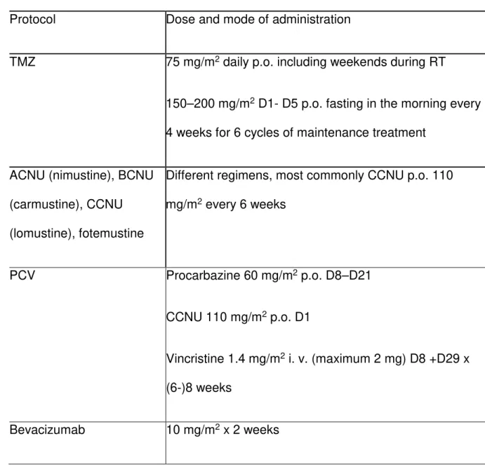
Table 2 - Chemotherapy protocols in malignant gliomas
Protocol Dose and mode of administration
TMZ 75 mg/m2 daily p.o. including weekends during RT
150–200 mg/m2 D1- D5 p.o. fasting in the morning every
4 weeks for 6 cycles of maintenance treatment
ACNU (nimustine), BCNU
(carmustine), CCNU
(lomustine), fotemustine
Different regimens, most commonly CCNU p.o. 110
mg/m2 every 6 weeks PCV Procarbazine 60 mg/m2 p.o. D8–D21 CCNU 110 mg/m2 p.o. D1 Vincristine 1.4 mg/m2 i. v. (maximum 2 mg) D8 +D29 x (6-)8 weeks Bevacizumab 10 mg/m2 x 2 weeks
45
Table 3 - Key recommendations*
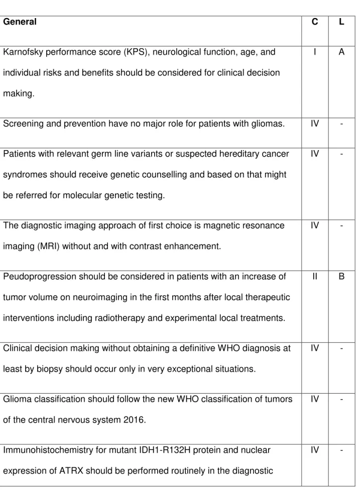
General C L
Karnofsky performance score (KPS), neurological function, age, and
individual risks and benefits should be considered for clinical decision
making.
I A
Screening and prevention have no major role for patients with gliomas. IV -
Patients with relevant germ line variants or suspected hereditary cancer
syndromes should receive genetic counselling and based on that might
be referred for molecular genetic testing.
IV -
The diagnostic imaging approach of first choice is magnetic resonance
imaging (MRI) without and with contrast enhancement.
IV -
Peudoprogression should be considered in patients with an increase of
tumor volume on neuroimaging in the first months after local therapeutic
interventions including radiotherapy and experimental local treatments.
II B
Clinical decision making without obtaining a definitive WHO diagnosis at
least by biopsy should occur only in very exceptional situations.
IV -
Glioma classification should follow the new WHO classification of tumors
of the central nervous system 2016.
IV -
Immunohistochemistry for mutant IDH1-R132H protein and nuclear
expression of ATRX should be performed routinely in the diagnostic