. . . .
. . . .
Native and reconstituted HDL protect
cardiomyocytes from doxorubicin-induced
apoptosis
Miguel A. Frias, Ursula Lang, Christine Gerber-Wicht, and Richard W. James
*
Faculty of Medicine, University of Geneva, Service of Endocrinology, Diabetology, and Nutrition, University Hospital, 4, rue Gabrielle-Perret-Gentil, CH-1211 Geneva 14, Switzerland
Received 5 May 2009; revised 28 July 2009; accepted 18 August 2009; online publish-ahead-of-print 21 August 2009
Time for primary review: 34 days
Aims We analysed the impact of native and reconstituted HDL on doxorubicin-induced cardiomyocyte apoptosis. While it is an effective anti-cancer agent, doxorubicin has serious cardiotoxic side effects. HDL has been shown to protect cardiomyocytes, notably against oxidative stress.
Methods and results
Cultured neonatal rat ventricular cardiomyocytes were subjected to doxorubicin-induced stress, monitored as caspase3 activation, apoptotic DNA fragmentation and cell viability. The protective effects of HDL and sphingo-sine-1-phosphate (S1P) were investigated using native HDL, reconstituted HDL of varied composition and agonists and antagonists of S1P receptors. Anti-apoptotic signalling pathways were identified with specific inhibitors. Native and reconstituted HDL significantly decreased doxorubicin-induced cardiomyocyte apoptosis, essentially due to the S1P component of HDL. The latter was mediated by the S1P2 receptor, but not the S1P1 or S1P3 receptors. The extracellular signal-regulated kinases 1 and 2 (ERK1/2) signalling pathway was required for the anti-apoptotic effects of HDL and S1P. The transcription factor Stat3 also played an important role, as inhibition of its activity com-promised the protective effects of HDL and S1P on doxorubicin-induced apoptosis.
Conclusion HDL and its sphingosine-1-phosphate component can protect cardiomyocytes against doxorubicin toxicity and may offer one means of reducing cardiotoxic side effects during doxorubicin therapy. The study identified anti-apoptotic pathways that could be exploited to improve cardiomyocyte survival.
-Keywords HDL † Oxidative stress † Signalling pathways † Cardiomyopathies † Apolipoprotein AI
1. Introduction
Doxorubicin (DOX) is an extremely effective antineoplastic agent used for the treatment of solid tumours and haematologic malig-nancies. However, its use is hampered by serious cardiotoxic side effects leading to cardiomyopathy and heart failure.1,2 The
mechanism of DOX-induced cardiac injury is not entirely clear. The free radical hypothesis of DOX-induced cardiotoxicity has garnered most support as DOX has been shown to generate reac-tive oxygen species.3 – 5 The increase in oxidative stress could cause membrane and macromolecular damage, including the acti-vation of intrinsic apoptotic pathways in cardiomyocytes,6 – 8
leading to heart injury. Large areas of myocardial apoptotic cells
have been observed in DOX-treated patients, and could contrib-ute to dilated cardiomyopathy and heart failure.9,10
High-density lipoprotein (HDL) promotes reverse cholesterol transport to the liver thereby reducing the risk of atherosclerosis. However, there is increasing evidence that HDL exerts other bio-logical effects corresponding to anti-inflammatory, anti-thrombotic, and anti-oxidative functions.11 – 13 In the heart, HDL and its sphingosine-1-phosphate (S1P) component have been shown to counteract ischaemia/reperfusion (I/R) injury.14,15As with DOX, the deleterious effects of I/R injury involve the generation of reactive oxygen species.16However, there is no information con-cerning direct protective effects of HDL or S1P on DOX-induced myocardial injury.
*Corresponding author. Tel: þ41 22 3729304, Fax: þ41 22 3729329, Email: [email protected]
The signal transducer and activator of transcription 3 (Stat3) appears to play a cardioprotective role in the heart.17 Mice with cardiomyocyte-specific deletion of Stat3 showed increased suscep-tibility to cardiac injury.18The cardioprotective role of Stat3 may also extend to DOX.19
At present, there is little information concerning the effect of HDL on Stat3 activity. We have recently demonstrated in ventricu-lar cardiomyocytes that HDL activates Stat3 mainly via its constitu-ent S1P. However, whether Stat3 acts as a signalling molecule in HDL-induced cell survival is unknown.
There are few therapeutic options currently available to increase HDL. One novel strategy, synthetic or reconstituted HDL (rHDL), is under development. HDL is a heterogeneous group of small lipoproteins whose basic elements are apolipoprotein (apo) AI and phospholipids. A recent innovation has been to complement rHDL with other bioactive molecules that can extend its physio-logical impact. Thus, rHDL containing in addition S1P was shown to induce endothelial cell tube formation.20
In the present study, we examined whether native and rHDL could counteract DOX-induced apoptosis in ventricular cardio-myocytes, determined the importance of S1P and investigated potential roles for the Stat3 and ERK1/2 (extracellular signal-regulated kinases 1 and 2) pathways.
2. Methods
2.1 Materials
S1P, DOX, and phosphatidylcholine were purchased from Sigma-Aldrich (Buchs, Switzerland). The S1P agonists and antagon-ists FTY720, VPC23019 were from Calbiochem (La Jolla, CA, USA) and Avanti Polar Lipids Inc. (Alabaster, AL, USA), respectively. SEW2871 and JTE013 were obtained from Tocris Bioscience (Ellisville, MO, USA). SB203580, AG490, and MTT (3-[4,5-dimethylthiazol-2-yl]-2,5-diphenyl tetrazolium bromide) were pur-chased from Calbiochem, while U0126 was from Biomol Research Laboratories (Plymouth Meeting, PA, USA).
2.2 Cell culture
The study was approved by the federal (Swiss) and cantonal (Geneva) veterinary authorities and conforms to the US guidelines on animal experimentation (NIH Publication No. 85-23, revised 1966). Neonatal cardiomyocytes were isolated from 1- to 2-day-old Wistar rat ventri-cles as previously described.21
2.3 Western blotting
After treatment as indicated in the figure legends, cardiomyocytes were washed with phosphate buffer saline (PBS) and lysed (50 mL of Tris – HCl (50 mM, pH 7.4), NaCl (150 mM), glycerol (10%v/v), EDTA (2 mM), EGTA (2 mM), Triton X-100 (1%v/v), b-glycerophosphate (40 mM), NaF (50 mM) containing a mixture of protease inhibitors (Roche, Mannheim, Germany), and phenyl-methyl-sulfonyl fluoride (1 mM)). Total cell proteins (20 mg) were separated by SDS – PAGE (12% acrylamide gel) and blotted onto nitrocellulose membrane. Membranes were probed for cleaved caspase3 (catalytically active 17 kDa fragment; Cell Signaling Technol-ogy, Denvers, MA, USA) to monitor apoptosis, and reprobed for glyceraldehyde-3-phosphate dehydrogenase (GAPDH) (Chemicon International Inc., Hampshire, UK). Specific bands were visualized with a chemiluminescence kit (Amersham, Zu¨rich, Switzerland).
2.4 Preparation of HDL and rHDL
HDL (d ¼ 1.063 – 1.21 g/mL) was isolated by cumulative flotation ultra-centrifugation22from a plasma pool provided by healthy volunteers, dialysed against PBS containing EDTA (1 mM) and stored at 48C.
ApoAI was isolated from delipidated human HDL.23Three different forms of rHDL were prepared by the cholate dialysis procedure:24 rHDL A contained phospholipids and S1P (for convenience labelled rHDL despite the absence of apoAI); rHDL B contained phospholipids, S1P and apoAI; rHDL C contained phospholipids and apoAI. The phos-pholipid:apoAI molar ratio was 100:1. For preparations containing S1P, aliquots from a stock solution (1.0 mg/mL in chloroform:methanol, 2:1 v/v) giving the required concentration (indicated in figure legends) were dried in glass tubes and the phospholipid/cholate mixture then apoAI added, as prescribed by the cholate dialysis procedure.24All preparations were dialysed against Tris 10 mM, pH 8.0, NaCl, 0.15 M and stored in sodium azide (0.05%w/v).
2.5 DNA fragmentation
DNA fragmentation was quantified by measuring the content of intracellu-lar nucleosomes. Cardiomyocytes (106cells per well) were deprived of serum for 24 h and subjected to treatment as indicated in the figure legends. After stimulation in serum-free DMEM, cells were washed with ice-cold PBS and lysed (60 mL of the buffer used for western blotting). Cellular lysates were used for protein determination and quantitative evaluation of histone-associated DNA fragments (mono- and oligonucleo-somes) by photometric enzyme immunoassay (Cell Death Detection ELISAPLUS, Roche Diagnostics, Germany). Results are reported as arbitrary absorbance units normalized to milligrams of proteins.
2.6 Determination of cardiomyocyte viability
Cell viability was evaluated by the colorimetric MTT assay (3-[4,5-dimethylthiazol-2-yl]-2,5-diphenyl tetrazolium bromide), as described previously.192.7 Statistical analysis
All values are expressed as mean (SEM). Differences between groups were determined using either two-tailed unpaired Student’s t-tests. P , 0.05 was accepted as statistically significant.
3. Results
3.1 HDL and S1P counteract
DOX-induced cell death
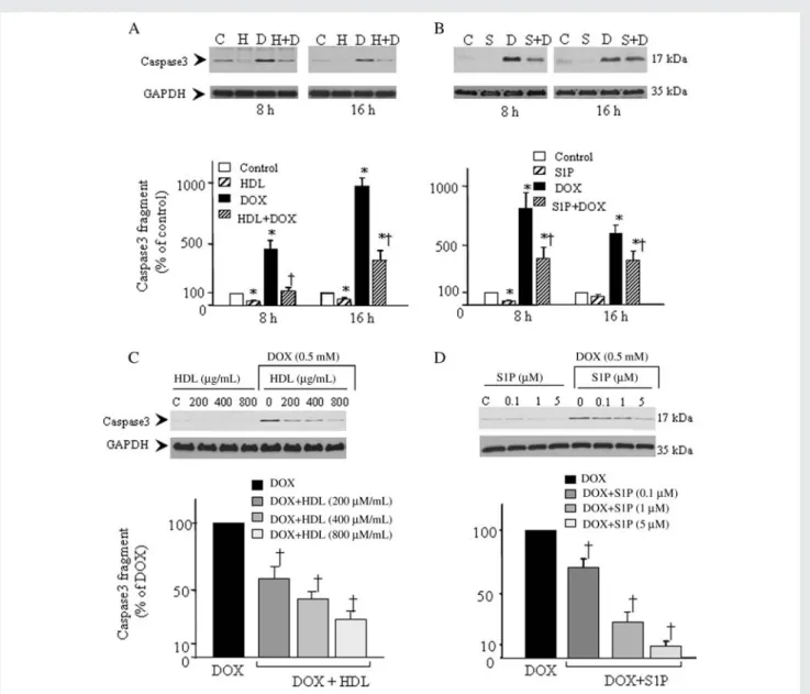
The anti-apoptotic effect of HDL and its constituent S1P was studied by investigating whether they could counteract DOX--induced caspase3 activation and DNA fragmentation. As illustrated in Figure 1A, HDL (400 mg protein/mL, corresponding to physio-logical extravascular concentrations25), caused a significant decrease in DOX-induced activation of caspase3 at 8 h (72 + 7%) and 16 h (62 + 7%). Likewise, S1P inhibited DOX-induced caspase3 activation by 54 + 4% (8 h) and 35 + 8% (16 h) (Figure 1B). The HDL- and S1P-induced inhibitory effects on DOX-induced caspase3 activation were concentration-dependent, with significant responses already observed at 200 mg/mL HDL (Figure 1C) and 100 nM S1P (Figure 1D). DOX also increased apop-totic DNA fragmentation (319 + 54% at 24 h) (Figure 2A) and reduced cardiomyocyte viability by 60% (40 + 5%; Figure 2B). The presence of native HDL or S1P inhibited DOX-induced
DNA fragmentation (64 + 5 and 58 + 6%, respectively) and increased cell viability of DOX-treated cells to 76 + 3 and 67 + 6%, respectively (Figure 2B). Interestingly, HDL and S1P also reduced basal DNA fragmentation (33 and 21%, respectively).
Our results indicate that in ventricular cardiomyocytes, both HDL and S1P counteract DOX-induced apoptotic effects.
3.2 Effect of HDL componants on
DOX-induced apoptosis in ventricular
cardiomyocytes
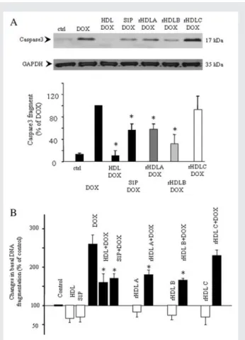
To investigate the specific role of the HDL constituents, S1P and apoAI, in the inhibitory action on DOX-induced apoptosis, we
pre-incubated cells with three different formulations of rHDL, con-taining S1P alone (rHDL A), S1PþapoAI (rHDL B), and apoAI (rHDL C). Cells were also pretreated with native, human HDL and S1P as positive controls. As illustrated in Figure 3A, the rHDLs containing S1P decreased DOX-induced caspase3 acti-vation to an extent comparable to control treatments. In contrast, rHDL C did not significantly affect the DOX-induced increase in caspase3 activation (Figure 3A). Interestingly, rHDL B containing S1P and apoAI appeared to induce a stronger (albeit non-significantly different) response than rHDL A containing only S1P. A comparable result was obtained for DOX-induced apoptotic DNA fragmentation (Figure 3B). Native HDL and its constituent S1P as well as the rHDLs containing S1P markedly decreased
Figure 1 HDL and S1P inhibit DOX-induced caspase3 activation in ventricular cardiomyocytes. (A and B) Cardiomyocytes were pretreated or not (control, C) with HDL (H, 400 mg/mL) or S1P (S, 1 mM), for 30 min prior to the incubation with DOX (D, 0.5 mM; 8 h and 16 h). (C and D) Cells were pretreated or not (control, C) with increasing concentrations of HDL (200 – 800 mg/mL) or S1P (S, 0.1 – 5 mM) for 30 min prior to the incubation with DOX (0.5 mM, 8 h). Caspase3 activation was analysed in cellular extracts by determining the level of the catalytically active 17 kDa fragment by western blot. Representative blots are shown at the top. Equal gel loading was assessed by immunoblotting GAPDH. Specific bands corresponding to activated caspase3 were quantified by densitometry and expressed as percentage of DOX-induced formation of the 17 kDa fragment. *P , 0.05 compared with control values,†P , 0.05 compared with values from DOX-treated cells (n ¼ 3 – 5).
DOX-induced DNA fragmentation, whereas rHDL C containing only apoAI had no significant effect.
Our results indicate that HDL exerts its anti-apoptotic action principally via its sphingolipid constituent S1P.
3.3 HDL and S1P counteract the
apoptotic effects of DOX via the S1P2
receptor
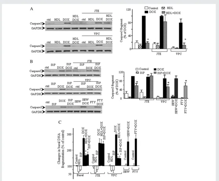
To determine which S1P receptors are involved in the anti-apoptotic action of HDL and S1P, we used available agonists and antagonists of the S1P receptors.26 – 28
The highly selective S1P2 antagonist, JTE013, abolished the ability of HDL and S1P to inhibit DOX-induced caspase3 activation (Figure 4A and B). In contrast, incubation of cardiomyocytes with VPC23019, a competitive antagonist of S1P1/S1P3 receptors, did not significantly affect the inhibitory action of HDL and S1P on DOX-induced caspase3 activation (Figure 4A and B). Similarly, SEW2871 (S1P1 receptor-specific agonist) and FTY720
(high-affinity agonist of S1P1, S1P3, S1P4, and S1P5 receptors) had no effect either on basal or DOX-induced caspase3 activation (Figure 4B). JTE013 also abolished the inhibitory action of HDL and S1P on DOX-induced DNA fragmentation, whereas VPC23019 had no effect (Figure 4C). Likewise, SEW2871 and FTY720 did not affect basal or DOX-induced DNA fragmentation. Thus our results indicate that in DOX-treated cardiomyocytes, HDL and S1P exert their anti-apoptotic action essentially through the receptor S1P2.
3.4 Critical role of Stat3 and ERK1/2 in
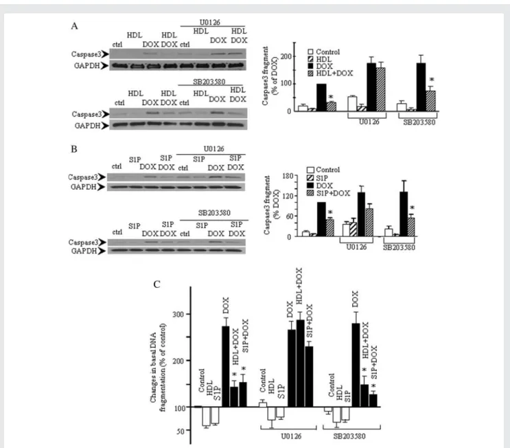
the anti-apoptotic effect of HDL and S1P
We have previously reported that in ventricular cardiomyocytes, ERK1/2 is critical for HDL- and S1P-induced Stat3 activation.29 Thus, we investigated the role of ERK1/2 and p38 MAPK in the anti-apoptotic responses induced by HDL and S1P using specific inhibitors of the ERK1/2 and p38 MAPK signalling pathways, U0126 and SB203580, respectively. U0126 strongly reduced the Figure 2 HDL and S1P inhibit DOX-induced apoptotic DNA
frag-mentation and increase cell viability in ventricular cardiomyocytes. Cells were pretreated or not (control) with HDL (400 mg/mL) or S1P (1 mM), for 30 min prior to incubation with DOX (0.5 mM, 24 h). (A) DNA fragmentation was analysed by measur-ing histone-associated DNA fragments. Changes in basal DNA fragmentation were calculated as arbitrary absorbance units nor-malized to protein and are expressed as percentage of the control (attributed 100%). (B) Cell viability determined using the MTT assay. Results are expressed as percentage of the control (attributed 100%). *P , 0.05 compared with control values, †P , 0.05 compared with values from DOX-treated cells (n ¼ 3 – 5).
Figure 3 Inhibition of DOX-induced caspase3 activation and DNA fragmentation by HDL, rHDL and S1P. (A and B) Cardio-myocytes were pretreated or not (control, ctrl) with HDL (400 mg/mL), S1P (1 mM), or three different rHDLs containing rHDL A (S1P, 1 mM), rHDL B (S1P, 1 mMþapoAI, 100 mg/mL), rHDL C (apoAI, 100 mg/mL) for 30 min prior to the incubation with DOX (0.5 mM) for 8 h (A) or 24 h (B). Caspase3 activation (A) was analysed as described in Figure 1 and DNA fragmentation (B) as described in Figure 2. Representative blots are shown. *P , 0.05 compared with DOX values (n ¼ 3 – 4).
anti-apoptotic effect of HDL and S1P on DOX-induced caspase3 activation (Figure 5A and B) and DNA fragmentation (Figure 5C), whereas SB203580 had no significant effect. We had previously shown that LY 294002, a specific inhibitor of the Akt pathway, had no effect on HDL or S1P-induced activation of Stat3.29 In
the present study, we confirmed that the protective effects of HDL and S1P against DOX-induced caspase-3 activation and DNA fragmentation are maintained in the presence of the inhibi-tor. The data are presented in Supplementary material online, Figures S1 and S2. It suggests that ERK1/2 is principally involved
in the protective influence of HDL and S1P against apoptosis in cardiomyocytes exposed to DOX.
To evaluate the role of Stat3 in the anti-apoptotic effect of HDL and S1P, we employed the Stat3 pathway inhibitor AG490.18 As shown in Figure 6A, the inhibitor decreased HDL- and S1P-induced Stat3 phosphorylation. In parallel, there were reductions in the ability of HDL and S1P to counter DOX-induced caspase3 acti-vation and DNA fragmentation (Figure 6B and C ).
Our results show that Stat3 is implicated in the protective effect of HDL and S1P against DOX-induced apoptosis.
Figure 4 HDL and S1P inhibit DOX-induced caspase3 activation and DNA fragmentation through S1P2. (A) Cardiomyocytes were untreated (basal) or incubated (20 min) with JTE013 (JTE, 5 mM; S1P2 antagonist) or VPC23019 (VPC, 5 mM; S1P1/S1P3 antagonist). A second pre-incubation period (30 min) +HDL was followed by addition of DOX (0.5 mM) for 8 h. Caspase3 activation was subsequently analysed in cel-lular extracts. (B) The same experimental procedure as for (A), except S1P replaced HDL (top two panels). For panel 3, cells were pre-incubated with S1P, SEW2871 (SEW, 1 mM; S1P1-specific receptor agonist), FTY720 (FTY, 1 mM; high affinity agonist of S1P1, S1P3, S1P4, and S1P5 receptors) or non-treated (control, ctrl) prior to addition of DOX (8 h) then analysis of caspase3 activation. (C ) Cells were pre-incubated (20 min) with JTE, VPC, SEW, FTY or untreated (basal) prior to addition of HDL or S1P (30 min) (to non-treated or JTE and VPC treated samples only). Subsequently DOX (0.5 mM) was added for 24 h before analysing measuring histone-associated DNA fragments. *P , 0.05 compared with corresponding DOX values (n ¼ 3 – 5).
4. Discussion
This is the first demonstration, to our knowledge, of the ability of HDL to reduce toxicity in ventricular cardiomyocytes by counteracting DOX-induced apoptosis. The anti-apoptotic action appears to be mediated essentially through the S1P component of HDL. Indeed, S1P and rHDLs containing S1P had a strong inhibitory action on DOX-induced caspase3 activation and apoptotic DNA fragmentation with, consequently, improved cell viability, comparable to HDL isolated from human plasma. In contrast, rHDL containing apoAI but no S1P did not significantly affect DOX-induced apoptosis.
Our studies further reveal that these anti-apoptotic activities are mediated by Stat3 and involve ERK1/2. Both the ERK1/2 antagonist, UO126 and the Stat3 pathway inhibitor, AG490, blunted the
anti-apoptotic effects of HDL and S1P on DOX-induced caspase3 acti-vation and DNA fragmentation. Our previous studies29 demon-strated that the ERK1/2 pathway inhibitor, U0126, suppressed HDL- and S1P-induced Stat3 activation in cardiomyocytes, indicat-ing that ERK1/2 activation occurs upstream of that of Stat3. In that study,29 we also analysed the influence of the PI3K/Akt pathway inhibitor, LY 294002 and showed that it had no impact on HDL or S1P activation of Stat3. It was confirmed in the present study with respect to the protective effects of HDL and S1P against DOX-induced apoptosis (see Supplementary material online, Figures S1 and S2). The results accord with several studies report-ing that Stat3 plays a role in the survival of cardiomyocytes in pathophysiological situations such as myocardial ischaemia, reper-fusion injury, and DOX treatment.30 – 32
Figure 5 ERK1/2 plays a role in the inhibitory effect of HDL and S1P on DOX-induced activation of caspase3 and DNA fragmentation. Car-diomyocytes were incubated (30 min) with U0126 (10 mM) or SB203580 (10 mM), prior to stimulation (30 min) with HDL (400 mg/mL) or S1P (1 mM). Subsequently, DOX (0.5 mM) was added and caspase3 activation (A and B) or DNA fragmentation (C ) analysed after 8 h (A and B) or 24 h (C ). *P , 0.05 compared with corresponding DOX values (n ¼ 4 – 5).
Also in agreement with our data, S1P and S1P associated with HDL have been shown to activate potent intracellular signalling pathways involved in cardiac remodelling.12 – 15 Conversely, the HDL structural peptide, apoAI, appears to confer cardioprotection primarily by influencing reverse cholesterol transport and reducing atherosclerosis as shown by studies using apoAI and mimetic pep-tides33or rHDL containing apoAI.34,35In this context, it has been established that increases of HDL in statin-treated patients corre-late in part with an inhibitory effect on atheroma progression.36 Interestingly, one study reported that rats treated with DOX showed a drop in serum HDL.37
Extracellular S1P functions as a ligand for a family of plasma mem-brane receptors designated S1P1 to S1P5. S1P1, S1P2, and S1P3 sub-types are expressed in the cardiovascular system.38Using various, well-characterized agonists and antagonists of the S1P receptor sub-types including two potent and specific inhibitors of S1P1/S1P3 and S1P2, respectively, our results clearly designate S1P2 as the receptor principally mediating the effects of HDL/S1P on apoptosis induced by DOX in cardiomyocytes. At the present time, there are few studies concerning the role of S1P receptors in cardiomyocytes. In rat-ventricular cardiomyocytes, S1P-induced hypertrophic and car-dioprotective effects during hypoxia were found to occur through Figure 6 Pharmacological inhibition of Stat3 reduces the inhibitory effect of HDL and S1P on DOX-induced caspase3 activation and apop-totic DNA fragmentation. Cells were incubated (16 h) with the Stat3 inhibitor AG490 (50 mM) prior to stimulation with HDL (400 mg/mL) or S1P (1 mM). (A) After HDL or S1P stimulation (90 min), Stat3 tyrosine phosphorylation (Stat3-P) was analysed in cellular extracts by western blotting. A representative blot is shown. (B) After stimulation (30 min) with HDL or S1P, cells were incubated with DOX (0.5 mM, 8 h), fol-lowed by analysis of caspase3. Representative blots are shown at the top of (B) (ctrl ¼ control). (C ) Cells were incubated (16 h) with the Stat3 inhibitor AG490 (10 mM) prior to stimulation with HDL or S1P. Thereafter, cells were incubated with DOX (0.5 mM, 24 h) and DNA fragmen-tation analysed. *P , 0.05 compared with values from corresponding DOX cells (n ¼ 3).
the S1P1 receptor.28,39In contrast, Theilmeier et al.15reported that HDL and its constituent S1P protect the heart against I/R injury via an S1P3 receptor mediated pathway. More recently, Means et al.40 showed that both S1P2 and S1P3 receptors contribute to protect cardiomyocytes from I/R damage in vivo. The same authors found that in cultured mouse cardiomyocytes, S1P acts predominantly through the S1P2 receptor, a result which is consistent with our findings. While our data provide no evidence for the involvement of S1P1 or S1P3 receptors, we cannot presently exclude that another receptor, perhaps activated through the apoAI component of HDL, might be involved in the anti-apoptotic action of HDL, poss-ibly by facilitating the action of S1P. Further studies are required to define the respective roles of the receptors S1P1, S1P2, and S1P3. Such studies should also address the influence of other receptors, such as the HDL binding scavenger receptor, SR-BI, that are involved in other intracellular pathways mediating specific functions.
In summary, the present study demonstrates that in ventricular cardiomyocytes: (i) HDL counteracts DOX-induced apoptosis mainly via its S1P constituent and the receptor S1P2; (ii) that Stat3 activation involving ERK1/2 plays an essential role in the anti-apoptotic effect of HDL. The study thus reveals pathways that could be explored as targets to limit cardiomyocyte death and poss-ible heart failure. The results also underline that the clinical relevance of HDL goes beyond facilitated removal of cholesterol from the blood vessel wall. Strategies designed to increase HDL levels in general and their S1P content in particular may improve the progno-sis of the myocardium at risk in DOX-treated patients. Infusion of rHDL has already been shown to increase transiently serum HDL levels. Whether such treatment concomitantly with DOX therapy could limit the cardiotoxic effects of the drug merits consideration.
Supplementary material
Supplementary material is available at Cardiovascular Research online.
Conflict of interest: none declared.
Funding
This work was supported by the Swiss National Science Foundation (grants 31-108342/1 and 31-105310), the Novartis Foundation for Medico-Biological Research, the Swiss University Conference Foun-dation, and the Swiss Romand Association for Diabetes Research.
References
1. Singal PK, Iliskovic N. Doxorubicin-induced cardiomyopathy. N Engl J Med 1998; 150:900 – 905.
2. Takemura G, Fujiwara H. Doxorubicin-induced cardiomyopathy from the cardio-toxic mechanisms to management. Prog Cardiovasc Dis 2007;49:330 – 352. 3. Davies KJ, Doroshow JH. Redox cycling of anthracyclines by cardiac
mitochondria. I. Anthracycline free radical formation by NADH dehydrogenase. J Biol Chem 1986;261:3060 – 3067.
4. Iliskovic N, Singal PK. Lipid lowering: an important factor in preventing adriamycin-induced heart failure. Am J Pathol 1997;150:727 – 734.
5. Siveski-Iliskovic N, Kaul N, Singal PK. Probucol promotes endogenous antioxi-dants and provides protection against adriamycin-induced cardiomyopathy in rats. Circulation 1994;89:2829 – 2835.
6. Arola OJ, Saraste A, Pulkki K, Kallajoki M, Parvinen M, Voipio-Pulkki LM. Acute doxorubicin cardiotoxicity involves cardiomyocyte apoptosis. Cancer Res 2000; 60:1789 – 1792.
7. Childs AC, Phaneuf SL, Dirks AJ, Phillips T, Leeuwenburgh C. Doxorubicin treat-ment in vivo causes cytochrome C release and cardiomyocyte apoptosis, as well as increased mitochondrial efficiency, superoxide dismutase activity, and Bcl-2:Bax ratio. Cancer Res 2002;62:4592 – 4598.
8. Kluza J, Marchetti P, Gallego MA, Lancel S, Fournier C, Loyens A et al. Mitochon-drial proliferation during apoptosis induced by anticancer agents: effects of dox-orubicin and mitoxantrone on cancer and cardiac cells. Oncogene 2004;23: 7018 – 7030.
9. Keizer HG, Pinedo HM, Schuuruis GJ, Joenje H. Doxorubicin (adriamycin): a criti-cal review of free radicriti-cal-dependent mechanisms of cytotoxicity. Pharmacol Ther 1990;47:219 – 231.
10. Singal PK, Li T, Kumar D, Danelisen D, Iliskovic N. Adriamycin-induced heart failure: mechanism and modulation. Mol Cell Biochem 2000;207:77 – 86. 11. Mineo C, Yuhanna IS, Quon MJ, Shaul PW. HDL-induced eNOS activation is
mediated by Akt and MAP kinases. J Biol Chem 2003;278:9142 – 9149. 12. Nofer JR, Assmann G. Atheroprotective effects of high-density
lipoprotein-associated lysosphingolipids. Trends Cardiovasc Med 2005;15:265 – 271. 13. O’Connell BJ, Genest JJ. High-density lipoproteins and endothelial function.
Circu-lation 2001;104:1978 – 1983.
14. Calabresi L, Rossoni G, Gomaraschi M, Sisto F, Berti F, Franceschini G. High-density lipoproteins protect isolated rat hearts from ischemia-reperfusion injury by reducing cardiac tumor necrosis factor-alpha content and enhancing prosta-glandin release. Circ Res 2003;92:330 – 337.
15. Theilmeier G, Schmidt C, Hermann J, Keul P, Scha¨fers M, Herrgott I et al. High-density lipoproteins and their constituent, sphingosine-1-phosphate, directly protect the heart against ischemia/reperfusion injury in vivo via the S1P3 lysopho-spholipid receptor. Circulation 2006;114:1403 – 1409.
16. Zhao ZQ. Oxidative stress-elicited myocardial apoptosis during reperfusion. Curr Opin Pharmacol 2004;4:159 – 165.
17. Barry SP, Townsend PA, Latchman DS, Stephanpou A. Role of the JAK-STAT pathway in myocardial injury. Trends Mol Med 2007;13:82 – 89.
18. Suleman N, Somers S, Smith R, Opie LH, Lecour SC. Dual activation of STAT-3 and Akt is required during the trigger phase of ischaemic preconditioning. Cardi-ovasc Res 2008;79:127 – 133.
19. Frias MA, Somers S, Gerber-Wicht C, Opie LH, Lecour S, Lang U. The PGE2-Stat3 interaction in doxorubicin-induced myocardial apoptosis. Cardiovasc Res 2008;80:69 – 77.
20. Matsuo Y, Miura SI, Kawamura A, Uehara Y, Rye KA, Saku K. Newly developed reconstituted high-density lipoprotein containing sphingosine-1-phosphate induces endothelial tube formation. Atherosclerosis 2007;194:159 – 168. 21. Frias MA, Rebsamen MC, Gerber-Wicht C, Lang U. Prostaglandin E2 activates
Stat3 in neonatal rat ventricular cardiomyocytes: A role in cardiac hypertrophy. Cardiovasc Res 2007;73:57 – 65.
22. James RW, Proudfoot A, Pometta D. Immunoaffinity fractionation of high density lipoprotein subclasses 2 and 3 using anti-apolipoprotein A-I and A-II immunosor-bent gels. Biochim Biophys Acta 1989;1002:292 – 301.
23. James RW, Hochstrasser D, Tissot J-D, Funk M, Appel R, Barja F et al. Protein heterogeneity of lipoprotein particles containing apolipoprotein A-I without apo-lipoprotein A-II and apoapo-lipoprotein A-I with apoapo-lipoprotein A-II, isolated from human plasma. J Lipid Res 1988;29:1557 – 1571.
24. Matz CE, Jonas A. Micellar complexes of human apolipoprotein A-I with phospha-tidylcholines and cholesterol prepared from cholate-lipid dispersions. J Biol Chem 1982;257:4535 – 4540.
25. Sloop CH, Dory L, Hamilton R, Krause BR, Roheim PS. Interstitial fluid lipopro-teins. J Lipid Res 1987;28:225 – 237.
26. Davis MD, Clemens JJ, Macdonald TL, Lynch KR. Sphingosine 1-phosphate analogs as receptor antagonists. J Biol Chem 2005;280:9833 – 9841.
27. Watterson KR, Ratz PH, Spiegel S. The role of sphingosine 1-phosphate in smooth muscle contraction. Cellular Signalling 2005;17:289 – 298.
28. Zhang J, Honbo N, Goetzl EJ, Chatterjee K, Karliner JS, Gray MO. Signals from type 1 sphingosine 1-phosphate receptors enhance adult mouse cardiac myocyte survival during hypoxia. Am J Physiol Heart Circ Physiol 2007;293: H3150 – H3158.
29. Frias MA, James RW, Gerber-Wicht C, Lang U. Native and reconstituted HDL activate Stat3 in ventricular cardiomyocytes via ERK1/2: role of sphingosine-1-phosphate. Cardiovasc Res 2009;82:313 – 323.
30. Hilfiker-Kleiner D, Hilfiker A, Fuchs M, Kaminski K, Schaefer A, Schieffer B et al. Signal transducer and activator of transcription 3 is required for myocardial capil-lary growth, control of interstitial matrix deposition, and heart protection from ischemic injury. Circ Res 2004;95:187 – 195.
31. Kunisada K, Negoro S, Tone E, Funamoto M, Osugi T, Yamada S et al. Signal trans-ducer and activator of transcription 3 in the heart transduces not only a hyper-trophic signal but a protective signal against doxorubicin-induced cardiomyopathy. Proc Natl Acad Sci USA 2000;97:315 – 319.
32. Negoro S, Kunisada K, Tone E, Funamoto M, Oh H, Kishimoto T et al. Activation of JAK/STAT pathway transduces cytoprotective signal in rat acute myocardial infarction. Cardiovasc Res 2000;47:797 – 805.
33. Garber DW, Datta G, Chaddha M, Palgunachari MN, Hama Y, Navab M et al. A new synthetic class A amphiphathic peptide analogue protects mice from diet-induced atherosclerosis. J Lipid Res 2001;42:545 – 552.
34. Nanjee MN, Doran JE, Lerch PG, Miller NE. Acute effects of intravenous infusion of apoAI/phosphatidylcholine discs on plasma lipoproteins in humans. Arterioscler Thromb Vasc Biol 1999;19:979 – 989.
35. Shaw JA, Bobil A, Murphy A, Kanellakis P, Blombery P, Muhamedova N et al. Infu-sion of reconstituted high-density lipoprotein leads to acute changes in human atherosclerotic plaque. Circ Res 2008;103:1084 – 1091.
36. Nicholls SJ, Tuzcu EM, Sipahi I, Grasso AW, Schoenhagen P, Hu T et al. Statins, High-density lipoprotein cholesterol, and regression of coronary atherosclerosis. JAMA 2007;297:499 – 508.
37. Subashini R, Ragavendran B, Ganapragasam A, Yogeeta SK, Devaki T. Biochemical study on the protective potential of Nardostachys jatamansi extract on lipid profile and lipid metabolizing enzymes in doxorubicin intoxicated rats. Pharmazi 2007;62: 382–387.
38. Alewijnse AE, Peters SLM, Michel MC. Cardiovasular effects of sphingosine-1-phosphate and other sphingomyelin metabolites. Br J Pharmacol 2004;143: 666 – 684.
39. Robert P, Tsui P, Laville MP, Livi GP, Sarau HM, Brill A et al. EDG1 receptor stimu-lation leads to cardiac hypertrophy in rat neonatal myocytes. J Mol Cell Cardiol 2001;33:1589 – 1606.
40. Means CK, Xiao CY, Li Z, Zhang T, Omens JH, Ishii I et al. Sphingosine 1-phosphate S1P2 and S1P3 receptor-mediated Akt activation protects against in vivo myocardial ischemia-reperfusion injury. Am J Physiol Heart Circ Physiol 2007; 292:H944 – H951.