Animal model to compare the effects of suture technique on
cross-sectional compliance on end-to-side anastomoses
q
P. Tozzi
a,*, D. Hayoz
b, P. Ruchat
a, A. Corno
a, C. Oedman
a, U. Botta
a, L.K. von Segesser
aaDepartment of Cardiovascular Surgery, Centre Hospitalier Universitaire Vaudois, Rue du Bugnon, 46, 1011 Lausanne, Switzerland bDepartment of Vascular Medecine, Centre Hospitalier Universitaire Vaudois, Rue du Bugnon, 46, 1011 Lausanne, Switzerland
Received 2 October 2000; received in revised form 21 January 2001; accepted 23 January 2001
Abstract
Objective: An animal model has been developed to compare the effects of suture technique on the luminal dimensions and compliance of end-to-side vascular anastomoses. Methods: Carotid and internal mammalian arteries (IMAs) were exposed in three pigs (90 kg). IMAs were sectioned distally to perform end-to-side anastomoses on carotid arteries. One anastomosis was performed with 7/0 polypropylene running suture. The other was performed with the automated suture delivery device (Perclose/Abbott Labs Inc.) that makes a 7/0 polypropylene interrupted suture. Four piezoelectric crystals were sutured on toe, heel and both lateral sides of each anastomosis to measure anastomotic axes. Anastomotic cross-sectional area (CSAA) was calculated with: CSAA p £ mM=4 where m and M are the minor and major axes of the elliptical anastomosis. Cross-sectional anastomotic compliance (CSAC) was calculated as CSAC DCSAA=DP where DP is the mean pulse pressure and DCSAA is the mean CSAA during cardiac cycle. Results: We collected a total of 1 200 000 pressure-length data per animal. For running suture we had a mean systolic CSAA of 26.94 ^ 0.4 mm2and a mean CSAA in diastole of 26.30 ^ 0.5 mm2(mean DCSAA was 0.64 mm2). CSAC for running suture was 4.5 £ 1026m2/kPa. For interrupted suture we had a mean CSAA in systole of 21.98 ^ 0.2 mm2and a mean CSAA in diastole of 17.38 ^ 0.3 mm2(mean DCSAA was 4.6 ^ 0.1 mm2). CSAC for interrupted suture was 11 £ 1026m2/kPa. Conclusions: This model, even with some limitations, can be a reliable source of information improving the outcome of vascular anasto-moses. The study demonstrates that suture technique has a substantial effect on cross-sectional anastomotic compliance of end-to-side anastomoses. Interrupted suture may maximise the anastomotic lumen and provides a considerably higher CSAC than continuous suture, that reduces ¯ow turbulence, shear stress and intimal hyperplasia. The Heart¯oe anastomosis device is a reliable instrument that facilitates performance of interrupted suture anastomoses. q 2001 Elsevier Science B.V. All rights reserved.
Keywords: Vascular anastomosis; Arterial compliance; Piezoelectric crystals
1. Introduction
Running versus interrupted suture can be reasonably considered one of the most frequent subject of discussion since vascular surgery moved its ®rst steps. To date, this is still an open issue despite many scienti®c works have been published all over the world. The suture material selected and the suture technique employed can in¯uence the size and the distensibility of the anastomotic lumen [1]. Cross-sectional compliance in the perianastomotic zone, wall shear stress, axial stress, and their relationship with intimal hyperplasia are the most frequently considered parameters to compare the two different techniques [1,2]. If we try to resume the pros and cons of each technique we conclude
that running suture is faster and somehow easier to do but it can produce a purstring effect that can impair the hemody-namic performance of the anastomosis. Multiple stitch tech-nique avoids purstring effect but requires more time and is often characterized with bleeding from the suture line.
The development of a surgical device that allows perfor-mance of a multiple stitch coronary sutures in an easier and faster way than usual, and the possibility to calculate the cross-sectional anastomotic area during each phase of the cardiac cycle using a brand new technology based on piezo-electric crystals, led us to develop an animal model to compare the effects of suture technique on luminal dimen-sions and compliance of end-to-side anastomoses.
2. Methods
Instrumentation: length measurements were obtained with small piezoelectric crystals that transmit and receive
1010-7940/01/$ - see front matter q 2001 Elsevier Science B.V. All rights reserved. PII: S1010-7940(01)00617-0
www.elsevier.com/locate/ejcts
qPresented at the 14th Annual Meeting of the European Association for
Cardio-thoracic Surgery, Frankfurt, Germany, October 7±11, 2000. * Corresponding author. Tel.:141-21-314-2280; fax: 141-21-314-2278.
short ultrasonic pulses. The crystals were sutured on toe, heel and both lateral sides of each anastomosis to measure distances as shown in Fig. 1. Under electric stimulation a crystal produces a sound wave that is detected by a second crystal, inducing an electrical response. A simple calcula-tion (Distance Velocity £ Time) yields the distance between the crystals. Sound velocity in pig's heparinized blood at 388C is 1.04 mm/ms [3]. The system setting is as follows: sampling rate 457 Hz; transmit pulse 357 m/s; sampling time 5 s. Extensive description of the device and validation of the technique have been reported previously [4].
Arterial pressure was obtained using high ®delity sure probe (Millar Mikro-Tip, model MPC-500) with a pres-sure range of 250 to 300 mmHg and a sensitivity of 5 mV/V per mmHg.
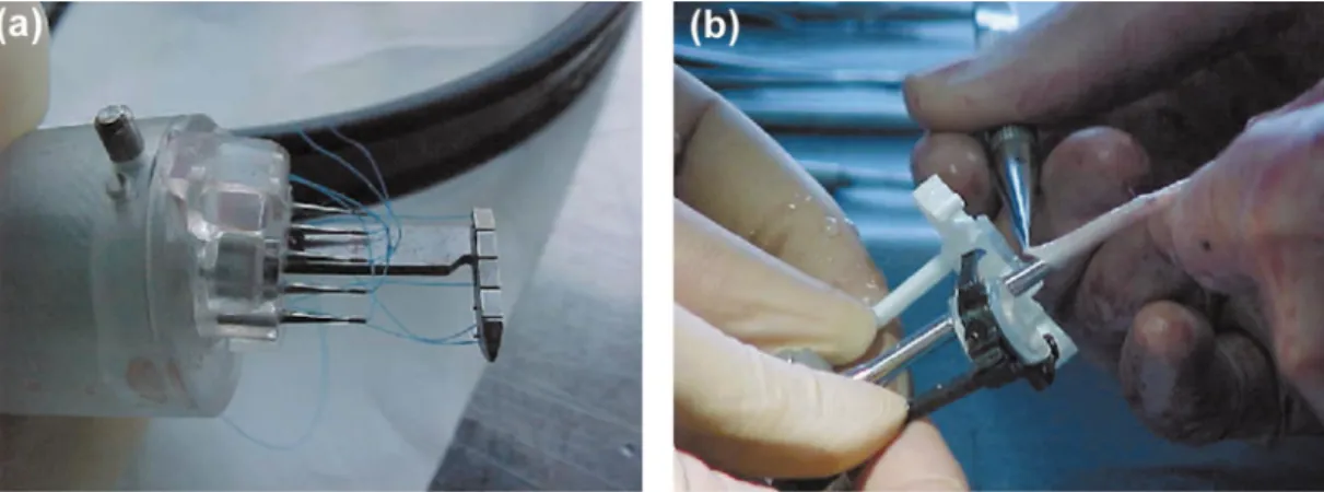
The Heart¯oe anastomosis device (Perclose/Abbott Labs, Inc.) was used to perform the end-to-side anastomosis with interrupted suture technique. It consists of a hydrauli-cally activated delivery mechanism, and two branches, with each branch housing needles and the opposite ends of ten
7-0 polypropylene sutures. The device ®rst simultaneously delivers ten sutures of one branch through the wall of the vessel (coronary), and then through the wall of the graft. The surgeon completes the anastomosis using conventional surgical knot tying techniques (Fig. 2).
The experiment was performed on three domestic pigs, 90 kg in weight. All animals received human care in compli-ance with the European Convention on Animal Care and the study has been approved by our ethics committee.
Surgical technique: pigs were given Ketamine 15 mg/kg, Azaperon 0.5 mg, Atropine 2 mg. General anaesthesia was induced and maintained with Fluotane 1.5%. EKG, SatO2
and pCO2 were continuously monitored. Pigs lay on the
back with a neck extension of 1608. Both carotid arteries were exposed. The pressure probe was inserted in the left common femoral artery and pushed up to the aortic arch. After median sternotomy, both internal mammalian arteries (IMAs) were isolated and 9000 U of Heparin were injected. IMAs were sectioned distally and rotate of 1808 to perform an end-to-side anastomoses on carotid arteries. The carotid arteriotomy was performed with the Heart¯oe scissors that
Fig. 1. Representation of end-to-side anastomosis between IMA and carotid artery. In yellow piezoelectric crystals that have been placed on toe, heel and lateral side of the anastomosis.
Fig. 2. The Hearth¯oe anastomosis device (Perclose/Abbott Labs, Inc.) was used to perform the end-to-side anastomosis with interrupted suture technique. It consists of a hydraulically activated delivery mechanism, and two branches, with each branch housing needles and the opposite ends of ten 7-0 polypropylene sutures. The device ®rst simultaneously delivers ten sutures of one branch through the wall of the coronary vessel (a), and then through the wall of the graft (b). The surgeon completes the anastomosis using conventional surgical knot tying techniques.
makes the correct arteriotomy length in which the device perfectly ®ts. One anastomosis was performed with 7-0 polypropylene running suture. The other was performed with the Heart¯oe anastomosis device. Four piezoelectric crystals were sutured on the carotid artery at toe, heel and sides of each anastomosis to measure major and minor anastomotic axes (Fig. 1). Carotid arteries were clamped proximally to the anastomosis. Finally, pressure probe, EKG and crystals were connected to our measurement system. Artery was irrigated with NaCl 0.9% solution at 378 every 10 min to prevent desiccation and to control its temperature. During data acquisition we carefully avoided any manipulation of the animal. In animal No. 3 blood ¯ow in both carotid arteries was assessed with a high ®delity ¯owmeter probe (Medi-Stim perivascular ¯owmeter probes, size 4 mm with ¯ow relative accuracy of 1%, resolution of 1 ml/min, ¯ow sample rates 333 Hz).
Data collection: both carotid arteries were clamped 2 cm proximally to the anastomoses and after 15 min of stabilisa-tion, data collection was carried out for a period of 5 s without interruption at least four times per minute for 1 h for each animal. During every second of acquisition anasto-motic diameters of both types of sutures and blood pressure were captured 457 times.
To avoid blood mass and pulse waves interference we switched the transmitter and receiver functions of the piezo-electric crystals.
Anastomotic cross-sectional area was calculated assum-ing that the shape of the anastomosis corresponds to a regu-lar ellipse and distances between crystals corresponds to major and minor axes of the considered ellipse (Fig. 1). If those hypotheses are accepted Cross-Sectional anastomotic area (CSAA) can be calculated as:
CSAA pmM4
where m and M are the minor and major axes of the anastomosis. CSAA is expressed in mm2. Cross-sectional
anastomotic compliance (CSAC) was calculated as the ratio between variations in anastomotic cross-sectional area (DCSAA) and blood pressure (DP):
CSAC DCSAADP
CSAC is expressed in m2/kPa.
Data are presented as mean ^ standard deviation (SD) 3. Results
We collected a total of 6 £ 105 simultaneous data for
blood pressure, and anastomotic axes for both anastomosis, per animal. For running suture we had a mean systolic CSAA of 26.94 ^ 0.4 mm2and a mean CSAA in diastole
of 26.30 ^ 0.5 mm2 (mean DCSAA was 0.64 ^ 0.0 mm2
that correspond to 2.4% incrementation of diastolic area during systole). CSAC for running suture was 4.5 £ 1026
m2/kPa. For interrupted suture we had a mean CSAA in
systole of 21.98 ^ 0.2 mm2and a mean CSAA in diastole
of 17.38 ^ 0.3 mm2(mean DCSAA was 4.6 ^ 0.1 mm2that
correspond to 20.9% incrementation of diastolic area during systole). CSAC for interrupted suture was 11 £ 1026 m2/
kPa. Table 1 reports anastomotic CSAA values in systole and diastole for both sutures.
Mean diastolic pressure was 60 ^ 13.2 mmHg; mean systolic pressure was 99 ^ 12.8 mmHg; pulse pressure was between 20 and 46 mmHg (mean 32 ^ 8 mmHg) and the mean heart rate was 88/min ^18.
Blood ¯ow in carotid was 54 ^ 12 ml/min for interrupted suture, and 62 ^ 13 ml/min for running suture.
IMAs had a mean diameter of 3 ^ 0.2 mm. Carotid arteries had a mean diameter of 5.2 ^ 0.2 mm.
The mean time to perform the interrupted suture was 10 ^ 2 min. The mean time to perform the continuous suture was 7 ^ 1 min.
4. Discussion
This study demonstrates that suture technique has a substantial effect on CSAC of end-to-side anastomoses. Interrupted suture provides a CSAC considerably higher than continuous suture and can be reasonably considered the most `physiologic' suture because it keeps the biome-chanical properties of arterial wall as close as possible to those of native vessel [2]. This anastomotic behavior appears to result mainly from the elastic recoil of the arterial wall constituents that is better preserved with interrupted suture [5]. Therefore, the notion that difference in
hemody-Table 1
Mean cross-sectional anastomotic areaa
Running suture Interrupted suture
Animal Diastole Systole %DCSAA Diastole Systole %DCSAA
1 25.68 ^ 0.2 26.38 ^ 0.2 2.7 17.23 ^ 0.1 21.71 ^ 0.2 20.6
2 27.12 ^ 0.3 27.67 ^ 0.2 2 17.04 ^ 0.2 21.89 ^ 0.1 22.1
3 26.11 ^ 0.3 26.79 ^ 0.3 2.6 17.87 ^ 0.2 22.34 ^ 0.2 20
Mean ^ SD 26.30 ^ 0.5 26.94 ^ 0.4 2.4 17.38 ^ 0.3 21.98 ^ 0.2 20.9
a Mean cross-sectional anastomotic area (CSAA) (mm2) ^ standard deviation, in systole and diastole for running and interrupted end-to-side anastomoses
namic property of end-to-side anastomoses done with the two considered techniques are negligible [1], deserves reap-praisal.
Furthermore, it is clear from the data provided in the study that systolic increase of cross-sectional anastomotic area (CSAA) is de®nitely bigger if interrupted suture is used and this behavior may theoretically improve the systolic ¯ow through the anastomosis.
The limitations of the study resides in the fact that the model represents a situation that doesn't exist in surgical practice. It should reproduce the hemodynamic condition of end-to-side anastomosis between IMA and Left Anterior Descending coronary artery, but carotid and coronary artery have a different histological pattern. Actually, muscular layer is much more represented in carotid than in coronary arteries. The surgical procedure may also have modi®ed the genu-ine elastic properties of the vessel wall. However, careful attention was paid not to severe the adventitia in the proxi-mity of the sutures, and in the translation of IMAs to avoid kinking and twisting.
Another limitation of the model is that we assume the anastomosis has a perfect elliptic shape and that distances we calculate with piezoelectric crystals correspond to the maximal and minimal diameters of this ellipse. Although other authors have done this assumption before [1,5] we are doing histological morphometric studies on end-to-side anastomoses performed with Heart¯oe device to verify their geometry.
The reference method for arterial diameter and cross-sectional compliance determination is the non-invasive ultrasound (NIUS 02) [6]. But, if we consider that end-to-side anastomosis doesn't lie on one cross-sectional plan we can assume that A-mode echotracking system is not a reliable method to calculate CSAC. Baumgartner proposes to measure anastomosis axes on the radiographs after anastomosis is removed, but it seems to be the less accurate method [1]. We chose sonometric technology to calculate CSAA and CSAC. Piezoelectric crystals have the highest resolution (15 mm) [4] and are easy to handle. This technique has been extensively used in vascular surgery mostly to validate Intra Vascular Ultra Sound measure-ments [3,4].
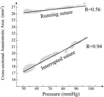
In Fig. 3 is plotted the correlation between pulse pressure and CSAA for interrupted and continuous sutures. The two parameters are directly correlated only if interrupted suture technique is used (Rinterrup 0:94 vs. Rrunning 0:56). The CSAA increase during systole causes a reduction of vascu-lar resistance and this can improve the blood ¯ow through the anastomosis as hypothesised the ®rst time in 1960 by Szilagyi [7]. However, when we measured the ¯ow through the anastomosis we did not ®nd any difference in systolic out¯ow between the two techniques and this is probably due to the sensibility of the ¯owmeter probe.
CSAA was slightly smaller for interrupted suture prob-ably because we used a dedicate Pot's scissors for the arter-iotomy so that the device can perfectly ®t in the arterarter-iotomy.
Better anastomotic compliance means less suture-line stresses [8,9], reduces ¯ow disturbances and may reduce the disposition toward the development of intimal hyperpla-sia or thrombosis [5,10]. Computer ¯owdynamic simulation demonstrates that a more compliant anastomosis is asso-ciated with a less stagnation point due to ¯ow separation (typically on heel, toe and the hood of the graft) giving rise to low wall shear stress that is associated with intimal hyper-plasia [11].
Despite 51 patented ideas describing vascular anastomo-tic devices, and the growing need for them in minimally invasive coronary bypass procedures, no data have been published concerning their clinical evaluation. In an elegant study, Scheltes and colleagues evaluate 11 most attractive end-to-side anastomotic devices and conclude that, in a coronary anastomotic device, the concept of using an anvil for the application of micromechanical bonding elements is not attractive, because excessive wall strain is likely to occur [12]. The Heart¯oe anasto-mosis device does not use bonding elements. This is a surgical instrument that automates the suture delivery process during the anastomosis procedure via the simulta-neous delivery of ten standard 7-0 polypropylene suture through the vessel wall. After the deployment of the device, the surgeon manually ties off the ten sutures to complete the anastomosis, similar to a hand-sewn inter-rupted anastomosis (Fig. 2). No signi®cative bleeding from the suture line has been observed.
The Heart¯oe anastomosis device reduces the time to perform an interrupted end-to-side anastomosis and it should facilitate a consistent and reproducible anastomosis for minimally invasive and beating heart surgery.
Fig. 3. The correlation between pulse pressure and cross-sectional anasto-motic area (CSAA) for running and interrupted sutures are plotted. The two parameters are directly correlated only if interrupted suture technique is used (Rinterrup 0:94 vs. Rrunning 0:56).
5. Conclusions
We believe this model, even with the limitations described above, can be a reliable source of information improving the outcome of coronary artery bypasses. This study demonstrates that suture technique has a substantial effect on cross-sectional anastomotic compliance of end-to-side anastomoses. Interrupted suture may maximise the anastomotic lumen and provides a considerably higher CSAC than continuous suture, that reduces ¯ow turbulence, shear stress and intimal hyperplasia. The Heart¯oe anasto-mosis device is a reliable instrument that facilitates perfor-mance of interrupted suture anastomoses.
Acknowledgements
We thank Perclose/Abbott Labs Inc. for its ®nancial and technical support in this study.
References
[1] Baungartner N, Dobrin PB, Morasch M, Quan-sheng Dong, Mrkvicka R. In¯uence of suture technique and suture material selection on the mechanics of end-to-end and end-to-side anastomoses. J Thor Cardi-ovasc Surg 1996;111:1063±1072.
[2] Klein SR, Goldberg L, Miranda RM. Effect of suture technique on arterial anastomotic compliance. Arch Surg 1982;117(1):45±47. [3] Tozzi P, Mueller XM, Mallabiabarrena I, von Segesser LK.
Intravas-cular ultrasound underestimates vessel dimensions. Eur J Vasc Endo-vas Surg 2000;19(5):501±503.
[4] Hardt SE, Just A, Bekeredjian R, Kubler W, Kirkchheim HR, Kuecherer HF. Aortic pressure-diameter relationship assessed by intravascular ultrasound: experimental validation in dogs. Am J Physiol 1999;276(3pt2):H1078±H1085.
[5] Dobrin PB. Mechanical factors associated with the development of intimal hyperplasia with respect to vascular grafts. In: Dobrin PB, editor. Intimal hyperplasia, Austin, TX: RG Landes, 1994. pp. 85± 109.
[6] Hayoz D, Tardy Y, Rutschmann B. Spontaneous diameter oscillations of the radial artery in humans. Am J Phys Jun; 1994;264(6Pt2):H2080± H2884.
[7] Szilagyi DE, Whitcomb J, Schenker W. The laws of ¯uid ¯ow and arterial grafting. Surgery 1960;47:55±67.
[8] Ballyk PD, Walsh C, Butany J, Ojha M. Compliance mismatch may promote graft-artery intimal hyperplasia by altering suture-line stres-ses. J Biomech 1998;31(3):229±237.
[9] Hasson JE, Megerman J, Abbot WM. Increased compliance near vascular anastomoses. J Vasc Surg May; 1985;2(3):419±423. [10] LoGerfo FW, Soncrant T, Teel T. Boundary layer separation in
models of side-to-end arterial anastomoses. Arch Surg 1979;114(12):1369±1373.
[11] Harris Peter, How T. Haemodynamics of cuffed arterial anastomoses. Crit Ischaemia 1999;9(1):20±26.
[12] Scheltes Jules S, Heikens M, Pistecky PV, van Andel C, Borst C. Assessment of patented coronary end-to-side anastomotic devices using micromechanical bonding. Ann Thorac Surg 2000;70(1):218± 221.