Significance of ventricular late potentials in non-ischaemic
dilated cardiomyopathy
D . D E N E R E A Z , M . ZIMMERMANN AND R. A D A M E C
Cardiology Center and Policlinic of Medicine, University Hospital, Geneva, Switzerland
KEY WORDS: Ventricular late potentials, signal-averaging, non-ischaemic dilated cardiomyopathy, ventricular tachycardia.
To assess the incidence and clinical significance of ventricular late potentials in non-ischaemic dilated cardiomyopathy, 51 consecutive (44 male, seven female, mean age 53±11 years) patients with dilated cardiomyopathy were studied. Twenty-eight patients (55%) were in New York Heart Association functional class III or IV, 34 out of 51 (76%) had a left ventricular ejection fraction of less than 40%, 10 out of 51 (20%) had a history of sustained ventricular tachycardia ( VT), 24 out of 37 (65%) had runs of non-sustained ventricular tachycardia during Holter monitoring and 15 out of 51 (29%) had a left bundle branch block. A signal-averaged electrocardiogram (gain 106 x , bipolar chest leads, filters 100-300 Hz)
was performed in all the patients; late potentials were considered present if the total filtered QRS duration was longer than 118 ms and the interval between the end of QRS and the voltage 40 fiVwas more than 40 ms in the absence of left bundle branch block (totalfiltered QRS duration > 140 ms and interval between the end of QRS and the voltage 40fiV>50ms in the presence of left bundle branch block).
Ventricular late potentials were detected in 22 out of 51 patients (43%). Late potentials were present in 80% (eight out of 10) of patients with sustained ventricular tachycardia but in only 34% (14 of 41) without sustained ventricular tachycardia (P < 001). This difference remained statistically significant even when patients with a left bundle branch block were excluded from the analysis (4 out of 6 vs 4 out of 30, P<001). To identify patients with dilated cardiomyopathy and sustained ventricular tachycardia, signal-averaging had a sensitivity of 80%, a specificity of 66%, a positive predictive
value of 36% and a negative predictive value of 93%.
It is concluded that, in non-ischaemic dilated cardiomyopathy, the signal-averaged electrocardiogram allows the identifi-cation of patients with sustained ventricular tachycardia, even in the presence of a left bundle branch block.
Introduction cardiomyopathy, (b) the association between late poten-tials and malignant ventricular tachyarrhythmias and (c) Patients suffering from non-ischaemic dilated cardio- the possible role of late potentials detection in assessing myopathy have a high incidence of serious ventricular the prognosis of patients with dilated cardiomyopathy. arrhythmias and most of the deaths in this syndrome are
sudden and unexpected, presumably related to malignant
ventricular arrhythmias1". However, until now, no clinical Methods
predictors of sudden death have been identified, and the TIFNTS
prognostic significance of Holter monitoring and pro- T, t . , .. . , . r . .
K O , . . . . . • . . , • • , • • The study population consisted of 51 consecutive,
grammed ventricular stimuation in this chnica entity is , t , .. . , . . , r .
. . [2.9] 3 unselected patients (44 male, seven female, mean age
still controversial . 53 ± 11 years, range 22-74 years) with non-ischaemic During the last few years, signal-averaging for the detec- d i,a t e d ^rixomyovrity. Each a t i e n t h a d a l e t e tion of ventricular late potentials has emerged as a pro- j c a, e x a m i n a t i o n > a s t a n d a r d ] 2.l e a d e l e c t r o c a r d i o.
mising tool for identifying patients at high risk of serious L * v J »• J • • i J . , , ., • . . . , r . . . gram, a chest X-ray and a time-domain signal-averaged
ventricular arrhythmias or sudden death after myocardial . . .. . ... . , , °f C1
• r •• no-Hi \r . • i i • . I. i electrocardiogram. In addition, 45 out of 51 patients
infarction1 JJ. Ventricular late potentials are abnormal, , . ,. ,, . • .. ... r . .
, .. , , . , r \ . ' underwent cardiac cathetenzation with coronary
arteri-low-amphtude, high-frequency signals, occurring in the , ._ , r c i u A-, r\ . J- U J „
. , . ° . , ^ r , ^ , ' .. . ography; 47 out of 51 had 2-D echocardiography and 37 terminal portion of the QRS complex and extending into , r c. .. . , , ,0 M, A~>A, , , ,. •; • ^^ i
, c_ K . . i • , . . out of 51 patients (73%) had 24 h Holter monitoring (Del
the ST segment: these late potentials are thought to rep- w A • • , , . , . . AA- •
, , . . . r ,, r L j - j Mar Avionics two-channel recorder, model 447,
semi-resent delayed activation of small areas of the diseased . . . , . • ., . . . _ ' ^
,. , , r . , , , automated analysis using the Avionics Trendsetter,
myocardium, and therefore, are considered as a marker . l l l m n. , ~, .. r. . . . , ,. ,
P I. i ~, . , , • model 9000 A). The diagnosis of dilated cardiomyopathy
for re-entry" " T h e purposes of the present study were to w a s b a s e d Qn ) e f t v e 6 n t r i c u l a r e j e c t i o n f r a c t i o^ QPf ^ determine (a) the mcidenoe of ventncular late potentials m t h a n 5 Q % & ventriculography) with no
a group of unselected patients with non-ischaemic di ated • c . ,, • • , • ™o^ \
b K y •- significant (luminal narrowing < 50%) coronary stenosis
Submitted for publication 30 April 1991, and in revised form 18 November ( ^ = 45 OUt of 51) Or (b) a left Ventricular ejection fraction 1991. • of less than 50% (by 2-D echocardiography), with diffuse
Correspondence: Marc Zimmermann. MD, Cardiology Center, University hypokinesia, no Segmental Wall motion abnormality and
Hospital, 24 rue Micheh-du-Crest, 1211 Geneva 4, Switzerland. no history of angina or myocardial infarction (A/=6 out
of 51). The presumed aetiology was chronic alcohol abuse in 13 cases, related to peripartum in two cases and idio-pathic in 36 cases. A history of sustained ventricular tachycardia was present in 10 cases (20%). At the time of recording, eight out of 51 patients were taking no medi-cation, 28 were receiving angiotensin converting-enzyme inhibitors, 22 digoxin, 21 diuretics, 13 amiodarone and three were receiving ^-blockers, alone or in combination.
SIGNAL AVERAGING
A time-domain signal-averaged electrocardiogram was performed in all patients using the signal averager used by our group and previously described'131. Briefly, band-pass
filters are set at 100 and 300Hz (12dB .octave"1), the
peak of the R wave is used as a trigger (jitter <0-5ms)and bipolar chest leads (between V2 and V4, between V4 and V6) are used. The averaging process is performed on 40 consecutive beats or more, in order to reduce the baseline noise to 0-4±0-2uV. The recording (2uV.cm~') is plotted on paper (paper speed 1000 mm. s"1) together with
a reference electrocardiogram (200uV.cm~')- Quanti-tative analysis includes (a) total filtered QRS duration (ms) and (b) the interval between the end of the total QRS complex and the point (determined in a retrograde manner) when QRS voltage falls below 40 uV (1-40, in ms). QRS onset and offset were manually determined and all tracings were interpreted by two different investigators without knowledge of the patient's condition.
DEFINITIONS
Non-sustained ventricular tachycardia was defined as
ventricular tachycardia lasting less than 30 s and terminat-ing spontaneously. Sustained ventricular tachycardia was defined as ventricular tachycardia lasting more than 30 s or requiring immediate DC shock because of haemodynamic compromise. Sudden death was defined as death occurring within 1 h of symptom onset in a clinically stable patient or unexpected death occurring during sleep. Ventricular
late potentials were defined as high-frequency, low
ampli-tude signals appearing during the terminal portion of the QRS or on the ST segment. Ventricular late potentials were considered to be present if (a) in the absence of left
bundle branch block, total filtered QRS duration is more
than 118mms and 1-40 is more than 45 ms (maximal value for normal subjects in the laboratory); and (b) in the
presence of left bundle branch block, total QRS duration is
more than 140 ms and 1-40 is at least 50 ms.
FOLLOW-UP
Follow-up assessment included clinical information obtained by questionnaire or telephone from the attend-ing physician and/or from any hospital the patient was admitted to during the follow-up period.
STATISTICAL ANALYSIS
Values are expressed as means ± standard deviation (SD). Results were analysed using the unpaired t-test and chi-square when appropriate. A P value of less than 005 was considered statistically significant.
Results
CLINICAL AND HAEMODYNAMIC DATA
Twenty-eight out of 51 patients (55%) showed clinical signs of heart failure (New York Heart Association func-tional class IV in 22 and class III in six). In patients for whom haemodynamic data were available (N=45), the mean left ventricular ejection fraction was 32±12%, range 10-50%, and the ejection fraction less than 40% in 34 cases (76%). The mean end-diastolic volume index was 152±49 ml. m~2 and the mean left ventricular
end-diastolic pressure 19±8mmHg. A left bundle branch block was present in 15 out of 51 patients (29%), and 17 out of 51 patients (33%) had non-specific intraventricular conduction disturbances. The mean standard QRS duration on the surface electrocardiogram was 105 ± 28 ms (range 80-180 ms) and the mean QTc interval was 357 ± 45 ms (range 280-480 ms). Permanent atrial fibrillation was present in 10 out of 51 patients (20%).
HOLTER MONITORING DATA ( t f = 3 7 )
Numerous (more than 30. h~') premature ventricular complexes were observed in 19 patients (51%), polymor-phous premature ventricular complexes in 25 (68%), ventricular couplets in 28 (76%) and episodes of non-sustained ventricular tachycardia in 24 (65%).
SIGNAL-AVERAGING DATA (N=5\)
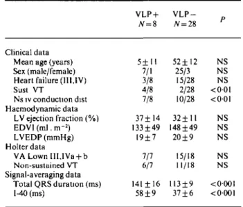
Using the aforementioned criteria, 22 patients (43%) had ventricular late potentials (Fig. 1,2). These were pres-ent in eight out of 36 patipres-ents with no left bundle branch block (22%) and in 14 out of 15 (93%) patients with left bundle branch block (Fig. 3). Compared to patients with-out late potentials, patients with late potentials more often had a left bundle branch block pattern and a history of sustained ventricular tachycardia (Table 1). When patients with a left bundle branch block were excluded from data analysis, the presence of ventricular late potentials was still associated with a history of sustained ventricular tachycardia (Table 2).
When comparing the clinical characteristics of patients with and without a history of sustained ventricular tachy-cardia, the only significant difference between the two groups was the presence of ventricular late potentials: eight out of 10 (80%) in the group with sustained ventricu-lar tachycardia vs 14 out of 41 (34%) in the group without sustained ventricular tachycardia (/><001) (Table 3). When patients with a left bundle branch block were excluded from data analysis, similar results were obtained and the quantitative parameters of the signal-averaged electrocardiogram allowed patients with and without sustained ventricular tachycardia to be distinguished (Table 4).
To identify patients who had sustained ventricular tachycardia, the presence of late potentials had a sensi-tivity of 80%, a specificity of 66%, a positive predictive value of 36% and a negative predictive value of 93%. These results compared favourably with the results of physical examination, 12-lead electrocardiogram, Holter
bip V 2 - V 4
110 ms 10 ms
Figure 1 Signal-averaged electrocardiogram obtained in a 63-year-old patient with non-ischaemic dilated cardiomyopathy, no bundle branch block and no history of sustained ventricular tachycardia. Bipolar chest lead between V2 and V4; filters 100-300 Hz; paper speed 1000 m m . s"'. Three successive high-gain record-ings (2 uV. cm"1) are displayed (each one obtained by averaging 40 consecutive cardiac cycles) together
with a reference electrocardiogram (200 uV . cm"'). The total filtered QRS duration is 110ms and no low-amplitude/high-frequency components are present in the terminal QRS complex.
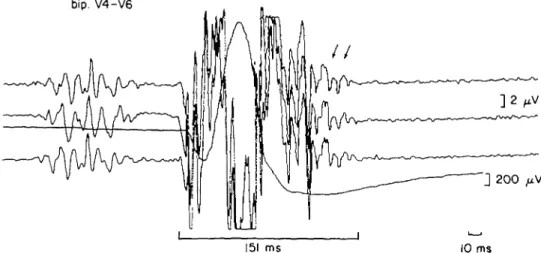
bip. V4-V6
151 ms 10 ms
Figure 2 Signal-averaged electrocardiogram obtained in a 38-year-old patient with idiopathic dilated cardiomyopathy, no bundle branch block but documented episodes of sustained ventricular tachycardia. Bipolar chest lead between V4 and V6; filters 100-300 Hz; paper speed 1000 mm . s"1. Three successive
high-gain recordings (2 uV . cm"1) are displayed (each one obtained by averaging 40 consecutive cardiac cycles) together with a reference electrocardiogram (200 uV. cm"'). The total filtered QRS duration is prolonged to 151 ms and ventricular late potentials (arrows) are identified in the terminal QRS complex.
recording or global left ventricular ejection fraction (Table 5).
FOLLOW-UP DATA ( # - 5 0 )
During a mean follow-up of 18 ±21 months (range 1-99 months), six patients (12%) had cardiac transplant, seven patients (14%) died, and three patients presented with sustained ventricular tachycardia (8%). The cause of death was due to post-transplant complications in three cases, and was non-cardiac in four cases (two were pul-monary embolisms, one stroke and one toxic hepatitis). The mortality rate was identical in patients with (four out of 22) and in patients without (three out of 28) ventricular late potentials but new episodes of sustained ventricular
tachycardia occurred only in patients with ventricular late potentials (three out of 22 patients with vs none out of 28 patients without late potentials, P<0-05).
Discussion
The value of signal-averaging for identifying patients with ventricular tachycardia has been established in coronary artery disease, and the prognostic significance of ventricular late potentials after myocardial infarction has been demonstrated in several prospective trials'12"141.
However, only few studies have been conducted in patients with non-ischaemic dilated cardiomyopathy, despite the fact that these patients have a high incidence of
bip V2-V4
185 ms 10 ms
Figure 3 Signal-averaged electrocardiogram obtained in a 49-year-old patient with non-ischaemic dilated
cardiomyopathy, complete left bundle branch block and a documented episode of sustained ventricular tachycardia. Bipolar chest lead between V2 and V4; filters 100-300 Hz; paper speed 1000 mm . s"'. Three successive high-gain recordings (2 uV . cm~') are displayed (each one obtained by averaging 40 consecutive cardiac cycles) together with a reference electrocardiogram (200 uV . cm"1). The total filtered QRS duration is prolonged to 185 ms and ventricular late potentials (arrows) are identified in the terminal portioji of the QRS complex despite the presence of complete left bundle branch block.
Table I Characteristics of patients with and without ventricular late potentials
Table 2 Characteristics of patients with and without ventricular late potentials in the absence of left bundle branch block
Clinical data Mean age (years) Sex (male/female) Heart failure (III,IV) History of sust. VT LBBB Ns iv conduction dist. Haemodynamic data LV ejection fraction (%) EDVI(ml.m-2) LVEDP(mmHg) Holter data VALownllI.IVa + b Non-sustained VT Signal-averaging data Total QRS duration (ms) 1-40 (ms) VLP + N = 22 55± 10 18/4 13/22 8/22 14/22 7/22 33±13 I55±5O 17±7 18/19 13/19 164±26 70 ±20 V L P -N=29 52±12 26/3 15/29 2/29 1/29 10/29 32±11 151±49 20±9 16/18 11/18 113±I0 37±6 P NS NS NS <001 <0001 NS NS NS NS NS NS <000i <0-O01 Clinical data Mean age (years) Sex (male/female) Heart failure (III,IV) Sust VT Ns iv conduction dist Haemodynamic data LV ejection fraction (%) EDVI (ml. m-:) LVEDP(mmHg) Holter data VALownIII,IVa + b Non-sustained VT Signal-averaging data Total QRS duration (ms) 1-40 (ms) VLP + N = i 5±H 7/1 3/8 4/8 7/8 37± 14 133±49 I9±7 7/7 6/7 141 ± 16 58±9 V L P -# = 28 52±12 25/3 15/28 2/28 10/28 32±11 I48±49 20±9 15/18 11/18 113±9 37±6 n r NS NS NS < 0 0 l <001 NS NS NS NS NS <0001 <0001
III,IV = functional class according to the New York State Heart Assocation, EDP = end-diastolic pressure, EDVl = end-diastolic volume index, 1-40 = interval between the end of the QRS complex and the voltage 40 uV, LV = left ventricular, LBBB-left bundle branch block, NS = not significant, ns iv conduction dist. = non-specific intraventncular conduction disturbance, sust. VT = sus-tained ventricular tachycardia, VA = ventricular arrhythmias, VLP =• ventricular late potentials.
ventricular tachyarrhythmias and sudden death1915"181. In the present study, it was found that 43% of patients with non-ischaemic dilated cardiomyopathy have ventricular
For key to abbreviations, see Table 1
late potentials and that the presence of late potentials may identify patients with sustained ventricular tachycardia. Thus, in non-ischaemic dilated cardiomyopathy, as in coronary artery disease, signal-averaging appears to be a useful non-invasive test to identify patients with severe ventricular arrhythmias.
Similar results have been reported by others: in a study published by Poll et a/.1'51, conducted in a group of 41 patients without bundle branch block, 83% of patients
Table 3 Characteristics of patients with and without documented sustained ventricular tachycardia
Clinical data Mean age (years) Sex (male/female) Heart failure (111,1V) LBBB Ns iv conduction dist. Haemodynamic data LV ejection fraction (%) EDVI(ml.m-2) LVEDP(mmHg) Holterdata VALownIII,IVa + b Non-sustained VT Signal-averaging data
Late potentials present Total QRS duration (ms) I^tO (ms) VT + JV=1O 50± 11 9/1 4/10 4/10 3/10 31 ± 1 2 % 154±52 19±8 9/9 6/9 8/10 142 ±29 61 ± 2 3 V T -JV=41 54±11 35/6 24/41 11/41 14/41 32 ± 1 2 % 152 ±49 19±9 26/28 18/28 14/41 129 ± 30 54±21 r NS NS NS NS NS NS NS NS NS NS <001 NS NS
For key to abbreviations, see Table 1.
Table 4 Characteristics of patients with and without documented sustained ventricular tachycardia in the absence of left bundle branch block
Clinical data Mean age (years) Sex (male/female) Heart failure (111,1V) Ns iv conduction dist. Haemodynamic data LV ejection fraction (%) EDVI (ml. m-2) LVEDP(mmHg) Holterdata VALownIII,IVa + b Non-sustained VT Signal-averaging data
Late potentials present Total QRS duration (ms) 1-40 (ms) VT + 46±10 5/1 0/6 3/6 38±11% 131 ±35 22±6 6/6 4/6 4/6 133±18 54± 15 V T -53±12 27/3 18/30 14/30 33 ±12% 147 ±52 19±9 16/19 13/19 4/30 114±16 45±9 P NS NS <0Ol NS NS NS NS NS NS <0-01 <005 <005
For key to abbreviations, see Table 1.
with sustained ventricular arrhythmias had an abnormal signal-averaged recording vs only 14% of patients with-out sustained ventriuclar arrhythmias. In another study1'6', parameters of the signal-averaged
electrocardio-gram seemed to correlate with the clinically documented arrhythmia: patients with a history of monomorphic ven-tricular tachycardia had a higher incidence of abnormal signal-averaged electrocardiograms than patients with a history of ventricular fibrillation. Results obtained by analysing the signal-averaged recording in the
frequency-domain have been comparable: nine out of 10 patients with sustained ventricular tachycardia had late potentials, vs only four out of 21 patients without sustained ventricu-lar tachycardia'19'. However, there are controversies
con-cerning the incidence of ventricular late potentials in non-ischaemic dilated cardiomyopathy'9', and a recent
study failed to show any correlation between the presence of late potentials and sudden death in patients with advanced heart failure1'81. The presence of ventricular late
potentials does not seem to be helpful in identifying patients with inducible ventricular tachyarrhythmias in electro-physiological study1'6-20'. The reason for this descrepancy is
unknown, but may be in part related to the low sensitivity and low specificity of programmed ventricular stimulation in non-ischaemic dilated cardiomyopathy19-2'1.
Ventricular late potentials are considered a marker for re-entrant ventricular arrhythmias and they are thought to represent delayed activation of some areas of the diseased myocardium1"'. The mechanism of ventricular
arrhythmias in dilated cardiomyopathy is not established, but recent studies suggest that re-entry is the underlying mechanism for monomorphic ventricular tachycardia in that setting12'22'. Therefore, late potentials may represent
the substrate for this type of ventricular arrhythmia in dilated cardiomyopathy. However, in non-ischaemic dilated cardiomyopathy, polymorphic sustained ven-tricular tachycardia and/or venven-tricular fibrillation are fre-quently the only documented ventricular arrhythmias and these arrhythmias are possibly due to other electrophysio-logical mechanisms, such as abnormal automaticity or triggered activity1'6'. This may explain some false-negative
results of the signal-averaged electrocardiogram in patients with sustained ventricular arrhythmias, and why fractionated ventricular electrograms are less frequently recorded during endocardial mapping in dilated cardio-myopathy122'. In fact, two patients from the present study
with documented sustained ventricular tachycardia had a normal signal-averaged electrocardiogram, and in these two cases, ventricular tachycardia was rapid (cycle length
< 270 ms) and poorly tolerated.
The results of the present study also applied to patients with bundle branch block. Most previous studies on signal-averaging have excluded patients with bundle branch block because intraventricular conduction defects pro-duce severe alterations of the signal-averaged electrocar-diogram, and may mimic late potentials1'0"'. However, it
has been shown that the value of late potentials recording in identifying patients with sustained ventricular arrhyth-mias is similar in the presence of bundle branch block, if the criteria to define an abnormal recording are adjusted1'7-23'. Such an adjustment has been made in the
present study for left bundle branch block, but the criteria proposed may not be applicable to other signal-averaging devices1'7-23'.
To identify patients with sustained ventricular tachy-cardia, late potentials were superior, in this study, to other recognized prognostic factors, such as a low ejection frac-tion or the presence of non-sustained runs of ventricular tachycardia during Holter monitoring (Table 5). Survival in non-ischaemic dilated cardiomyopathy is strongly
Table 5 Value of the presence of heart failure, left bundle branch block, ejection fraction less than 40%, non-sustained ventricular tachycardia during Holler monitoring and ventricular late potentials to identify patients with sustained ventricular tachycardia
NYHA 111,1V LBBB E F < 4 0 % Non-sustained VT Late potentials Sensitivity (%) Specificity (%) + Predictive value (%) — Predictive value (%) 40 41 14 74 40 73 27 83 70 23 21 73 67 36 33 77 80 66 36 93 VT = ventricular tachycardia.
influenced by the left ventricular function, but the prog-nostic values of a left bundle branch block, and of Holter monitoring are controversial'2"5'71. In patients of this
present study no relation was found between the presence of late potentials and the occurrence of non-sustained ventricular tachycardia during Holter monitoring, poss-ibly because different mechanisms underlie these two phenomena.
LIMITATION OF THE STUDY
The number of patients is too small and the follow-up period too short in the present study to determine the value of signal-averaging in predicting prognosis of patients with non-ischaemic dilated cardiomyopathy. Several patients in the study group were on medication at the time of record-ing and the use of drugs may have influenced both the signal-averaged electrocardiogram and the outcome. Criteria used to define late potentials in the presence of left bundle branch block are difficult to establish and further studies are required, with or without frequency analysis, to make further adjustments of criteria.
CONCLUSION
In non-ischaemic dilated cardiomyopathy, the signal-averaged electrocardiogram allows the identification of patients with sustained monomorphic ventricular tachy-cardia even in the presence of left bundle branch block. The exact prognostic significance of this finding, however,
r e m a i n s t o be d e t e r m i n e d .
This study was supported by a grant (No. 3.805-0.86) from the Swiss National Foundation for Scientific Research, Bern, Switzerland.
References
[1] Johnson RA, Palacios I. Dilated cardiomyopathies of the adult. Part I. N Engl J Med 1982; 307: 1051-8.
[2] Huang SK, Messer JV, Denes P. Significance of ventricular tachycardia in idiopathic dilated cardiomyopathy: obser-vations in 35 patients. Am J Cardiol 1983; 51: 507-12. [3] von Olshausen K, Schafer A, Mehmel HC, Schwarz F, Senges
J, Kubler W. Ventricular arrhythmias in idiopathic dilated cardiomyopathy. Br Heart J 1984; 51: 195-201.
[4] Neri R, Mestroni L, Salvi A, Camcrini F. Arrhythmias in dilated cardiomyopathy. Postgrad Med J 1986; 62: 593-7. [5] Romeo F, Pellicia F, Cianfrocca C, Cristofani R, Reale A.
Pre-dictors of sudden death in idiopathic dilated cardiomyopathy. Am J Cardiol 1989; 63: 138-45.
[6] Meinertz T, Hofmann T, Kasper W et al. Significance of ventricular arrhythmias in idiopathic dilated cardiomyopathy. Am J Cardiol 1984; 53: 902-7.
[7] Unverferth DV, Magorien RD, Moeschberger ML, Baker PB, Fetters SK, Leier CV. Factors influencing the one-year mor-tality of dilated cardiomyopathy. Am J Cardiol 1984; 54:
147-52.
[8] Holmes J, Kubo SH, Cody RJ, Kligfield P. Arrhythmias in ischemic and nonischmemic dilated cardiomyopathy: predic-tion of mortality by ambulatory electrocardiography. Am J Cardiol 1985; 55: 146-51.
[9] Meinertz T, Treese N, Kasper W et al. Determinants of prog-nosis in idiopathic dilated cardiomyopathy as determined by programmed electrical stimulation. Am J Cardiol 1985; 56: 337-41.
[10] Simson MB. Use of signals in the terminal QRS complex to identify patients with ventricular tachycardia after myocardial infarction. Circulation 1981; 64: 235-41.
[11] Breithardt G, Borggrefe M. Pathophysiological mechanisms and clinical significance of ventricular late potentials. Eur Heart J 1986; 7: 364-85.
[12] NalosPC,GangES, MandelWJ, Ladenheim ML, LassY, Peter T. The signal-averaged electrocardiogram as a screening test for inducibility of sustained ventricular tachycardia in high risk patients: a prospective study. J Am Coll Cardiol 1987; 9: 539-48.
[13] Zimmermann M, Adamec R, Simonin P, Richez J. Prognostic significance of ventricular late potentials in coronary artery disease. Am Heart J 1985; 109: 725-32.
[14] Kanovsky MS, Falcone RA, Dresden CA, Josephson ME, Simson MB. Identification of patients with ventricular tachy-cardia after myotachy-cardial infarction: signal-averaged electro-cardiogram, Holter monitoring and cardiac catheterization. Circulation 1984; 70: 264-70.
[15] Poll DS, Marchlinski FE, Falcone RA, Josephson ME, Simson MB. Abnormal signal-averaged electrocardiograms in patients with nonischemic congestive cardiomyopathy: relationship to sustained ventricular tachyarrhythmias. Circulation 1985; 72: 1308-13.
[16] BorggrefeM, EglofTJ, BuddeT, FetschT, Karbenn U, Breithardt G. Clinical value of late potentials as a marker of electrical instability in dilative cardiomyopathy. In: Santini M, Pistolese M, Alliegro A, eds. Progress in Clinical Pacing. Amsterdam: Excerpta Medica 1988; 247-55.
[17] Brembilla-Perrot B, Terrier de la Chaise A, Suty-Selton C, Marcon F. Effet d'un bloc de branche complet sur le signal moyenne de 1'ECG a haute amplification. Arch Mai Coeur 1990; 83: 907-12.
[18] Middlekauff HR, Stevenson WG, Woo MA, Moser DK, Stevenson LW. Comparison of frequency of late potentials in idiopathic dilated cardiomyopathy and ischemic cardiomyo-pathy with advanced congestive heart failure and their useful-ness in predicting sudden death. Am J C a r d i o l l 9 9 0 ; 6 6 : l l l 3-7. [19] Lindsay BD, Fischer AE, Ambos HD, Markham J, Cain ME. Detection of patients with nonischemic cardiomyopathy prone to sustained ventricular arrhythmias by frequency analysis of
signal-averaged electrocardiograms. Circulation 1988; 7: [22] Cassidy D, Vassallo JA, Miller JM el al Endocardial catheter IV-345(Abstr). mapping in patients in sinus rhythm: relationship to underlying [20] Gonksa BD, Bethge Kp, Figulla HR, Kreuzer H. Occurrence heart disease and ventricular arrhythmias. Circulation 1986:73:
and clinical significance of endocardial late potentials and frac- 645-52.
tionations in idiopathic dilated cardiomyopathy. Br Heart J [23] Buckingham TA, Thessen CC, Stevens LL, Redd RM, Kennedy 1988; 59: 39-46. HL. Effect of conduction defects on the signal-averaged electrc-[21] Poll DS, Marchlinski FE, Buxton AF, Josephson ME. Useful- cardiographic determination of late potentials. Am J Cardiol
ness of programmed stimulation in idiopathic dilated cardio- 1988; 61: 1265-71. myopathy. Am J Cardiol 1986; 58: 992-7.