2006/283
Parrado
and Tad A. Holak
*
1
Max-Planck-Institut fu¨r Biochemie, D-82152
Martinsried, Germany
2
Abteilung fu¨r Klinische Chemie und Klinische
Biochemie, Chirurgische Klinik Innenstadt, Klinikum der
Ludwig-Maximilians-Universita¨t Mu¨nchen, D-80336
Mu¨nchen, Germany
3
Adolf-Butenandt-Institut der
Ludwig-Maximilians-Universita¨t Mu¨nchen, D-80336 Mu¨nchen, Germany
* Corresponding author
e-mail: holak@biochem.mpg.de
Abstract
Calpains are a large family of Ca
2q-dependent cysteine
proteases that are ubiquitously distributed across most
cell types and vertebrate species. Calpains play a role in
cell differentiation, apoptosis, cytoskeletal remodeling,
signal transduction and the cell cycle. The cell cycle
pro-teins cyclin D1 and p21
KIP1, for example, have been
shown to be affected by calpains. However, the rules that
govern calpain cleavage specificity are poorly
under-stood. We report here studies on the pattern of
m
-calpain
proteolysis of the p19
INK4dprotein, a cyclin-dependent
kinase 4/6 inhibitor that negatively regulates the
mam-malian cell cycle. Our data show new characteristics of
calpain action:
m
-calpain cleaves p19
INK4dimmediately
after the first and second ankyrin repeats that are
struc-turally less stable compared to the other repeats. This is
in contrast to features observed so far in the specificity
of calpains for their substrates. These results imply that
calpain may be involved in the cell cycle by regulating
the cell cycle regulatory protein turnover through CDK
inhibitors and cyclins.
Keywords: calpain; calpastatin; cyclin-dependent
kinases; nuclear magnetic resonance spectroscopy;
proteolysis; p19
INK4d.
Introduction
Calpains are a family of non-lysosomal, cysteine
protein-ases that show Ca
2qdependent papain-like cysteine
pro-tease activity (Goll et al., 2003). The two ubiquitously
expressed calpain forms, with proteolytic activities
requiring
m
Mand m
Mcalcium, were identified and named
These authors contributed equally to this work.
a
Present address: Novartis Pharma AG, Werk Klybeck, CH-4002
b
Basel, Switzerland.
domains (I–IV) and a common, small, 30-kDa regulatory
subunit organized in two domains (V and VI). Domain II
is a cysteine protease domain and contains the catalytic
cysteine, histidine and asparagine residues. Domain IV is
a Ca
2q-binding domain in which five EF-hand motifs are
present. The small regulatory subunit is composed of
an N-terminal glycine-clustering hydrophobic region
(domain V) and a C-terminal Ca
2q-binding domain
(domain VI). The hydrophobicity of the N-terminal domain
(domain V) has been taken as an indication of its role in
membrane anchoring (Kuboki et al., 1987, 1990; Inomata
et al., 1989, 1990; Lee et al., 1990; Molinari et al., 1994;
Moldoveanu et al., 2002, 2003; Khorchid and Ikura, 2002;
Pal et al., 2003). Calpains have potential biological
func-tions in apoptosis, the pathology of degenerative
dis-eases,
and
mediating
intracellular
calcium signals
(Nicotera et al., 1986; Du et al., 1995; Spencer et al.,
1995; Arora et al., 1996; Huang and Wang, 2001; Glading
et al., 2002). A number of studies indicated that calpains
have a role in the cell cycle, specifically in the G
1to S
transition (reviewed in Goll et al., 2003). For example,
rapid loss of cyclin D levels in serum-starved NIH 3T3
cells is restored by synthetic calpain inhibitors or by
over-expression of an endogenous inhibitor of calpain,
calpastatin (Choi et al., 1997). Calpain-mediated
de-gradation of p21
KIP1, which is a member of CIP1/KIP1
family of CDKIs, has been reported in preadipocyte cell
cycle progression and differentiation (Patel and Lane,
2000). However, the biological role of calpain in cell cycle
regulation is still poorly understood.
Although the rules that govern calpain specificity have
not yet been determined, experimental reports published
so far indicate that proteolysis by calpains is limited and
does not lead to small peptides, suggesting that calpains
may modulate the functions of substrate proteins by
cut-ting their interdomain regions (Croall and Demartino,
1991). In this study, we attempted to characterize
cal-pain-preferred cleavage positions in p19
INK4d. p19
INK4dis
a 165-aa protein that comprises 10 a-helices assembled
sequentially in five ankyrin repeats and it shares
struc-tural and biochemical properties of the other three INK4
family proteins, p16
INK4a, p15
INK4band p18
INK4c(Baumgart-ner et al., 1998; Sherr and Roberts, 1999). The four INK4
family proteins negatively regulate the mammalian cell
cycle by specifically binding and inhibiting CDK4/CDK6,
which are involved in phosphorylation of the
retinoblas-toma tumor suppressor protein and thereby in G
1-S
con-trol (Morgan, 1995; Harper and Elledge, 1996; Pines,
1996; Bartek et al., 1997). CDK inhibitors have also been
implicated in terminal differentiation and senescence
(Bartek et al., 1997; Serrano et al., 1997; Ruas and
Figure 1 SDS-PAGE analysis of fragments generated by cal-pain cleavage.
(A)
m
-Calpain-mediated proteolysis of p19. Lane M, prestained molecular mass protein marker; lane 1, p19 incubated in the calpain assay buffer; lane 2, p19 digested with calpain in the presence of Ca2qat a molar ratio of 50:1; lane 3, p19 digested with calpain in the presence of Ca2q
at a molar ratio of 100:1. (B) Calpastatin inhibitory and Ca2q
-dependent protease assay. Lane M, prestained molecular mass protein marker; lane 1, p19 incubated in the calpain assay buffer; lanes 2 and 3 show p19 digested with calpain in the absence of calpastatin and presence of 1 and 5 mM Ca2q, respectively; lanes 4 and 5 show p19
digested with calpain in the absence of calpastatin and Ca2q,
and in the presence of calpastatin and Ca2q.
Figure 2 Western blot of p19 fragments generated by
m
-cal-pain protease cleavage.p19 was digested with calpain in the presence or absence of calcium and calpastatin, resolved by SDS-PAGE and transferred onto nitrocellulose membranes. Non-cleaved and cleaved frag-ments of p19 were detected by immunoassaying with p19 poly-clonal antibodies. Loading of lanes was as for Figure 1B.
Peters, 1998; Sherr and Roberts, 1999). p16 inactivation
by mutations is one of the frequent defects contributing
to tumorigenesis (Bartek et al., 1997; Serrano, 1997;
Ruas and Peters, 1998; Sherr and Roberts, 1999).
Inac-tivation of p19 through mutations contributes to bladder
cancer (Tsutsumi et al., 1998) and spermatogenesis
defects in mice (Zindy et al., 2001). We have chosen to
study p19 as a model protein because the structure of
this protein is known and, in contrast to the calpain
sub-strate proteins studied so far, it is not composed of
glob-ular domains linked by large solvent-exposed flexible
fragments.
Results
Calpain-mediated proteolysis of p19 and calpastatin
inhibitory actions
Calpain is an autolytic enzyme. Experiments by Tompa
and Friedrich (2000) and Li et al. (2004) have shown that
calpain autolyses within the first 30 min, which results in
protease activation. Our previous studies on calpain
action on IGFBPs (insulin-like growth factor binding
pro-teins) showed the same cleavage pattern, for 1, 2 and
even 4 h (Ghosh et al., 2005). This suggested that an
increased time interval would not result in any extra
cleavage sites. On the basis of this, we carried out our
experiments on calpain-mediated proteolysis of p19
INK4dand performed all our experiments for 1 h. p19
INK4dwas
recombinantly expressed and purified (Figure 1A, lane 1).
SDS-PAGE analysis of the in vitro assay carried out by
incubating p19 with
m
-calpain in the presence of Ca
2qresulted in p19 fragmentation. The reaction results were
not dependent on protein concentration, because an
equal amount of p19 was cleaved in the presence of Ca
2qat molar ratios of 50:1 and 100:1 (Figure 1A, lanes 2 and
3). The protein was not cleaved in the calpain assay
buf-fer in the absence of
m
-calpain (Figure 1B, lane 1), which
acted as a control for the experiment. No fragmentation
of p19
INK4dwas observed in the presence of the
endog-enous calpain inhibitor, calpastatin, and calcium (Figure
1B, lane 5), or in the absence of calcium and calpastatin
(Figure 1B, lane 4). Hence, calpastatin efficiently blocked
the cleavage of p19
INK4d, as no cleavage fragments of the
protein were observed. In addition, no cleavage was
observed in the absence of calcium, which is in
agree-ment with the fact that calcium is necessary to induce
the autocatalytic activity of calpain. On the other hand,
an almost equal amount of p19 was cleaved by
m
-cal-pain in the presence of 5 m
MCaCl
2(Figure 1B, lane 3)
compared to 1 m
MCaCl
2(Figure 1B, lane 2).
Analysis of cleaved p19
INK4dproducts by Western
blotting
To further assay the products after
m
-calpain digestion,
Western blot analysis was carried on the p19
INK4dcleav-age products. The digested products were resolved by
SDS-PAGE and transferred onto a nitrocellulose
mem-brane and were further detected by p19 polyclonal
anti-bodies, as shown in Figure 2.
Figure 3 Representation of calpain cleavage sites in p19 and the tertiary structure of p19.
(A) Schematic representation of domain organization and calpain cleavage sites of p19. Peptides generated from p19 in the calpain cleavage reaction were analyzed by N-terminal amino acid sequencing. Downward and upward arrows indicate major and minor calpain cleavage sites, respectively. Major fragments generated by calpain are schematically represented below the full-length sequence of p19. (B) Ribbon diagram schematically depicting p19 tertiary structure and its binding region to CDK4/CDK6. p19 consists of 10 a-helices assembled sequentially as five ankyrin repeats, forming an elongated structure. Ankyrin repeats 1 and 2 bind to CDK4/CDK6, represented by a circle. The calpain cleavage sites of p19 are denoted with gray spheres. Flexible amino acid residues detected by NMR relaxation studies (H34, E59 and S66) are labeled and the regions corresponding to the respective amino acid residues are marked in black.
Identification of the major calpain cleavage sites in
p19 by Edman degradation
Characterization of the cleavage sites in p19
INK4dwas
accomplished by subjecting proteolytic products to NH
2-sequencing by Edman degradation. The amino acid
sequences obtained for p19
INK4dwere: R
26
LLHRELV,
R
30ELVHPDA, V
48MMFGST, A
65SPNVQDT, A
114VQEGHTA,
and A
128ESDLHRR. The results of the fragments
gener-ated from polyvinylidene fluoride (PVDF) analysis and
reverse-phase HPLC were mapped on p19
INK4d. From
these sequences, the calpain cleavage sites in p19
INK4dwere identified and are schematically shown in Figure 3A.
Fragments containing R
30ELVHPDA and A
65SPNVQDT as
N-terminal amino acid sequences were obtained in
detectable amounts from reverse-phase HPLC, and
hence we have concluded that the two cleavage sites
located between histidine 29 and arginine 30, and
between glycine 64 and alanine 65 represent major
cal-pain cleavage sites. The remaining four fragments were
obtained in low quantities and thus probably correspond
to minor cleavage sites. Figure 3B shows the positions
of the major calpain cleavage sites in the
three-dimen-sional structure of p19.
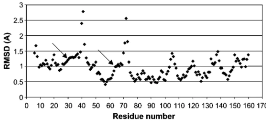
The RMSD values for each residue from the 20 NMR
structures of p19 (PDB ID 1AP7) were calculated and are
shown in Figure 4. It is clearly evident from the RMSD
plot that the residues after the first and second ankyrin
repeats (His29/Arg30 and Gly64/Ala65, respectively)
show higher RMSD than the preceding residues.
Discussion
Transition from the G
1to the S phase of the mammalian
cell cycle is regulated by the Rb/E2F pathway (Weinberg,
1995). Cyclin D/cyclin-dependent kinase-4/6 complexes
phosphorylate the retinoblastoma protein (pRb), which
frees E2F from the Rb/E2F complex. The freed E2F
acti-vates the transcription of genes involved in cell
proli-feration (Weinberg, 1995; Leone et al., 1998), and
p53-dependent (De Gregore et al., 1997; Bates et al.,
Figure 4 Atomic RMS deviation (A˚) of the backbone atoms of p19.
A total of 20 NMR structures (PDB ID 1AP7) were used to calculate the RMSD with respect to the mean. The major calpain cleavage sites His29xArg30 and Gly64xAla65 are marked with arrows. Note that these residues show higher RMSD than the preceding residues.
1998) and p53-independent apoptosis (Irwin et al., 2000).
INK4 inhibitors specifically inhibit the G
1cyclin-depend-ent kinase-mediated phosphorylation of pRb, and thus
the normal function of the Rb/E2F pathway is
deregulat-ed. INK4 inhibitors exert their action by binding directly
to CDK4/6 through their two N-terminal ankyrin repeats
(Brotherton et al., 1998; Russo et al., 1998). It is
inter-esting to note that the major calpain cleavage sites in
p19
INK4dare located exactly in these two ankyrin repeats.
Calpains should therefore influence p19
INK4dinhibitor
binding to CDK4/6 and thus may take part in the
regu-lation of this binding in vivo. This proposition is further
supported by the observation that CDK6 is resistant to
calpain proteolysis. We treated CDK6 with
m
-caplain and
observed that calpain does not affect CDK6. In addition,
we performed cleavage studies on the CDK6/p19
com-plex. Calpain did not cleave p19 when complexed to
CDK6 (data not shown). This is because the major
cleav-age sites on p19 described above are not accessible to
calpain in the CDK6-bound p19. CDK4 and CDK6 have
a typical overall bilobal fold found in many eukaryotic
protein kinases, with the smaller N-terminal domain
con-sisting predominantly of b-sheet structure and the larger
C-terminal domain consisting primarily of a-helices
(Pav-letich, 1999; Cheek et al., 2002). The structure is globular,
in contrast to the elongated rod-like structure of p19
INK4d.
Resistance of CDK6 and CDK6/p19 to calpain
proteoly-sis, as compared to the propensity of p19
INK4dand cyclin
D1 (Choi et al., 1997) for calpain degradation, suggests
that calpain may be involved in the cell cycle by
regulat-ing the cell cycle regulatory protein turnover. p19
INK4dmay
therefore be a substrate for calpains in these in vivo
situations.
There are a limited number of reports on substrate
specificity of calpains and these have been reviewed by
Croall and Demartino (1991), and more recently by Goll
et al. (2003). Early studies suggested that calpains
pre-ferentially cleave peptide bonds with a Leu or a Val
res-idue in the P2 position. More complete data, however,
indicated that substrate specificity of the calpains is
con-trolled by the conformation of a polypeptide chain and
not by an amino acid sequence (Harris et al., 1988;
Sta-bach et al., 1997; Ghosh et al., 2005). In general, the
literature data indicate that the calpains cleave target
proteins at a limited number of sites and produce large
polypeptide fragments rather than small peptides or
ami-no acids (Sasaki et al., 1984; Croall and Demartiami-no, 1991;
Goll et al., 2003). A typical example is provided by the
m-calpain proteolysis of vimentin. Vimentin belongs to
the intermediate filament (IF) family of proteins (Strelkov
et al., 2002). All IF proteins share a common structural
organization of the dimer that includes a central
coiled-coil ‘rod’ domain flanked by ‘head’ and ‘tail’ domains at
both ends (Fuchs and Weber, 1994; Strelkov et al., 2002).
The a-helical core part is not continuous, however, but
interrupted in several places, resulting in four
consecu-tive a-helical segments that are connected by linkers.
The major calpain proteolytic fragments in vimentin arise
from cleavage in the unordered amino-terminal
head-piece and the tail domain, followed by cleavage in the
linker that separates two major coiled-coil domains
(Fischer et al., 1986). Another example, related to our
a-helical p19, is provided by a-tropomyosin, a polypeptide
that is 100% a-helical. Nine of the 11 calpain cleavages
in the a-tropomyosin polypeptide are in the
COOH-ter-minal half of the molecule (Croall and Demartino, 1991).
The COOH-terminal half of the helix is significantly less
stable than the NH
2-terminal half, again suggesting that
the substrate specificity of the calpains depends on the
conformation of the polypeptide, with a more open
struc-ture favoring cleavage.
The p19 calpain cleavage seems to show yet more
fea-tures. The major calpain cleavage sites are located at the
end of the second helix (a-2) of the first ankyrin repeat
(His 29) and again at the end of the second helix (a-4) of
the second ankyrin repeat (Gly 64), as evident from
Figure 3B. It is clearly apparent from the RMSD plot
(Fig-ure 4) that the residues after the first and second ankyrin
repeats (His29/Arg30 and Gly64/Ala65, respectively)
show higher RMSD than the preceding residues,
indicat-ing higher variability and probable flexibility. Our previous
relaxation measurements (Renner et al., 1998) on p19
agree with these results. The relaxation measurements
and the RMSD values clearly indicate that the first two
ankyrin repeats exhibit increased instability. The
de-formed second ankyrin repeat is dynamically most
het-erogeneous. It exhibits high flexibility around Val69 on
fast time scales. The loop between the first and the
sec-ond ankyrin repeats shows strong exchange broadening
for many residues (His34, Gly42, Thr44, Gln47). Thus, it
Chemicals, plasmid, expression host strain and
growth media
BL21 (DE3), DH5-a competent cells and pET-15 vector were purchased from Novagen (Darmstadt, Germany). Luria broth, agar, ampicillin, thrombin, urea were obtained from Sigma (Tauf-kirchen, Germany). PVDF and nitrocellulose membranes were bought from Amersham Pharmacia Biotech (Freiburg, Germany). The restriction enzymes Nde1, BamH1 and T4 DNA ligase were purchased from New England Biolabs (Beverly, MA, USA). Plas-mid miniprep kit and Ni-NTA resin were purchased from Qiagen (Hilden, Germany). p19 anti-rabbit antibody and isopropyl b-D-thiogalactoside were obtained from Santa Cruz Biotechnology (Santa Cruz, USA) and PeqLabs (Erlangen, Germany), respec-tively. All other chemicals used were of analytical grade.
Proteolytic cleavage of p19 by
m
-calpain
Human p19 was expressed and purified in BL21 (DE 3) as a His-tag fusion protein (Kalus et al., 1997) using pET15b as an expression vector. The His-tag was removed using 5 U of throm-bin per mg of protein. CDK6 was expressed as a GST fusion protein in Spodoptera frugiperda strain Sf9 and purified as pre-viously described (Smialowski et al., 2005). The CDK6/p19 complex (1:3) was purified on a HiLoad 26/60 Superdex 75 pre-parative grade column using an AKTA explorer gel filtration chro-matographic system. Proteolytic cleavage studies were carried out at 258C in 20
m
l of reaction mixture containingm
-calpain purified from human erythrocytes (Gabrijelcic-Geiger et al., 2001) and p19 at a molar ratio of 1:50, and a calpain assay buffer (25 mMTris-HCl, pH 7.3, 150 mMNaCl, 1 mMCaCl2). A similarproteolytic assay was conducted on CDK6/p19 complex. After 1-h incubation, the reactions were terminated by addition of 10 mMEDTA. The fragments were then separated by SDS-PAGE and visualized by Coomassie brilliant blue staining.
Calpain-mediated proteolytic assays of p19 in the
presence or absence of calcium and calpastatin
The inhibitory role of calpastatin on calpainolytic digestion, the calcium requirement for calpain-mediated proteolysis of p19, and the stability of p19 in the calpain assay buffer were inves-tigated by performing three separate proteolytic cleavage assays: (1)
m
-calpain, p19 and calpastatin domain 1 were incu-bated in the calpain assay buffer (25 mM Tris-HCl, pH 7.3, 150 mMNaCl, 1 mMCaCl2) at a molar ratio of 1:50:1; (2) p19was incubated with
m
-calpain in the calpain assay buffer (25 mM Tris-HCl, pH 7.3, 150 mMNaCl) at a molar ratio of 50:1; and (3) p19 was digested withm
-calpain in the absence of calpastatin and presence of 1 and 5 mMCaCl2. All reactions were carriedout for 1 h at 258C and then stopped by addition of 10 mMEDTA. Calpain-treated samples were resolved by SDS-PAGE and then transferred onto a nitrocellulose membrane with the aid of a semi-dry electroblotting apparatus. Protein transfer from the gel onto the nitrocellulose membrane was carried out at con-stant power supply of 125 mV for 1 h. The membrane was
ini-Coomassie brilliant blue. ini-Coomassie-stained bands were cut out from the membrane and then used for N-terminal amino acid analysis by Edman degradation. Sequences obtained were used to map calpain cleavage sites and fragments generated from p19 by calpain-mediated proteolysis.
Acknowledgments
We thank Isabel Winkelmann and Lourdes Garcı´a-Ruı´z for excel-lent technical assistance. This work was supported by DFG grant (SFB 469).
References
Arora, A.S., DeGroen, P.C., Croall, D.E., Emori, Y., and Gores, G.J. (1996). Hepatocellular carcinoma cells resist necrosis during anoxia by preventing phospholipase-mediated calpain activation. J. Cell. Physiol. 167, 434–442.
Bates, S., Phillips, A.C., Clark, P.A., Stott, F., Peters, G., Ludwig, R.L., and Vousden, K.H. (1998). p14(ARF) links the tumour suppressors RB and p53. Nature 395, 124–125.
Bartek, J., Bartkova, J., and Lukas, J. (1997). The retinoblastoma protein pathway in cell cycle control and cancer. Exp. Cell Res. 237, 1–6.
Baumgartner, R., Fernandez-Catalan, C., Winoto, A., Huber, R., Engh, R.A., and Holak, T.A. (1998). Structure of human cyclin-dependent kinase inhibitor p19 (INK4d): comparison to known ankyrin-repeat-containing structures and implications for the dysfunction of tumor suppressor p16 (INK4a). Struc-ture 6, 1279–1290.
Brotherton, D.H., Dhanaraj, V., Wick, S., Brizuela, L., Domaille, P.J., Volyanik, E., Xu, X., Parisini, E., Smith, B.O., Archer, S.J., et al. (1998). Crystal structure of the complex of the cyclin D dependent kinase Cdk6 bound to the cell-cycle inhibitor p19 (INK4d). Nature 395, 244–250.
Cheek, S., Zhang, H., and Grishin, N.V. (2002). Sequence and structure classification of kinases. J. Mol. Biol. 320, 855–881. Choi, Y.H., Lee, S.J., Nguyen, P., Jang, J.S., Lee, J., Wu, M.L., Takano, E., Maki, M., Henkart, P.A., and Trepel, J.B. (1997). Regulation of cyclin D1 by calpain protease. J. Biol. Chem. 272, 28479–28484.
Croall, D.E. and Demartino, G.N. (1991). Calcium-activated neu-tral protease (calpain) system – structure, function, and reg-ulation. Physiol. Rev. 71, 813–847.
Dayton, W.R., Schollmeyer, J.V., Lepley, R.A., and Cortes, L.R. (1981). A calcium-activated protease possibly involved in myofibrillar protein-turnover-isolation of a low-calcium-re-quiring form of the protease. Biochim. Biophys. Acta 659, 48–61.
De Gregore, J., Leone, G., Miron, A., Jakoi, L., and Nevins, J.R. (1997). Distinct roles for E2F proteins in cell growth control and apoptosis. Proc. Natl. Acad. Sci. USA 94, 7245–7250. Du, X.P., Saido, T.C., Tsubuki, S., Indig, F.E., Williams, M.J., and
Ginsberg, M.H. (1995). Calpain cleavage of the cytoplasmic domain of the integrin b3 subunit. J. Biol. Chem. 270, 26146–26151.
Fischer, S., Vandekerckhove, J., Ampe, C., Traub, P., and Weber, K. (1986). Protein-chemical identification of the major cleav-age sites of the Ca2q proteinase on murine vimentin, the
mesenchymal intermediate filament protein. Biol. Chem. Hoppe-Seyler 367, 1147–1152.
Fuchs, E. and Weber, K. (1994). Intermediate filaments – struc-ture, dynamics, function, and disease. Annu. Rev. Biochem. 63, 345–382.
Gabrijelcic-Geiger, D., Mentele, R., Meisel, B., Hinz, H., Assfalg-Machleidt, L., Assfalg-Machleidt, W., Moller, A., and Auerswald, E.A. (2001). Human
m
-calpain: simple isolation from erythrocytes and characterization of autolysis fragments. Biol. Chem. 382, 1733–1737.Ghosh, M., Shanker, S., Siwanowicz, I., Mann, K., Machleidt, W., and Holak, T.A. (2005). Proteolysis of insulin-like growth fac-tor binding proteins (IGFBPs) by calpain. Biol. Chem. 386, 85–93.
Glading, A., Lauffenburger, D.A., and Wells, A. (2002). Cutting to the chase: calpain proteases in cell motility. Trends Cell Biol. 12, 46–54.
Goll, D.E., Thompson, V.F., Li, H.Q., Wei, W., and Cong, J.Y. (2003). The calpain system. Physiol. Rev. 83, 731–801. Harper, J.W. and Elledge, S.J. (1996). Cdk inhibitors in
devel-opment and cancer. Curr. Opin. Genet. Dev. 6, 56–64. Harris, A.S., Croall, D.E., and Morrow, J.S. (1988). The
calmo-dulin-binding site in a-fodrin is near the calcium-dependent protease-I cleavage site. J. Biol. Chem. 263, 15754–15761. Huang, Y. and Wang, K.K. (2001). The calpain family and human
disease. Trends Mol. Med. 7, 355–362.
Inomata, M., Hayashi, M., Nakamura, M., Saito, Y., and Kawa-shima, S. (1989). Properties of erythrocyte-membrane bind-ing and autolytic activation of calcium-activated neutral protease. J. Biol. Chem. 264, 18838–18843.
Inomata, M., Saito, Y., Kon, K., and Kawashima, S. (1990). Bind-ing-sites for calcium-activated neutral protease on erythro-cyte membranes are not membrane phospholipids. Biochem. Biophys. Res. Commun. 171, 625–632.
Irwin, M., Marin, M.C., Phillips, A.C., Seelan, R.S., Smith, D.I., Liu, W.G., Flores, E.R., Tsai, K.Y., Jacks, T., Vousden, K.H., and Kaelin, W.G. (2000). Role for the p53 homologue p73 in E2F-1-induced apoptosis. Nature 407, 645–648.
Kalus, W., Baumgartner, R., and Renner, C., Noegel, A., Chan, F.K.M., Winoto, A., and Holak, T.A. (1997). NMR structural characterization of the CDK inhibitor p19 (INK4d). FEBS Lett. 401, 127–132.
Khorchid, A. and Ikura, M. (2002). How calpain is activated by calcium. Nat. Struct. Biol. 9, 239–241.
Kuboki, M., Ishii, H., and Kazama, M. (1987). Procalpain is acti-vated on the plasma-membrane and the calpain acts on the membrane. Biochim. Biophys. Acta 929, 164–172.
Kuboki, M., Ishii, H., and Kazama, M. (1990). Characterization of calpain-I binding-proteins in human erythrocyte plasma-membrane. J. Biochem. 107, 776–780.
Lee, W.J., Adachi, Y., Maki, M., Hatanaka, M., and Murachi, T. (1990). Factors influencing the binding of calpain-I to human erythrocyte inside-out vesicles. Biochem. Int. 22, 163–171. Leone, G., DeGregori, J., Yan, Z., Jakoi, L., Ishida, S., Williams,
R.S., and Nevins, J.R. (1998). E2F3 activity is regulated dur-ing the cell cycle and is required for the induction of S phase. Genes Dev. 12, 2120–2130.
Li, H., Thompson, V.F., and Goll, D.E. (2004). Effects of autolysis and properties of
m
-and m-calpain. Biochim. Biophys. Acta 1691, 91–103.Mellgren, R.L. (1980). Canine cardiac calcium-dependent pro-teases – resolution of two forms with different requirements for calcium. FEBS Lett. 109, 129–133.
Moldoveanu, T., Hosfield, C.M., Lim, D., Elce, J.S., Jia, Z.C., and Davies, P.L. (2002). A Ca2q switch aligns the active site of
calpain. Cell 108, 649–660.
Moldoveanu, T., Hosfield, C.M., Lim, D., Jia, Z., and Davies, P.L. (2003). Calpain silencing by a reversible intrinsic mechanism. Nat. Struct. Biol. 10, 371–378.
Molinari, M., Anagli, J., and Carafoli, E. (1994). Ca2q-activated
neutral protease is active in the erythrocyte-membrane in its nonautolyzed 80-kDa form. J. Biol. Chem. 269, 27992–27995.
Morgan, D.O. (1995). Principles of Cdk regulation. Nature 374, 131–134.
Nicotera, P., Hartzell, P., Baldi, C., Svensson, S.A., Bellomo, G., and Orrenius, S. (1986). Cystamine induces toxicity in hepa-tocytes through the elevation of cytosolic Ca2q
and the stim-ulation of a nonlysosomal proteolytic system. J. Biol. Chem. 261, 4628–4635.
Pal, G.P., De Veyra, T., Elce, J.S., and Jia, Z.C. (2003). Crystal structure of a
m
-like calpain reveals a partially activated conformation with low Ca2q requirement. Structure 11,1521–1526.
Patel, Y.M. and Lane, M.D. (2000). Mitotic clonal expansion dur-ing preadipocyte differentiation: calpain-mediated turnover of p27. J. Biol. Chem. 275, 17653–17660.
Pavletich, N.P. (1999). Mechanisms of cyclin-dependent kinase regulation: structures of Cdks, their cyclin activators, and Cip and INK4 inhibitors. J. Mol. Biol. 287, 821–828.
Pines, J. (1996). Cell cycle: reaching for a role for the CDKs proteins. Curr. Biol. 6, 1399–1402.
Renner, C., Baumgartner, R., Noegel, A.A., and Holak, T.A. (1998). Backbone dynamics of the CDK inhibitor p19INK4d
studied by N-15 NMR relaxation experiments at two field strengths. J. Mol. Biol. 283, 221–229.
Ruas, M. and Peters, G. (1998). The p16INK4a/CDKN2A tumor
suppressor and its relatives. Biochim. Biophys. Acta 1378, 115–177.
Russo, A.A., Tong, L., Lee, J.O., Jeffrey, P.D., and Pavletich, N.P. (1998). Structural basis for inhibition of the cyclin-dependent kinase Cdk6 by the tumour suppressor p16INK4a. Nature 395,
237–243.
Sasaki, T., Kikuchi, T., Yumoto, N., Yoshimura, N., and Murachi, T. (1984). Comparative specificity and kinetic-studies on por-cine calpain-I and calpain-Ii with naturally-occurring peptides and synthetic fluorogenic substrates. J. Biol. Chem. 259, 2489–2494.
Serrano, M. (1997). The tumor suppressor protein p16INK4a. Exp.
Cell Res. 237, 7–13.
Serrano, M., Lin, A.W., McCurrach, M.E., Beach, D., and Lowe, S.W. (1997). Oncogenic ras provokes premature cell senes-cence associated with accumulation of p53 and p16INK4a. Cell 88, 593–602.
Sherr, C.J. and Roberts, J.M. 1999. CDK inhibitors: positive and negative regulators of G1-phase progression. Genes Dev. 13,
1501–1512.
Smialowski, P., Singh, M., Mikolajka, A., Majumdar, S., Joy, J.K., Nalabothula, N., Krajewski, M., Degenkolbe, R., Bernard, H.U., and Holak, T.A (2005). NMR and mass spectrometry studies of putative interactions of cell cycle proteins pRb and CDK6 with cell differentiation proteins MyoD and ID-2. Bio-chim. Biophys. Acta 1750, 48–60.
Spencer, M.J., Croall, D.E., and Tidball, J.G. (1995). Calpains are activated in necrotic fibers from Mdx dystrophic mice. J. Biol. Chem. 270, 10909–10914.
Stabach, P.R., Cianci, C.D., Glantz, S.B., Zhang, Z.S., and Mor-row, J.S. (1997). Site-directed mutagenesis of aII spectrin at codon 1175 modulates its
m
-calpain susceptibility. Biochem-istry 36, 57–65.Strelkov, S.V., Herrmann, H., Geisler, N., Wedig, T., Zimbelmann, R., Aebi, U., and Burkhard, P. (2002). Conserved segments 1A and 2B of the intermediate filament dimer: their atomic structures and role in filament assembly. EMBO J. 21, 1255–1266.
Tompa, P. and Friedrich, P. (2000). Kinetic analysis of human
m
-calpain autolysis. In: Calpain Methods and Protocols, J.S. Elce, ed. (Totowa, NJ, USA: Humana Press), pp. 129–136. Tsutsumi, M., Tsai, Y.C., Gonzalgo, M.L., Nichols, P.W., andJones, P.A. (1998). Early acquisition of homozygous deletions of p16/p19 during squamous cell carcinogenesis and genetic mosaicism in bladder cancer. Oncogene 17, 3021–3027.