diffuse out from the matrix and the hydrogel will have to be replaced regularly.[4] Here, we propose an in situ cross-linking liposome-containing mechanoresponsive hydrogel combining the advantages of each system, liposomes and hydrogels, to yield a proof-of-concept intra-articular on-demand drug delivery system.
Liposomes are self-assembled water filled spheres made of a phospholipid bilayer.[5] This bilayer can be for-mulated from a wide variety of phospholipids that may even bear targeting moieties or that react to external stimuli such as temperature or shear stress.[6] Virtually any water soluble molecule can be encapsulated inside a liposome, making it a versatile drug delivery technology. In this paper, we have formulated 100 nm extruded large unilamellar vesicles (LUVEt100) from 1,2-dipalmitoyl-sn-glycero-3-phosphatidylcholine (DPPC) and 20 mol% of admixed 1,2-dipalmitoyl-sn-glycero-3-phosphatidyl-ethanolamine (DPPE). The addition of DPPE will yield liposomes with an amine-covered nucleophilic surface that could then react with electrophilic functional groups such as aldehydes (Figure 1).
Various liposome-in-hydrogel systems have been introduced in the past years. In its most straightforward forms, the hydrogels are simply made by increasing the viscosity of the liposomal suspensions via the addition of a polymer.[7] Alternatively, Schmidt et al. showed how suppressing the vesicle undulation movement by stiff-ening the membrane will lead to adhesive vesicle con-tacts and therefore vesicle hydrogels.[8]
The direct injection of a drug into a joint can relieve
osteoar-thritic pain for a short period of time. The problem is that the
drug will not stay at the allocated location. Therefore, a
proof-of-concept in situ is designed forming hydrogel containing
liposomes that are covalently linked to the hydrogel
net-work. When the liposomes are filled with a cargo, the formed
hydrogel is thus loaded with this cargo, too. Due to the link
between the hydrogel and the liposomes, a compression or
other mechanical force applied to the hydrogel will rupture
the liposomes and release a small percentage of the cargo.
Overall, a long-term intra-articular drug release is feasible.
Liposome-Containing Mechanoresponsive
Hydrogels
E. Stalder, A. Zumbuehl*
E. Stalder, Prof. A. Zumbuehl University of Fribourg Department of Chemistry
Swiss National Centres of Competence in Research in Chemical Biology (Geneva) and Bio-Inspired Materials (Fribourg) Chemin du Musée 9, 1700 Fribourg, Switzerland E-mail: [email protected]
1. Introduction
Osteoarthritis is the most common form of arthritis and is a degenerative disease affecting the body’s synovial joints, mainly in the hand, knee, and hips.[1] The age-related disease leads to chronic pain and loss of mobility of the affected joints. By 2030 up to 25% of the adult popu-lation will be affected by doctor-diagnosed arthritis, i.e., almost 67 million people alone in the USA.[1,2] Current treatment varies from physiotherapy to total joint replace-ment and concomitant pain managereplace-ment using topical, oral, and parenteral administration of pain killers.[3] Con-sidering the side effects of the long-term administration of off-site pain killers, the need for a localized intra-artic-ular drug delivery system arises. Pain medication can be incorporated into liposomes but these are not suitable for long-term localized treatment as they are liquid suspen-sions that will rapidly diffuse away from the site of the joint. Hydrogels on the other hand can be cross-linked in situ in a joint and do not move away from the point of injection. However, any hydrogel-loaded cargo will rapidly
http://doc.rero.ch
Published in "Macromolecular Materials and Engineering (): , 2017"
which should be cited to refer to this work.
Liposomes can also be grafted physically onto poly-mers using either electrostatic interactions[9] or polymers grafted with long hydrophobic chains that would reach into the hydrophobic liposomal membranes and retain the vesicles in a gel matrix.[10] For surfactant vesicles, electrostatic interactions between charged vesicles and polymers led to longer lived cross-links but the density of cross-links proofed to be larger between vesicles and hydrophobic polymers. Additionally, a too high charge density was found to be nonideal because then the polymer would adhere too strongly to the vesicles and they might be disrupted.[10]
Linking vesicles to chitosan containing n-dodecyl chains restrains vesicles in a hydrogel network. A simple dilution or the action of chitosanase will lead to the release of the vesicles.[11] The gelation can be reversed by the addition of α-cyclodextrin that takes up the hydro-phobic chain of the modified chitosan in its hydrohydro-phobic pocket.[12] The incorporation of a hydrophobic chain into a vesicle bilayer will increase its main phase transition temperature and will induce the formation of faceted ves-icles.[13] These types of hydrogels are well advanced and a vesicle coated with a hydrogel containing hydrophobic chains was used for a proof-of-concept for combined cancer therapy and diagnosis.[14]
Far less advanced is the chemical cross-linking of vesi-cles and polymers. Similarly to the work presented here, hydrogels were formed from xanthan aldehyde and pure PE liposomes leading to an interesting self-healable and injectable 3D hydrogel scaffold.[15] In another
approach, aldehyde functionalized polymeric micelles made from poly(ethylene-glycol)-b-poly(lactic acid) were cross-linking a polyethyleneimine (PEI) polymer within seconds.[16]
The release of a compound from a liposome in a hydrogel is expected to be slower than the release of the compound in a pure hydrogel.[17] Indeed, the release of 5(6)-carboxyfluorescein from liposomes entrapped inside a chitosan hydrogel was found to be delayed, compared to free 5(6)-carboxyfluorescein inside a hydrogel.[18] This is of course interesting for long-term drug delivery as was demonstrated using doxorubicin-loaded vesicles inside chitosan hydrogels after an intra-tumor injection of a hydrogel.[19]
2. Experimental Section
2.1. Vesicle Formulation
The large unilamellar vesicles were formulated using standard
LUV extrusion protocol.[20] Briefly, the desired lipid mixture was
dissolved in chloroform, then the solvent was removed under reduced pressure to form a lipid thin film which was then dried under high vacuum overnight. The film was hydrated with the
desired buffer (10 × 10−3M HEPES, pH = 7.4; or 10 × 10−3M HEPES,
50 × 10−3M sulforhodamine B, pH = 7.4) for 30 min at 65 °C (for
HG2, HG5) and 70 °C (for HG3, HG6). After five freeze-thaw cycles
(liquid nitrogen and 65 °C (for HG2, HG5) or 70 °C (for HG3, HG6)) the multilamellar vesicle suspension was extruded through track-edged membranes (5 times each for 400 nm, 200 nm, and
Figure 1. Molecular representation of 1,2-dipalmitoyl-sn-glycero-3-phosphatidylcholine (DPPC),
1,2-dipalmitoyl-sn-glycero-3-phosphatidyle-thanolamine (DPPE), oxidized dextran DextCHOx, and polyethyleneimine (PEI). Below, an artist’s rendering is shown of the concept where
drug-loaded liposomes are A) physically trapped inside a hydrogel, or B) physically and chemically attached to the hydrogel. C) In the second case, a mechanical force applied to the hydrogel will rupture the vesicle and release the entrapped cargo.
15 times for 100 nm) with a pressure extruder (Liposofast LF-50, Avestin, Canada). The suspensions containing sulforhodamine B were then purified using size exclusion chromatography (PD-10 columns, GE Healthcare, Great Britain). The resulting liposomal suspension was finally mixed with a solution of PEI (2.5 weight% in HEPES buffer).
2.2. Dextan Oxidation
A stock solution of 10 g dextran (high fraction, Acros Organics, USA) was dissolved in ultrapure water (1 L, 18.2 MΩ cm at 25 °C) then sodium metaperiodate (Fluka, Switzerland) was added (1.0 g,
4.7 mmol for DextCHO5; 2.0 g, 9.4 mmol for DextCHO10) and the
solution was stirred at room temperature for 2 h. Glycerol (25 mL, 216 mmol, Reactolab, Switzerland) was added to quench the reaction and the solution was stirred for another hour at room
temperature. The DextCHOx was precipitated from water with
ethanol (80% v/v) and centrifuged at 8000 rpm for 15 min. The precipitate was redissolved in water at 40 °C before being precip-itated again with ethanol (80% v/v) and centrifuged at 8000 rpm for 30 min. The resulting precipitate was dried under vacuum yielding a white glass-like solid (65%–85%). The degree of oxi-dation was determined using the hydroxylamine hydrochloride
titration method (see the Supporting Information).[21]
2.3. Hydrogel Formation
The oxidized dextran DextCHOx was fully dissolved in HEPES
buffer (10 × 10−3M HEPES, pH = 7.4, 20% w/v). 850 μL of the
oxi-dized dextran was then added to one barrel of a double barrel syringe (L-System, Medmix, Switzerland). The second barrel was
filled with 850 μL of a liposomal suspension (10 mg mL−1) and
admixed PEI (1.25% w/v, 750 kDa, Sigma-Aldrich, Germany). The solutions were pushed through the double barrel syringe into a custom-made round teflon mold (d = 14 mm, h = 10 mm) forming a hydrogel within seconds at room temperature. The hydrogel disk was extracted from the mold after 5 min.
2.4. Elastic Modulus
The hydrogel samples were formulated as described above using
pure HEPES buffer (10 × 10−3M HEPES, pH = 7.4) and were then
put on the mechanical dynamical analyzer (Z010, Zwick/Roell,
Germany). The samples were gradually compressed (2 mm min−1)
and the standard force was recorded as a function of the com-pression. The Young’s modulus was calculated using the slope of the first 5% compression.
2.5. Diffusion Controlled Release
The hydrogel samples were formulated as described above using HEPES buffer containing sulforhodamine B loaded liposomes. The samples were placed into a Transwell permeable sup-port (Corning, USA) and the supsup-port was placed into the cor-responding 6 well plate containing 10 mL of HEPES buffer. The fluorescence release over time was measured after removing the permeable support and directly inserting the plate into a plate reader (Sense 425-301, Hidex, Finland). The sample was excited
at 544 nm and the fluorescence was recorded at 622 nm. The 100% fluorescence was obtained after disrupting the liposomes with Triton X100 (200 μL of a 10% v/v solution, Sigma-Aldrich, Germany). The fluorescence was normalized by dividing the value by the mass of the sample.
2.6. Mechanoresponsive Release
The hydrogel samples were formulated as described above using HEPES buffer containing sulforhodamine B loaded liposomes and were then put on the mechanical dynamical analyzer (Z010,
Zwick/Roell, Germany; compression speed 2 mm s−1, 5 s holding
time). The samples were compressed at 10% or 25% compres-sion, respectively for 50, 100, or 200 cycles, respectively. The control sample was not compressed. Then the hydrogel samples were transferred to a Transwell plate and the fluorescence was recorded after 36 h following the same protocol as for the diffu-sion controlled release above.
2.7. Release of Intact Liposomes from the Hydrogels
An untouched hydrogel sample as prepared for the mechanore-sponsive release experiment (details see above) was placed in
pure HEPES buffer for 36 h. Then 200 μL aliquots of the buffer
were transferred into 96 well plates (TPP, Switzerland) and to
half of the samples was added Triton X100 (4 μL of a 10% v/v
solution) to destroy any vesicles that were present in the solu-tion. After 30 min incubation the fluorescence was measured as stated in the diffusion controlled release above.
3. Results and Discussion
Here, we use dextran as the basis for a hydrogel. Dex-tran is a polysacharide made of repeating glucose units that can be oxidized by sodium periodate to get an aldehyde-containing polymer (Figure 1B).[22] The oxidized dextran will be named DextCHOx with the
subscript number representing the percentage of oxidized glucose units (see the Supporting Informa-tion). The formed aldehyde group reacts with both the amines on the surface of the liposomes and the added copolymer PEI (Figure 1). The Schiff base formation was chosen because it tolerates an aqueous environment at physiological pH, the reaction leads to no byproducts, and the formed chemical bond is dynamic, allowing the hydrogel to degrade over time.
Two hydrogels were formed from oxidized dextran, PEI, and vesicles. The first hydrogel contained pure DPPC vesi-cles, the second hydrogel contained DPPC/DPPE vesicles. This creates two systems in which the vesicles are only physically attached to the hydrogel (Figure 1A) or both physically and chemically attached (Figure 1B). In both systems, the vesicles and their loaded cargo stay intact. If a mechanical force is applied to the first type of hydrogel, e.g., inside a joint, this force is transmitted to the vesicles.
Because these vesicles are not chemically connected to the hydrogel matrix they are able to evade the stress applied to the hydrogel and no cargo is released (Figure 1A). How-ever, if the vesicles are covalently attached to the hydrogel (Figure 1B) the deformation of the hydrogel under com-pression will lead to a rupture of the vesicle membrane and will release the cargo (Figure 1C). The released mole-cules diffuse through the hydrogel into the surrounding joint tissue, possibly allowing for a local, on-demand delivery of a drug, reducing its side effects.
Each hydrogel system was made from either DextCHO5
or DextCHO10. With the negative controls of vesicle-free
hydrogels all sums up a total of six different hydrogels tested in this paper (see Table 1). The hydrogels were pre-pared in a double barrel syringe. One barrel contained the oxidized dextran, the other barrel contained the PEI and the vesicles. This set-up allows for a ready-to-use fluid, in situ gelating system that would toughen in under 5 min. The liposomes could be filled with a cargo. Here, we used the fluorescent marker sulforhodamine B but this could be replaced by a drug, leading to a drug-filled hydrogel.
The Young’s modulus (elastic modulus) was determined for all six systems (see Figure 2A,B). Using a dynamic mechanical analyzer in compression mode the hydrogels were continually compressed at room temperature until they structurally failed. All systems showed a Young’s modulus of 1 kPa calculated from the linear domain of the compression curve. In this linear domain the addition of a vesicle to the hydrogel had no influence on the mate-rial properties. However, the degree of oxidation of the dextran did affect the general properties of the hydrogels. The DextCHO5 systems showed the expected behavior for
a soft hydrogel (see Figure 2A).[23] Here, the linear domain was followed by an elastic domain where the material stiffened until the system structurally failed at around 35%
Table 1. Composition of the six hydrogels discussed. Each hydrogel
was formed in a double barrel syringe at room temp erature and gelated as soon as the components came into contact with each other. The systems all toughened in under 5 min.
Hydrogel Composition
HG1 DextCHO5; PEI
HG2 DextCHO5; PEI + DPPC
HG3 DextCHO5; PEI + DPPC + DPPE
HG4 DextCHO10; PEI
HG5 DextCHO10; PEI + DPPC
HG6 DextCHO10; PEI + DPPC + DPPE
Figure 2. Top row: elastic moduli of the different hydrogel formulations used. A) Elastic moduli of HG1 (blue), HG2 (green), and HG3 (red).
B) Elastic moduli of HG4 (blue), HG5 (green), and HG6 (red). Bottom row: spontaneous release of a vesicle-entrapped dye from the hydrogel into the surrounding fluid. C) Spontaneous release of sulforhodamine B from HG1 (blue), HG2 (green), and HG3 (red). D) Spontaneous release of sulforhodamine B from HG4 (blue), HG5 (green), and HG6 (red).
compression. Going to a higher degree of dextran oxida-tion, the DextCHO10 system showed only a linear
rela-tionship between stress and compression and quickly lost its structural integrity above 10% compression (see Figure 2B). The higher degree of dextran oxidation might mean that the hydrogels are more likely to form smaller gel domains that are only weakly linked together. No influence was found when unreactive vesicles were added to the hydrogel (HG5, green) but a significantly
weakened hydrogel formed by adding reactive vesi-cles (HG6, red). This could be explained by the presence of a higher number of reactive aldehydes in DextCHO10
compared to DextCHO5, which would allow more
cova-lent bonds between the reactive vesicles in HG6 and the dextran, leaving less aldehyde groups to react with the crosslinking PEI and thus reducing the chance of two strands of dextran to be chemically connected. The overall result would be smaller gel domains in DextCHO10
hydro-gels compared to DextCHO5 and even smaller domains
for HG6. This would make the resulting hydrogels more brittle.
The diffusion controlled release profile of sulforho-damine B is the same for both pure DextCHO5 and
DextCHO10 hydrogels HG1 and HG4 (see Figure 2C,D, blue
curves). The release is rapid for the first couple of hours
and stalls at 70% after 20 h. When the sulforhodamine B is not admixed directly within the hydrogel but encapsu-lated in an inert liposome (HG2 and HG5 see Figure 2C,D, green curve), only around 5% of the total dye content is released over several days. When the hydrogel is loaded with reactive liposomes (HG3 and HG6, see Figure 2C,D, red curves) some of the vesicles will break during the gelation process and around 20% of the dye is released during the first day whereupon the system stays tight for the remainder of days tested (see Figure 2C,D, red curves).
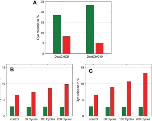
Next, we determined how the liposomes themselves were diffusing through the hydrogel network into the buffer solution. The addition of the surfactant Triton X100 to the neat buffer destroyed any free floating vesicles and the increase in fluorescence measured was directly linked to the amount of vesicles that escaped from the hydro-gels (see Figure 3A). Covalently linking the vesicles to the hydrogels resulted in a 50% reduction of intact liposomes in the buffer for the DextCHO5 and over four times
reduc-tion for DextCHO10.
In order to show the mechanoresponse of the hydrogels in a simulated joint system, the samples were compressed repeatedly for 10% (see Figure 3B) and 25% (see Figure 3C) of their height in a dynamic mechanical analyzer. The
DextCHO10 sample was not analyzed because it failed
Figure 3. Release of vesicle-entrapped sulforhodamine B under various conditions. The release from inert DPPC vesicles is depicted in green
and the release from the reactive DPPC/DPPE vesicles in red. A) Control experiment quantifying the amount of intact vesicles released from the hydrogel samples after 36 h. (B) and (C) depict the mechanically induced dye-release as a function of the number of compression cycles.
B) DextCHO5 hydrogels compressed by 10% of their height. C) DextCHO5 hydrogels compressed by 25% of their height.
at around 10% compression (see Figure 2B). After the compression cycles, the samples were placed in a buffer for 36 h and the fluorescence increase was recorded. Interestingly, the compression of hydrogel samples (Figure 3B,C) showed that compressing the samples had no effect on the release from the inert pure DPPC liposomes (HG2, green bars in Figure 3B,C). With the reac-tive DPPC/DPPE liposomes there was a small increase in release with increasing compression (HG3, red bars in Figure 3B,C). For the 200 cycles experiment we calculated a 0.016% dye release per cycle for the 10% compression and 0.034% dye release for the 25% compression. This small per-cycle release observed here is interesting in the drug delivery point of view as it will allow the system to work for a longer time before running out of drug.
4. Conclusions
In conclusion, we have presented a proof-of-concept of a mechanoresponsive hydrogel containing covalently bound liposomes. The hydrogels can be formed in situ using a double barrel syringe. Once formed, the hydrogels can har-vest the mechanical forces applied to them and transfer them to the bound liposomes, thus triggering the release of the liposomal cargo. The system’s very low rate of dye release makes it interesting for prolonged drug delivery applications, e.g., to mitigate osteoarthritic pain. Applied intra-articularly to an inflamed joint, a baseline drug release and a higher peak release during high effort could be the hallmarks of a new type of long-term on-demand pain therapy.
Supporting Information
Supporting Information is available from the Wiley Online Library or from the author.
Acknowledgements: The authors thank the Swiss National Science Foundation (PP00P2_138926/1) and the University of Fribourg for financial support and Dr. Marco Lüchinger for Gel Permeation measurements.
Received: December 15, 2016; Published online: ; DOI: 10.1002/ mame.201600549
Keywords: arthritis; hydrogels; liposomes; mechanoresponsive drug delivery; on demand drug delivery
[1] R. C. Lawrence, D. T. Felson, C. G. Helmick, L. M. Arnold, H. Choi, R. A. Deyo, S. Gabriel, R. Hirsch, M. C. Hochberg, G. G. Hunder, J. M. Jordan, J. N. Katz, H. M. Kremers, F. Wolfe, for the National Arthritis Data Workgroup, Arthritis Rheum.
2008, 58, 26.
[2] J. M. Hootman, C. G. Helmick, Arthritis Rheum. 2006, 54, 226. [3] M. Hochberg, R. Altman, K. April, M. Benkhalti, G. Guyatt,
M. Jessie, T. Towheed, V. Welch, G. Wells, P. Tugwell, Arthritis
Care Res. 2012, 64, 465.
[4] D. Caccavo, S. Cascone, G. Lamberti, A. A. Barba, Int. J. Pharm.
2015, 486, 144.
[5] T. M. Allen, P. R. Cullis, Adv. Drug Delivery Rev. 2013, 65, 36. [6] D. Mellal, A. Zumbuehl, J. Mater. Chem. B 2014, 2, 247.
[7] S. Mourtas, C. A. Aggelopoulos, P. Klepetsanis,
C. D. Tsakiroglou, S. G. Antimisiaris, Langmuir 2009, 25, 8480. [8] D. Grabner, S. Hoffmann, H. Forster, S. Rosenfeldt, J. Linders,
C. Mayer, Y. Talon, J. Schmidt, Adv. Colloid Interface Sci. 2014,
208, 252.
[9] E. F. Marques, O. Regev, A. Khan, M. G. Miguel, B. Lindman,
Macromolecules 1999, 32, 6626.
[10] F. E. Antunes, E. F. Marques, R. Gomes, K. Thuresson, B. Lindman, M. G. Miguel, Langmuir 2004, 20, 4647.
[11] C. Zhu, J.-H. Lee, S. R. Raghavan, G. F. Payne, Langmuir 2006, 2951.
[12] Y. Chen, V. Javvaji, I. C. MacIntire, S. R. Raghavan, Langmuir
2013, 29, 15302.
[13] F. E. Antunes, R. O. Brito, E. F. Marques, B. Lindman, M. Miguel, J. Phys. Chem. B 2007, 111, 116.
[14] J.-H. Park, H.-J. Cho, H. Y. Yoon, I.-S. Yoon, S.-H. Ko, J.-S. Shim, J.-H. Cho, J. H. Park, K. Kim, I. C. Kwon, D.-D. Kim, J. Controlled
Release 2014, 174, 98.
[15] Y.-H. Ma, J. Yang, B. Li, Y.-W. Jiang, X. Lu, Z. Chen, Polym.
Chem. 2016, 7, 2037.
[16] Y. Uchida, K. Fukuda, Y. Murakami, J. Polym. Sci., Part B:
Polym. Phys. 2012, 51, 124.
[17] S. Mourtas, S. Fotopoulou, S. Duraj, V. Sfika, C. Tsakiroglou, S. G. Antimisiaris, Colloids Surf., B 2007, 55, 212.
[18] A. Billard, L. Pourchet, S. Malaise, P. Alcouffe,
A. Montembault, C. Ladaviere, Carbohydr. Polym. 2015, 115, 651.
[19] S. Ren, Y. Dai, C. Li, Z. Qiu, X. Wang, F. Tian, S. Zhou, Q. Liu, H. Xing, Y. Lu, X. Chen, N. Li, Eur. J. Pharm. Sci. 2016, 92, 137. [20] F. Olson, C. A. Hunt, F. C. Szoka, W. J. Vail, D. Papahadjopoulos,
Biochim. Biophys. Acta 1979, 557, 9.
[21] D. Bruneel, E. Schacht, Polymer 1993, 34, 2628.
[22] S. P. Hudson, R. Langer, G. R. Fink, D. S. Kohane, Biomaterials
2010, 31, 1444.
[23] R. G. Larson, The Structure and Rheology of Complex Fluids, Oxford University Press, New York 1999.