HAL Id: hal-02299909
https://hal.archives-ouvertes.fr/hal-02299909
Submitted on 9 Dec 2020
HAL is a multi-disciplinary open access archive for the deposit and dissemination of sci-entific research documents, whether they are pub-lished or not. The documents may come from teaching and research institutions in France or abroad, or from public or private research centers.
L’archive ouverte pluridisciplinaire HAL, est destinée au dépôt et à la diffusion de documents scientifiques de niveau recherche, publiés ou non, émanant des établissements d’enseignement et de recherche français ou étrangers, des laboratoires publics ou privés.
Detection of Brain Activation in Unresponsive Patients
with Acute Brain Injury
Jan Claassen, Kevin Doyle, Adu Matory, Caroline Couch, Kelly Burger,
Angela Velazquez, Joshua Okonkwo, Jean-Rémi King, Soojin Park, Sachin
Agarwal, et al.
To cite this version:
Jan Claassen, Kevin Doyle, Adu Matory, Caroline Couch, Kelly Burger, et al.. Detection of Brain Activation in Unresponsive Patients with Acute Brain Injury. New England Journal of Medicine, Mas-sachusetts Medical Society, 2019, 380 (26), pp.2497-2505. �10.1056/NEJMoa1812757�. �hal-02299909�
The
new england
journal
of
medicine
established in 1812 June 27, 2019 vol. 380 no. 26
From the Departments of Neurology (J.C., K.D., A.M., C.C., K.M.B., A.V., J.U.O., S.P., S.A., D.R., M.M., A.E., B.R.) and Neurosurgery (E.S.C.), Columbia Univer-sity, and the Department of Psychology, New York University (J.-R.K.) — both in New York. Address reprint requests to Dr. Claassen at the Neurological Institute, Co-lumbia University, 177 Fort Washington Ave., MHB 8 Center, Rm. 300, New York, NY 10032, or at jc1439@ columbia . edu. This article was updated on June 27, 2019, at NEJM.org.
N Engl J Med 2019;380:2497-505. DOI: 10.1056/NEJMoa1812757
Copyright © 2019 Massachusetts Medical Society. BACKGROUND
Brain activation in response to spoken motor commands can be detected by elec-troencephalography (EEG) in clinically unresponsive patients. The prevalence and prognostic importance of a dissociation between commanded motor behavior and brain activation in the first few days after brain injury are not well understood.
METHODS
We studied a prospective, consecutive series of patients in a single intensive care unit who had acute brain injury from a variety of causes and who were unrespon-sive to spoken commands, including some patients with the ability to localize painful stimuli or to fixate on or track visual stimuli. Machine learning was ap-plied to EEG recordings to detect brain activation in response to commands that patients move their hands. The functional outcome at 12 months was determined with the Glasgow Outcome Scale–Extended (GOS-E; levels range from 1 to 8, with higher levels indicating better outcomes).
RESULTS
A total of 16 of 104 unresponsive patients (15%) had brain activation detected by EEG at a median of 4 days after injury. The condition in 8 of these 16 patients (50%) and in 23 of 88 patients (26%) without brain activation improved such that they were able to follow commands before discharge. At 12 months, 7 of 16 pa-tients (44%) with brain activation and 12 of 84 papa-tients (14%) without brain activa-tion had a GOS-E level of 4 or higher, denoting the ability to funcactiva-tion indepen-dently for 8 hours (odds ratio, 4.6; 95% confidence interval, 1.2 to 17.1).
CONCLUSIONS
A dissociation between the absence of behavioral responses to motor commands and the evidence of brain activation in response to these commands in EEG re-cordings was found in 15% of patients in a consecutive series of patients with acute brain injury. (Supported by the Dana Foundation and the James S. McDonnell Foundation.)
abs tr act
Detection of Brain Activation in Unresponsive Patients
with Acute Brain Injury
Jan Claassen, M.D., Kevin Doyle, M.A., Adu Matory, B.A., Caroline Couch, B.A., Kelly M. Burger, B.A., R.E.E.G.T., Angela Velazquez, M.D., Joshua U. Okonkwo, M.D., Jean-Rémi King, Ph.D., Soojin Park, M.D.,
Sachin Agarwal, M.D., David Roh, M.D., Murad Megjhani, Ph.D., Andrey Eliseyev, Ph.D., E. Sander Connolly, M.D., and Benjamin Rohaut, M.D.
T h e ne w e ngl a nd jou r na l o f m e dicine
C
linically unresponsive patients can have electroencephalographic (EEG) or magnetic resonance imaging (MRI) evidence of brain activation in response to spo-ken commands.1-4 A meta-analysis has reportedthat 14% of chronically unresponsive patients may have a dissociation between behavior and brain activation (cognitive–motor dissociation5)
months or years after injury.6 However, the
prevalence and prognostic relevance of this dis-sociation, if detected in the days soon after brain injury, are not well understood. The absence of an ability to follow commands shortly after brain injury may have an effect on decisions regarding the withdrawal of life-sustaining therapies.7,8
We studied the prevalence and prognostic im-portance of brain activation detected by EEG in response to spoken commands to perform a motor task. We used a machine-learning tech-nique3 to analyze EEG recordings obtained at
the bedside in a prospective cohort of unrespon-sive patients with acute brain injury in a single intensive care unit (ICU).
Methods
Patients
From July 2014 through September 2017, we pro-spectively screened all patients who were admit-ted with acute brain injury to the neuroscience ICU of our hospital; screening was performed within 3 days after admission. Patients were screened for the absence of the ability to follow spoken commands — for example, “stick out your tongue” or “show me two fingers with your right hand” (details are provided in the Supple-mentary Appendix, available with the full text of this article at NEJM.org). In keeping with our routine practice and in accordance with guide-lines regarding EEG monitoring of patients in the ICU,9 unresponsive patients either were
moni-tored by continuous EEG or were anticipated to be connected to monitoring within 12 hours after screening, unless imminent death was ex-pected. We enrolled all patients who were in a coma, vegetative state, or minimally conscious state–minus (defined as unresponsiveness with preserved visual fixation, visual pursuit, or local-ization to noxious stimuli); who had an acute brain injury of any type; and who were undergo-ing or were expected to undergo imminent
con-tinuous EEG monitoring. The presence of the minimally conscious state–minus was determined with the use of the Coma Recovery Scale–Revised (CRS-R,10 a six-dimension, 23-point scale of
hier-archically arranged items [with no cutoff score used for enrollment]), which we assessed among patients who were not receiving deep sedation or neuromuscular blockade. The exclusion criteria were an age of less than 18 years, a preexisting disorder of consciousness before the onset of the acute brain injury that resulted in the current admission, pregnancy, deafness before the acute brain injury, clinical recovery of the ability to follow commands before enrollment, patients or families who did not want to participate in the study, or logistic reasons (details are provided in the Supplementary Appendix).
Patients, families, and treating physicians were unaware of the results of the EEG recordings, and these results were not made available to treat-ing clinicians in relation to decisions regardtreat-ing the withdrawal of care. Demographic data and data on complications that occurred during the hospital stay and on outcomes were prospectively collected. In addition, we recorded EEGs from 10 healthy volunteers with a mean age of 31 years, using the same EEG protocol as in the patients (see the Supplementary Appendix).
Study Oversight
The study was approved for patients and healthy volunteers by the local institutional review board. Written informed consent was obtained from the patients’ surrogates and from the healthy volunteers; patients who recovered consciousness were given the opportunity to withdraw from the study.
The first and last authors are responsible for the study design and drafting of the manuscript. There was no industry involvement in or support for the study. The authors vouch for the accuracy and completeness of the data and for the fidelity of the trial to the protocol, available at NEJM.org. The results are reported in accordance with the Strengthening the Reporting of Observational Studies in Epidemiology (STROBE) guidelines for reporting observational studies.11
Study Procedures
Daily neurologic examinations, including a clin-ical assessment of the ability or inability of the
A Quick Take is available at NEJM.org
patient to follow spoken commands (“stick out your tongue,” “show me two fingers with your right hand,” and “wiggle your toes”), were per-formed during morning rounds, and the results were recorded.12 Each EEG assessment was
pre-ceded by a clinical examination that included the CRS-R10 in order to categorize the clinical
state of consciousness at the time of the record-ing (details are provided in the Supplementary Appendix).
Functional outcome was assessed with the Glasgow Outcome Scale–Extended (GOS-E; levels range from 0 to 8, with higher levels indicating better outcomes), with data obtained in a struc-tured telephone interview at 12 months after the injury.13,14 Both the patient and the interviewer
who performed the outcome assessments were unaware of the results of the above-noted exami-nations during routine rounds and were unaware of the EEG categorization. Outcomes were dichot-omized at a GOS-E level of 4, a level that signi-fies the ability to be left up to 8 hours during the day without assistance.
At the time of the EEG and clinical assess-ments, all patients were evaluated to ensure the absence of the following complications: seizures, hyperglycemia (serum glucose level, >11.1 mmol per liter [>200 mg per deciliter]), hyponatremia or hypernatremia (serum sodium level <133 and >150 mmol per liter, respectively), and renal or fulminant liver failure. For the daily neurologic assessment, sedated patients underwent inter-ruption or reduction of sedation if it was deemed safe by the attending physician during rounds.15
Both the behavioral and the EEG assessments were performed during interruption of sedation whenever possible. To account for cases in which stopping sedation was unsafe, during the study we developed a post hoc method to explore the effect of sedative and analgesic medications on EEG responses by collecting information on the doses of administered medications at the time of the assessments as well as the cumulative doses received within the two preceding elimination half-lives of each agent.3 Sedation was categorized
as “minimal” for discontinuous (e.g., single-push) administration and as “low” or “moderate” accord-ing to the cumulative doses administered through continuous drip during the two previous half-lives (details are provided in Table S3 in the Supple-mentary Appendix).
Motor Command Protocol
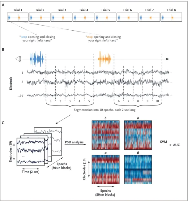
Spoken command instructions during EEG record-ing alternated between “keep openrecord-ing and clos-ing your right hand” and “stop openclos-ing and closing your right hand.”16 A total of six blocks
each with eight consecutive trials of “keep open-ing . . .” and “stop openopen-ing . . .” commands were recorded (Fig. 1A). For each patient, we recorded three blocks in which the patient was asked to move the right hand and three blocks in which the patient was asked to move the left hand (recordings of the right-hand and left-hand blocks were alternated; see the Supplementary Appendix). The total duration of the motor com-mand session was approximately 25 minutes.
EEG Acquisition and Processing
Digital bedside EEG monitoring was performed with a standard 21-electrode montage.9 EEG
re-cording quality (e.g., lead maintenance and move-ment artifact) was determined by bedside visual observation at the time of recording in addition to twice-daily lead maintenance by EEG techni-cians (see the Supplementary Appendix). For each EEG recording, power in predefined frequency ranges was calculated3,12,17 and used to train a
machine-learning algorithm (support vector ma-chine [SVM] with a linear kernel) to distinguish between the EEG responses that followed the commands “keep opening . . .” and “stop opening . . . .”
Statistical Analysis
The performance of the machine-learning algo-rithm for each EEG recording was estimated as the area under the receiver-operating-character-istic curve (AUC). To evaluate the significance of the AUC, a one-tailed permutation test was per-formed (training and evaluation of the classifier 500 times after random shuffling of the “keep opening . . .” and “stop opening . . .” com-mands18,19). Recordings were considered to show
evidence of brain activation that was temporally concordant with spoken commands if the AUC was significantly greater than 0.5 (corresponding to the level that would be expected by chance), after application of the Benjamini–Hochberg false-discovery-rate method in cases of multiple recordings in a given patient.20,21 All EEG
analy-ses were performed with the use of open-source packages, including MNE-Python (www . martinos
T h e ne w e ngl a nd jou r na l o f m e dicine
. org/ mne/ stable/ index . html)22 and Scikit-learn
(http://scikit - learn . org/ stable/ index . html).23
Categorical variables were expressed as num-bers and percentages and were compared with
the use of Fisher’s exact or ordinal chi-square tests, as appropriate. Continuous variables were expressed as medians and interquartile ranges or as means and standard deviations, as
appro-Figure 1. Motor Command Protocol and Data Processing.
Each block in the motor command protocol consisted of eight trials alternating between the instructions “keep open-ing and closopen-ing your right (left) hand” and “stop openopen-ing and closopen-ing your right (left) hand” (Panel A). The 10 sec-onds of electroencephalographic (EEG) recording after the instructions were given were extracted and segmented in five epochs, each 2 seconds long, for further analysis (Panel B). This procedure resulted in 480 epochs in patients (5 epochs × 2 instructions × 8 trials × 6 blocks) and 240 epochs in controls (5 epochs × 2 instructions × 8 trials × 3 blocks). Power spectral density (PSD) analysis was applied to the obtained EEG matrix in four frequency bands (δ [1 to 3 Hz], θ [4 to 7 Hz], α [8 to 13 Hz], and β [14 to 30 Hz]) (Panel C). The resulting features were used to train and test a sup-port vector machine (SVM). The classification performance of the SVM for a given recording was assessed as the area under the receiver-operating-characteristic curve (AUC).
B
C A
Trial 1 Trial 2 Trial 3 Trial 4 Trial 5 Trial 6 Trial 7 Trial 8
Segmentation into 10 epochs, each 2 sec long 4 2 ...19 Electrodes (19) Electrodes (19) Time (2 sec) PSD analysis α β θ δ Epochs (80×n blocks) Epochs (80×n blocks) SVM AUC Electrode 5 1 2 3 6 7 8 9 10 1
“keep opening and closing
your right (left) hand” “stop your right (left) hand”opening and closing
...
... ... ...
priate, and were compared with the use of Wil-coxon signed-rank tests. All tests (other than the permutation test applied on the SVM output) were two-sided. Statistical analyses were per-formed with R statistical software, version 3.4.1 (R Project for Statistical Computing).24
R esults
Patients and Volunteers
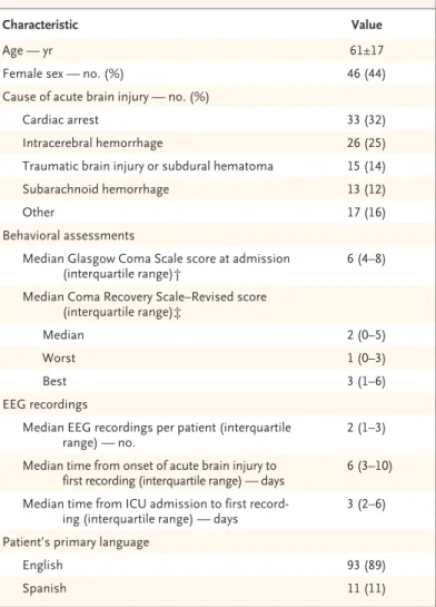
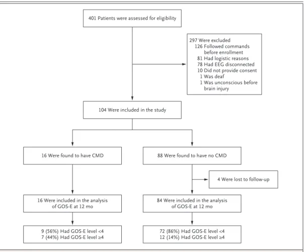
A total of 401 unresponsive patients with acute brain injury were screened, and 104 met the criteria for inclusion and were enrolled (Table 1 and Fig. 2). Enrolled patients were similar to those who were not enrolled with respect to age, sex, and admission Glasgow Coma Scale score (Table S1 in the Supplementary Appendix).
The healthy volunteers all had EEG evidence of brain activation in response to motor com-mands. We obtained a total of 240 EEG record-ings from the 104 patients (median number of recordings per patient, 2; interquartile range, 1 to 3) a median of 6 days (interquartile range, 3 to 10) after the injury. Of the 240 EEG recordings, 126 (52%) were acquired while patients were coma-tose, 54 (22%) while patients were in the vegeta-tive state, and 60 (25%) while patients were in the minimally conscious state–minus category.
Of the 104 patients, 16 (15%) had cognitive– motor dissociation detected on at least one re-cording. This dissociation was detected at a median of 4.0 days (interquartile range, 2.0 to 5.3) after admission to the ICU. Among these 16 patients, the causes of the acute brain injury were subarachnoid hemorrhage (5 patients), trau-matic brain injury (3), intracerebral hemorrhage (4), cardiac arrest (2), neurosarcoidosis (1), and bupropion overdose (1) (Table 2). The group-classification performance of the machine-learn-ing algorithm among patients and healthy volun-teers over time is shown in Figure 3.
The condition in 8 (50%) of the patients with cognitive–motor dissociation improved such that they were able to follow the spoken commands used during the daily clinical assessment by the time of hospital discharge, 6.0 days (interquar-tile range, 4.5 to 8.3) after cognitive–motor dis-sociation was first documented by EEG (Table S2 in the Supplementary Appendix). The condi-tion in 2 addicondi-tional patients (12%) with cogni-tive–motor dissociation improved after hospital
discharge such that they were able to follow the spoken commands used during the daily clinical assessment. In comparison, 26% of the patients who did not have cognitive–motor dissociation (23 patients) were able to follow commands be-fore hospital discharge (median ICU day, 12.0; interquartile range, 9.5 to 19.5).
Outcomes
The GOS-E level at 12 months after the acute brain injury was obtained for 100 of the 104 patients (4 patients were lost to follow-up). A total of 7 of 16 patients (44%) with and 12 of 84
Characteristic Value
Age — yr 61±17
Female sex — no. (%) 46 (44)
Cause of acute brain injury — no. (%)
Cardiac arrest 33 (32)
Intracerebral hemorrhage 26 (25) Traumatic brain injury or subdural hematoma 15 (14) Subarachnoid hemorrhage 13 (12)
Other 17 (16)
Behavioral assessments
Median Glasgow Coma Scale score at admission
(interquartile range)† 6 (4–8) Median Coma Recovery Scale–Revised score
(interquartile range)‡
Median 2 (0–5)
Worst 1 (0–3)
Best 3 (1–6)
EEG recordings
Median EEG recordings per patient (interquartile
range) — no. 2 (1–3)
Median time from onset of acute brain injury to
first recording (interquartile range) — days 6 (3–10) Median time from ICU admission to first
record-ing (interquartile range) — days 3 (2–6) Patient’s primary language
English 93 (89)
Spanish 11 (11)
* Plus–minus values are means ±SD. Percentages may not total 100 because of rounding. EEG denotes electroencephalographic, and ICU intensive care unit. † Scores on the Glasgow Coma Scale range from 3 to 15, with higher scores
in-dicating less neurologic dysfunction.
‡ Scores on the Coma Recovery Scale–Revised range from 0 to 23, with higher scores indicating a higher level of consciousness.
T h e ne w e ngl a nd jou r na l o f m e dicine
patients (14%) without cognitive–motor dissoci-ation had a GOS-E level of 4 or greater (odds ratio, 4.6; 95% confidence interval [CI], 1.2 to 17.1), and 6 (38%) with cognitive–motor disso-ciation and 50 (60%) without cognitive–motor dissociation were dead at 12 months. Among the 6 patients with cognitive–motor dissociation who were dead at 12 months, 4 had died in the context of withdrawal of life-sustaining therapy (Table 2, and Table S2 in the Supplementary Ap-pendix). After the 28 patients who underwent withdrawal of life-sustaining therapy were re-moved from the analysis, cognitive–motor dis-sociation remained predictive of a GOS-E level of 4 or greater (odds ratio, 5.4; 95% CI, 1.2 to 26.0).
Discussion
We found that 15% of patients in a consecutive series of patients with acute brain injury who were clinically unresponsive — some of whom had motor localization to pain stimuli, visual fixation, or visual tracking — had evidence of brain activation in response to spoken motor commands, as determined on the basis of EEG activity. This dissociation between behavior and EEG responses to spoken motor commands has been referred to as cognitive–motor dissocia-tion.5 In our study, this state was seen more
frequently in patients with trauma or brain hem-orrhages than in patients with hypoxic–ischemic
Figure 2. Enrollment and Follow-up.
The group of 126 patients who were excluded from the analysis because they followed commands before enrollment includes patients with reproducible movements in response to commands or with intentional communication defined according to the Coma Recovery Scale–Revised. Glasgow Outcome Scale–Extended (GOS-E) levels range from 1 to 8, with higher levels indicating better outcomes; a level of 4 indicates the ability to be left up to 8 hours during the day without assistance. CMD denotes cognitive–motor dissociation.
401 Patients were assessed for eligibility
104 Were included in the study
297 Were excluded 126 Followed commands
before enrollment 81 Had logistic reasons 78 Had EEG disconnected 10 Did not provide consent
1 Was deaf
1 Was unconscious before brain injury
16 Were found to have CMD 88 Were found to have no CMD
4 Were lost to follow-up
16 Were included in the analysis
of GOS-E at 12 mo 84 Were included in the analysisof GOS-E at 12 mo
9 (56%) Had GOS-E level <4
Characteristic Cognitive–Motor Dissociation Odds Ratio (95% CI)† Present (N = 16) (N = 88)Absent no. of patients (%) Age >63 yr 5 (31) 40 (45) 0.5 (0.1–1.9) Female sex 7 (44) 39 (44) 1.0 (0.3–3.3)
Cause of acute brain injury
Subarachnoid hemorrhage 5 (31) 8 (9) 4.5 (1.0–19.0) Intracerebral hemorrhage 4 (25) 22 (25) 1.0 (0.2–3.8) Traumatic brain injury or subdural hematoma 3 (19) 12 (14) 1.5 (0.2–6.5)
Cardiac arrest 2 (12) 31 (35) 0.3 (0.0–1.3)
Other 2 (12) 15 (17) 0.7 (0.1–3.6)
Behavioral assessments
Glasgow Coma Scale score at admission <8 12 (75) 58 (66) 1.5 (0.4–7.1) Median Coma Recovery Scale–Revised score ≥2 10 (62) 47 (53) 1.4 (0.4–5.3) Worst Coma Recovery Scale–Revised score ≥1 10 (62) 56 (64) 1.0 (0.3–3.5) Best Coma Recovery Scale–Revised score ≥3 11 (69) 49 (56) 1.7 (0.5–6.9) Behavioral category at enrollment
Coma 8 (50) 48 (55) 0.8 (0.2–2.8)
Unresponsive wakefulness syndrome 3 (19) 20 (23) 0.8 (0.1–3.3) Minimally conscious state–minus 5 (31) 20 (23) 1.5 (0.4–5.5) Mechanical ventilation 15 (94) 81 (92) 1.3 (0.1–62.3) EEG studies
No. of recordings ≥3‡ 8 (50) 28 (32) 2.1 (0.6–7.3)
Time from onset of acute brain injury to first recording
≥6 days 6 (38) 50 (57) 0.5 (0.1–1.5)
Time from ICU admission to first recording ≥3 days 7 (44) 52 (59) 0.5 (0.2–1.8) Length of stay
ICU stay ≥13 days 11 (69) 43 (49) 2.3 (0.7–9.1)
Hospital stay ≥14 days 11 (69) 41 (47) 2.5 (0.7–10.0) Outcomes
Following clinical commands before discharge 8 (50) 23 (26) 2.8 (1.0–8.4) Withdrawal of life-sustaining therapy 4 (25) 24 (27) 0.9 (0.2 – 3.3) Death before discharge 6 (38) 32 (36) 1.0 (0.3–3.5) Glasgow Outcome Scale–Extended level ≥4 at 12 months§ 7 (44) 12 (14)¶ 4.6 (1.2–17.1) * Percentages may not total 100 because of rounding. CI denotes confidence interval.
† Odds ratios for the categorical variables were computed using Fisher’s exact test. Data for continuous and ordinal vari-ables were split according to the median.
‡ A median of 3 EEG studies (interquartile range, 1 to 4) per patient was performed in the group of patients with cognitive– motor dissociation, and a median of 2 EEG studies (interquartile range, 1 to 3) was performed in the group of patients without cognitive–motor dissociation.
§ Glasgow Outcome Scale–Extended levels range from 1 to 8, with higher levels indicating better outcomes. A level of 4 in-dicates the ability to be left up to 8 hours during the day without assistance.
¶ Outcomes were missing for 4 patients.
T h e ne w e ngl a nd jou r na l o f m e dicine
injury, but it was detected in patients with other acute brain injuries and, in some instances, in patients who had been lightly sedated. The fre-quency of cognitive–motor dissociation and the prognostic associations in our single-center de-scriptive study require validation in larger, multi-center studies that are powered to detect differ-ences in long-term outcomes.
Our findings support those of previous stud-ies that have shown that EEG or functional MRI can in some cases reflect activation of parts of the brain in response to spoken commands in unresponsive patients,1,4,26 but whether the
de-tected signal represents recognition or compre-hension of commands is uncertain. Patients who had an EEG response to spoken commands more often had later recovery than those who did not have this pattern. It is possible that these pa-tients had overall greater functional integrity of the brain stem, thalamus, and cortex and of the connections among these structures, similar to findings in previous studies in which metabolic measurements such as fluorodeoxyglucose posi-tron-emission tomography were used.27
In chronic brain injury, cognitive–motor dis-sociation has been studied most often in patients with traumatic brain injury2,4,16,17,28,29 and has been
estimated to have a prevalence of 14%.6 Our
study shows that cognitive–motor dissociation can be detected in the ICU early after brain in-jury in a similar percentage of patients.3 The use
of methods such as functional MRI to detect cognitive–motor dissociation may result in more frequent detection than EEG, but functional MRI is challenging to perform in a critical care setting.30
Limitations of our study include the varied causes of brain injuries among the patients. In addition, the withdrawal of life-sustaining ther-apies confounds studies of the natural history of acute brain injury.31 However, even after the
ex-clusion of patients who underwent withdrawal of life-sustaining therapy, our study continued to show a difference in long-term functional out-comes between patients with and patients with-out cognitive–motor dissociation. We did not perform in-person 12-month follow-up assess-ments but recorded functional outcomes (GOS-E) by telephone. Sedation is a potential confounder in the classification of patients as being in a comatose, vegetative, or minimally conscious state; however, we were able to detect brain activation in response to motor commands in some lightly sedated patients.
Figure 3. Temporal Pattern in Healthy Volunteers and in Patients with and Patients without Cognitive–Motor Dissociation. The y axis indicates the decoding prediction, based on the EEG response, that a given epoch corresponds to a “move” instruction (higher number) or a “rest” instruction (lower number). The graph is shown for descriptive purposes only; the displayed averaged decoding prediction curves are related to the AUCs used to diagnose cognitive–motor dissociation.25 10 sec Decoding Prediction 1.00 0.70 0.60 0.50 0.45 0.30 0.55 0.40 0.00 1 2 3 4 5 6 7 8 Time (trial) Move Rest Healthy controls Patients with CMD Patients with no CMD Move period Rest period
In conclusion, early after brain injury, 15% of clinically unresponsive patients who did not fol-low commands had EEG evidence of brain acti-vation in response to spoken motor commands recorded at the bedside in the ICU.
A data sharing statement provided by the authors is available with the full text of this article at NEJM.org.
Supported by the Dana Foundation (to Dr. Claassen) and the James S. McDonnell Foundation. The study was conceptualized with the use of instruments that were developed and standard-ized as part of an international collaborative funded by James S. McDonnell Foundation (principal investigator, Nicholas D. Schiff;
details are provided in the Supplementary Appendix). Dr. Rohaut received postdoctoral grants from Amicale des Anciens Internes des Hôpitaux de Paris and Syndicat des Chefs de Cliniques et Assistants des Hôpitaux de Paris, Assistance Publique–Hôpitaux de Paris, and the Philippe Foundation.
Disclosure forms provided by the authors are available with the full text of this article at NEJM.org.
We thank the nurses, attending physicians, fellows, and neu-rology and neurosurgery residents of the Neuroscience ICU and Epilepsy Division for their overall support of this project; Denis A. Engemann and Federico Raimondo for their support with MNE-Python; Amelia K. Boehme for statistical advice; Jennifer A. Egbebike and Anna A. Calderon for additional data collec-tion; and Nicholas D. Schiff for providing the auditory recording of spoken commands.
References
1. Owen AM, Coleman MR, Boly M, Davis MH, Laureys S, Pickard JD. Detecting awareness in the vegetative state. Science 2006; 313: 1402.
2. Goldfine AM, Victor JD, Conte MM, Bardin JC, Schiff ND. Determination of awareness in patients with severe brain injury using EEG power spectral analysis. Clin Neurophysiol 2011; 122: 2157-68.
3. Edlow BL, Chatelle C, Spencer CA, et al. Early detection of consciousness in patients with acute severe traumatic brain injury. Brain 2017; 140: 2399-414.
4. Monti MM, Vanhaudenhuyse A, Cole-man MR, et al. Willful modulation of brain activity in disorders of conscious-ness. N Engl J Med 2010; 362: 579-89.
5. Schiff ND. Cognitive motor dissocia-tion following severe brain injuries. JAMA Neurol 2015; 72: 1413-5.
6. Kondziella D, Friberg CK, Frokjaer VG, Fabricius M, Møller K. Preserved sciousness in vegetative and minimal con-scious states: systematic review and meta-analysis. J Neurol Neurosurg Psychiatry 2016; 87: 485-92.
7. Turgeon AF, Lauzier F, Simard J-F, et al. Mortality associated with withdrawal of life-sustaining therapy for patients with severe traumatic brain injury: a Canadian multicentre cohort study. CMAJ 2011; 183: 1581-8.
8. Elmer J, Torres C, Aufderheide TP, et al. Association of early withdrawal of life-sustaining therapy for perceived neuro-logical prognosis with mortality after cardiac arrest. Resuscitation 2016; 102: 127-35.
9. Claassen J, Taccone FS, Horn P, Holt-kamp M, Stocchetti N, Oddo M. Recom-mendations on the use of EEG monitor-ing in critically ill patients: consensus statement from the neurointensive care section of the ESICM. Intensive Care Med 2013; 39: 1337-51.
10. Giacino JT, Kalmar K, Whyte J. The JFK Coma Recovery Scale-Revised: mea-surement characteristics and diagnostic
utility. Arch Phys Med Rehabil 2004; 85: 2020-9.
11. von Elm E, Altman DG, Egger M, Po-cock SJ, Gøtzsche PC, Vandenbroucke JP. The Strengthening the Reporting of Observational Studies in Epidemiology (STROBE) statement: guidelines for re-porting observational studies. Int J Surg 2014; 12: 1495-9.
12. Claassen J, Velazquez A, Meyers E, et al. Bedside quantitative electroenceph-alography improves assessment of con-sciousness in comatose subarachnoid hem-orrhage patients. Ann Neurol 2016; 80: 541-53.
13. Jennett B, Snoek J, Bond MR, Brooks N. Disability after severe head injury: ob-servations on the use of the Glasgow Out-come Scale. J Neurol Neurosurg Psychia-try 1981; 44: 285-93.
14. Lu J, Marmarou A, Lapane K, Turf E, Wilson L. A method for reducing misclas-sification in the extended Glasgow Out-come Score. J Neurotrauma 2010; 27: 843-52.
15. Oddo M, Crippa IA, Mehta S, et al. Optimizing sedation in patients with acute brain injury. Crit Care 2016; 20: 128.
16. Curley WH, Forgacs PB, Voss HU, Conte MM, Schiff ND. Characterization of EEG signals revealing covert cognition in the injured brain. Brain 2018; 141: 1404-21.
17. Cruse D, Chennu S, Chatelle C, et al. Bedside detection of awareness in the veg-etative state: a cohort study. Lancet 2011; 378: 2088-94.
18. Noirhomme Q, Lesenfants D, Gomez F, et al. Biased binomial assessment of cross-validated estimation of classification accuracies illustrated in diagnosis predic-tions. Neuroimage Clin 2014; 4: 687-94.
19. Good P. Permutation, parametric, and bootstrap tests of hypotheses. 3rd ed. New York: Springer Science+Business Media, 2005.
20. Ojala M, Garriga GC. Permutation tests for studying classifier performance. J Mach Learn Res 2010; 11: 1833-63.
21. Benjamini Y, Hochberg Y. Control-ling the false discovery rate: a practical and powerful approach to multiple test-ing. J R Stat Soc [B] 1995; 57: 289-300.
22. Gramfort A, Luessi M, Larson E, et al. MEG and EEG data analysis with MNE-Python. Front Neurosci 2013; 7: 267.
23. Pedregosa F, Varoquaux G, Gramfort A, et al. Scikit-learn: machine learning in Python. J Mach Learn Res 2011; 12: 2825-30.
24. R Development Core Team. R: a lan-guage and environment for statistical computing. R Project for Statistical Com-puting. 2017 (http://www .R - project .org/ ).
25. Kriegeskorte N, Simmons WK, Bell-gowan PSF, Baker CI. Circular analysis in systems neuroscience: the dangers of dou-ble dipping. Nat Neurosci 2009; 12: 535-40.
26. Naccache L. Psychology: is she con-scious? Science 2006; 313: 1395-6.
27. Stender J, Mortensen KN, Thibaut A, et al. The minimal energetic requirement of sustained awareness after brain injury. Curr Biol 2016; 26: 1494-9.
28. Giacino JT, Katz DI, Schiff ND, et al. Practice guideline update recommenda-tions summary: disorders of conscious-ness: report of the Guideline Develop-ment, Dissemination, and Implementation Subcommittee of the American Academy of Neurology; the American Congress of Rehabilitation Medicine; and the National Institute on Disability, Independent Liv-ing, and Rehabilitation Research. Neurol-ogy 2018; 91: 450-60.
29. Cruse D, Chennu S, Chatelle C, et al. Relationship between etiology and covert cognition in the minimally conscious state. Neurology 2012; 78: 816-22.
30. Weijer C, Bruni T, Gofton T, et al. Ethical considerations in functional mag-netic resonance imaging research in acute-ly comatose patients. Brain 2016; 139: 292-9.
31. Rohaut B, Claassen J. Decision mak-ing in perceived devastatmak-ing brain injury: a call to explore the impact of cognitive biases. Br J Anaesth 2018; 120: 5-9. Copyright © 2019 Massachusetts Medical Society.