HAL Id: hal-01216176
https://hal-amu.archives-ouvertes.fr/hal-01216176
Submitted on 15 Oct 2015
HAL is a multi-disciplinary open access
archive for the deposit and dissemination of sci-entific research documents, whether they are pub-lished or not. The documents may come from teaching and research institutions in France or abroad, or from public or private research centers.
L’archive ouverte pluridisciplinaire HAL, est destinée au dépôt et à la diffusion de documents scientifiques de niveau recherche, publiés ou non, émanant des établissements d’enseignement et de recherche français ou étrangers, des laboratoires publics ou privés.
Hildenborough under Continuous Low Oxygen
Concentration Sparging: Impact of the
Membrane-Bound Oxygen Reductases
Fanny Ramel, Gael Brasseur, Laetitia Pieulle, Odile Valette, Agnès
Hirschler-Réa, Marie Laure Fardeau, Alain Dolla
To cite this version:
Fanny Ramel, Gael Brasseur, Laetitia Pieulle, Odile Valette, Agnès Hirschler-Réa, et al.. Growth of the Obligate Anaerobe Desulfovibrio vulgaris Hildenborough under Continuous Low Oxygen Concen-tration Sparging: Impact of the Membrane-Bound Oxygen Reductases. PLoS ONE, Public Library of Science, 2015, 10, pp.e0123455. �10.1371/journal.pone.0123455�. �hal-01216176�
Growth of the Obligate Anaerobe
Desulfovibrio vulgaris Hildenborough under
Continuous Low Oxygen Concentration
Sparging: Impact of the Membrane-Bound
Oxygen Reductases
Fanny Ramel1, Gael Brasseur1, Laetitia Pieulle1, Odile Valette1, Agnès Hirschler-Réa2,
Marie Laure Fardeau2, Alain Dolla1*
1 Aix-Marseille Université, CNRS, LCB-UMR7283, Marseille, France, 2 Aix-Marseille Université, Université de Toulon, CNRS, IRD, MIO, UM110, 13288 Marseille, Cedex 09, France
*dolla@imm.cnrs.fr
Abstract
Although obligate anaerobe, the sulfate-reducing bacterium Desulfovibrio vulgaris Hilden-borough (DvH) exhibits high aerotolerance that involves several enzymatic systems, includ-ing two membrane-bound oxygen reductases, a bd-quinol oxidase and a cc(b/o)o3
cytochrome oxidase. Effect of constant low oxygen concentration on growth and morpholo-gy of the wild-type, single (Δbd, Δcox) and double deletion (Δcoxbd) mutant strains of the genes encoding these oxygen reductases was studied. When both wild-type and deletion mutant strains were cultured in lactate/sulfate medium under constant 0.02% O2sparging, they were able to grow but the final biomasses and the growth yield were lower than that ob-tained under anaerobic conditions. At the end of the growth, lactate was not completely con-sumed and when conditions were then switched to anaerobic, growth recon-sumed. Time-lapse microscopy revealed that a large majority of the cells were then able to divide (over 97%) but the time to recover a complete division event was longer for single deletion mutantΔbd than for the three other strains. Determination of the molar growth yields on lactate sug-gested that a part of the energy gained from lactate oxidation was derived toward cells pro-tection/repairing against oxidative conditions rather than biosynthesis, and that this part was higher in the single deletion mutantΔbd and, to a lesser extent, Δcox strains. Our data show that when DvH encounters oxidative conditions, it is able to stop growing and to rapid-ly resume growing when conditions are switched to anaerobic, suggesting that it enters ac-tive dormancy sate under oxidaac-tive conditions. We propose that the pyruvate-ferredoxin oxidoreductase (PFOR) plays a central role in this phenomenon by reversibly switching from an oxidative-sensitive fully active state to an oxidative-insensitive inactive state. The oxygen reductases, and especially the bd-quinol oxidase, would have a crucial function by maintaining reducing conditions that permit PFOR to stay in its active state.
OPEN ACCESS
Citation: Ramel F, Brasseur G, Pieulle L, Valette O, Hirschler-Réa A, Fardeau ML, et al. (2015) Growth of the Obligate Anaerobe Desulfovibrio vulgaris Hildenborough under Continuous Low Oxygen Concentration Sparging: Impact of the Membrane-Bound Oxygen Reductases. PLoS ONE 10(4): e0123455. doi:10.1371/journal.pone.0123455 Academic Editor: Franck Chauvat, CEA-Saclay, FRANCE
Received: February 2, 2015 Accepted: March 4, 2015 Published: April 2, 2015
Copyright: © 2015 Ramel et al. This is an open access article distributed under the terms of the
Creative Commons Attribution License, which permits unrestricted use, distribution, and reproduction in any medium, provided the original author and source are credited.
Data Availability Statement: All relevant data are within the paper and its Supporting Information files. Funding: AD, GB, LP and FR were funded by the French national Research Agency (ANR grant #2011BSV601201). The funder had no role in study design, data collection and analysis, decision to publish, or preparation of the manuscript. Competing Interests: The authors have declared that no competing interests exist.
Introduction
Sulfate-reducing bacteria (SRB) are anaerobic microorganisms ubiquitously distributed, even in atypical environments for this physiological group, e.g. in aerobic layer of a stratified fjord [1], in aerobic wastewater biofilms [2–3], in oxic layers of microbial mats [4–8] or in oxic ma-rine sediment layers close to the sediment surface [9]. Microscopy of roots and rhizomes also revealed the presence of SRB in the sea grass rhizosphere sediments [10], on the surfaces [11], inside epidermal and exodermal cells [12], even deep into the cortex cells of aquatic plants roots [13]. All these ecological niches can temporary be exposed to oxygen concentration up to saturation [4,14–15] and thus force SRB to cope with elevated oxygen tension.
Although SRB are classified as strict anaerobes [16], these examples above suggest that they exhibit high aerotolerance capabilities. Numerous laboratory works have tried to evaluate this aerotolerance, by studying impact of temporary oxygen stresses on various sulfate-reducing bacteria species. The effects of oxygen are visible at either protein [17], transcript [18–21], me-tabolite [22–24] and morphology levels [25] even at low concentration (0.1% O2) or
short-term exposure. But SRB appear not to be killed by a simple contact with oxygen and are able to cope with oxygen for several hours or even days in pure culture [26–29]. Together with ecologi-cal data [8,30–31], these studies reveal an aerotolerance variability in the various strains of SRB tested, with a higher oxygen tolerance for the members of the genus Desulfovibrio [28]. Several studies have even suggested a possible advantage of oxygenic conditions. Jonhson et al. [32] succeeded to observe the formation of a focused band of Desulfovibrio vulgaris Hildenborough (DvH) cells in an oxygen gradient, at an O2concentration between 0.02 and 0.04% O2. Other
artificial oxygen gradient experiments revealed growth of SRB close to the oxic-anoxic interface [27,33]. These data point out a positive aerotaxis that would enable bacteria to find environ-mental conditions favourable for their metabolic lifestyle. In homogeneously aerated pure cul-tures of several sulfate-reducing bacteria, it has been shown that rate of sulfide formation from the reduction of either sulfate, sulfite or thiosulfate decreased as the oxygen concentration in-creased, and was abolished above 15μM [27]. In a lactate/sulfate medium, growth of DvH was shown to be not significantly affected by up to constant 0.04% O2in a N2-H2-CO2gas mixture
sparging while it was inhibited at 0.08% O2[32]. Sigalevich and Cohen [34] reported that when
an initially established chemostat coculture of Desulfovibrio oxyclinae and the facultative het-erotrophic aerobe Marinobacter sp. strain MB grown under anaerobic conditions in lactate/sul-fate medium was exposed to an oxygen flux, the sullactate/sul-fate reducing bacterium performed an incomplete oxidation of lactate to acetate. The authors suggested thus that in these steady-state continuous culture, D. oxyclinae was able of oxygen-dependent growth [34]. Even if none of the SRB isolated so far can either grow aerobically or reduce sulfate under high oxygen concen-trations, several strains of Desulfovibrio species have been demonstrated to have the capability to couple oxygen reduction with proton translocation and energy conservation [35].
The ability for oxygen reduction is widespread among the SRB. The substrates used for this reduction are normally the same as those used for sulfate reduction. So far, H2, formate, lactate,
ethanol and pyruvate have been shown to be oxidized in the presence of oxygen [36]; however, the highest oxygen uptake rates of several Desulfovibrio species were obtained with H2as
elec-tron donor [37]. In Desulfovibrio termitidis, an oxygen reduction rate of about 1570 nmol of O2min-1mg-1of protein was found, higher than the value found in most aerobic bacteria
(~700 nmol of O2min-1mg-1) [38]. It has been proposed that enzymes involved in the energy
metabolism during the anaerobic sulfate respiration, i.e., hydrogenases and c-type cyto-chromes, also are enable of H2-dependent oxygen reduction [18,39–40]. In Desulfovibrio
spe-cies, a rubredoxin-oxygen oxidoreductase (ROO) has been shown to be the terminal enzyme of a cytoplasmic NADH-linked non-energy-conserving chain that reduced oxygen in water
[41–43]. Wildschut [44] pointed out that O2reduction activity by ROO accounted for 20 to
40% of the total specific oxygen reduction rate of DvH. The authors also highlighted the impor-tance of ROO in the survival of the cells to microaerophilic conditions, but not under fully aer-obic conditions. Sulfate reducers have also the ability to consume oxygen at the membrane level. Several isolates of Desulfovibrio and Desulfomicrobium from salt-marsh sediments were found to contain cytochrome bd oxidase and/or cytochrome c oxidase encoding genes [45]. In D. gigas, a high affinity bd-quinol oxidase was isolated, characterized, and shown to completely reduce oxygen to water [46–47]. In DvH, two membrane-bound terminal oxidases have been characterized, a bd-quinol oxidase and a cytochrome c oxidase that was of a new cc(o/b)o3
type, using the monohaem cytochrome c553as electron donor [48]. The DvH cyd genes,
encod-ing the bd-quinol oxidase, were 36-fold more transcribed than the cox genes, encodencod-ing the cc (b/o)o3oxidase, which could be related with the high oxygen reduction rate in the presence of
menadiol. Analysis of a cyd deletion mutant strain of DvH pointed out the existence of an elec-tronic link between the periplasmic H2oxidation by hydrogenases and the membranous
reduc-tion of O2by the bd-quinol oxidase [26]. Even if the two membrane terminal oxidases
appeared involved in the survival of DvH under low (0.1%) and saturated oxygen conditions, the cox deletion mutant was slightly more sensitive, pointing out the importance of the cc(b/o) o3cytochrome oxidase in oxygen protection [26]. In addition to the O2-reduction capability,
Desulfovibrio strains have developed other strategies to cope with oxygen and protect enzymes from oxidative damages. One of them is a specific reversible thiol-disulfide redox switch, effi-cient in the pyruvate-ferredoxin oxidoreductase (PFOR) that catalyses the oxidative decarbox-ylation of pyruvate forming acetyl-coenzyme A [49]. This mechanism involves the reversible formation of a disulfide bond in the C-terminal domain of the PFOR. During the oxidative time period, PFOR switches to an inactive but O2-stable form triggered by the formation of an
intramolecular disulfide bridge, this specific conformation allowing the protection of a [4Fe-4S] cluster from oxidative damages [24]. Once conditions return to reductive, the disulfide bond is reduced by using a thioredoxin/thioredoxin reductase system [50] and the PFOR is fully reactivated, leading to the restoration of a high rate of pyruvate oxidation in the cells with-out de novo synthesis of this key enzyme. This mechanism is highly valuable for an enzyme that catalyses a crucial step in carbon and energy metabolism of SRB.
As mentioned above, most of the aerotolerance capabilities description of Desulfovibrio strains are from temporarily oxygen exposed cells studies. In the present study, the effect of a continuous low oxygen concentration gas mixture sparging on growth, morphology, metabolic activities and cell viability of Desulfovibrio vulgaris Hildenborough is reported. In addition, the importance of each of the membrane bound oxygen reductase under these growth conditions is described.
Materials and Methods
Bacterial growth conditions
Desulfovibrio vulgaris Hildenborough (DvH) was grown at 33°C in liquid lactate (37mM)/sul-fate (32mM) medium (medium C) under anaerobic conditions in 10 ml Hungate tubes, inocu-lated at 10% (vol/vol) as previously described [16]. It should be noted that in this medium, lactate is the limiting factor for growth since two lactate molecules are required to reduce one sulfate in H2S [16]. A system using a gas mixer (PEGAS 4000 MF) was developed for a constant
gas sparging of the cultures in Hungate tubes with very low concentration of oxygen (from 0% to 0.1%) and high accuracy. In this case, cultures in Hungate tubes (10 ml medium C) were in-oculated at 15% (vol/vol) with a pre-culture at OD600~0.6, and continuously sparged with
Mettler-Toledo M700 recorder equipped with an O2module ppb 4700 and a calibrated Inpro
6900 O2probe. Single DvH deletion mutant strains of the genes encoding the bd-quinol
oxi-dase (Δbd) and the cytochrome cc(o/b)o3oxidase encoding genes (Δcox) as well as the double
deletions mutant strain (Δcoxbd) [26] were cultured under the same conditions as the wild-type strain. Growth resumption studies after constant 0.02% O2gas mixture sparging were
achieved by bubbling the cultures for 25 min with 100% N2to remove traces of oxygen and
then incubating the closed Hungate tubes for 20 hours more at 33°C. Growth was monitored by following the optical density at 600 nm. Growth rates were estimated by fitting the scatter plots of logarithmic optical density at 600nm with a linear regression from five independent experiments.
To determine the number of cells per volume unit of culture media, an aliquot of each cul-ture was sampled and placed on a Thoma cell counting chamber (0.0025 mm2–0.01 mm depth, Prolabo). At least 20 squares were observed to have a robust measure of the number of bacteria per ml of culture. A correlation factor between the optical density at 600 nm and the number of cells in cultures was determined under both anaerobic and constant 0.02% O2gas mixture
sparging conditions.
Organic acids quantification
Lactate and acetate were quantified by high-performance liquid chromatography (HPLC). Samples of cultures (500μL) were collected and centrifuged 10 min at 10000g. The superna-tants were analysed by high-performance liquid chromatography using a SpectraSERIES P100 pump equipped with a SpectraSYSTEM RI-150 detector and an Aminex HPX-87H-300x7.8 mm column C18 (Bio-Rad). Column temperature was 35°C and eluant (H2SO4, 0.005N) was
used at a flow rate of 0.6 mL/min. 20μl supernatant was injected.
Microscopy analyses
Microscopy analyses were performed on a temperature-controlled (33°C) TE2000-E-PFS in-verted epifluorescence microscope (Nikon, France). Images were recorded with a CoolSNAP HQ2 (Roper Scientific, Roper Scientific SARL, France) and a 100x/1.4 DLL objective coupled with a camera (Hamamatsu Orca-R2). Images were viewed using NIS-Elements Viewer 4.0. The impact of oxidative conditions on cell morphology was determined by measuring the length of at least 300 cells for each strain and condition (anaerobic/oxidative) from two inde-pendent experiments using the software Fiji [51]. For cell division analysis by time-lapse mi-croscopy, cells were cultured with constant 0.02% O2gas mixture sparging until they stopped
to grow (about 25 h) and conditions were switched to anaerobic by bubbling with N2for 25
min to remove traces of oxygen from the medium. In an anaerobic Coy chamber (5% H2, 95%
N2gas atmosphere), 3μL of the culture was then placed between the coverslip and a thin layer
of medium C supplemented with 1.5% of Phytagel in an hermetic chamber and sealed (Fievet, unpublished). This observation chamber was then transferred to the TE2000-E-PFS micro-scope. Phase-contrast images were acquired every 10 min for at least 16 hours. The number of cells able to divide, times to recover a complete division event and second division times were determined manually.
PFOR activity assays
Pyruvate-Ferredoxin Oxidoreductase (PFOR) activity was determined spectrophotometrically at 30°C by measuring the reduction of methyl viologen as previously described [49]. Briefly, the reaction mixture containing 50 mM Tris-HCl (pH 8.5), 10 mM sodium pyruvate, 0.1 mM sodium coenzyme A and 2 mM methyl viologen, was bubbled with argon for 25 min. Then,
10μl Triton X100 (1/10 in Tris-HCl buffer, pH 8.5), kept under anaerobic conditions in an an-aerobic chamber (Jacomex BS531NMT), was added and the reaction was started by injection of 50μl of cells culture into the cuvette using a gastight syringe. Absorbance at 604 nm was moni-tored and when variation of absorbance over time appeared linear, 15mM dithioerythritol (DTE) was added anaerobically into the cuvette to reach the maximum of activity which corre-sponded to a totally reactivated enzyme. The rate of inactive and thus protected enzyme was calculated by the difference of the slope before and after the addition of DTE, reported to the optical density at 600 nm of the culture. As PFOR in cells cultured under anaerobic conditions is fully active [24], the corresponding value of the PFOR protected rate for the wild-type strain was arbitrary set to 1 and all other values were related to this unit.
Results
Effect of constant low oxygen concentration sparging on growth
Wild-type DvH and both single (Δbd, Δcox) and double (Δcoxbd) mutants were cultured in lac-tate/sulfate medium in Hungate tubes with constant sparging with 0.02% O2in order to
evalu-ate the role of the membrane bound oxygen reductases in the growth under these oxidative conditions. The 0.02% O2sparging conditions led to a constant dissolved oxygen concentration
of 0.23μM in the tubes as measured with the Mettler-Toledo O2probe.Fig 1shows that all
strains were able to grow (about 2.5, 1.8, 2 and 2.4 division events for the wild-type,Δcox, Δbdand Δcoxbd strains, respectively). The oxygen concentration of 0.02% was determined as the highest that permitted growth of both the wild-type and the deletion mutants strains. The growth parameters, determined from the growth curves, are shown inTable 1. Under anaero-bic conditions, the doubling time of the wild-type strain was found in the same range (about 5 h) as that of the deletion mutant strains, except for theΔcox strain which exhibited a slightly increased doubling time (5.97 h). In the same way, the final biomass was similar for all the strains, except for theΔcox strain which showed a lower final biomass (0.45 vs 0.55 109 cells ml-1). When cultured with constant 0.02% O2gas mixture sparging, the doubling time of
the wild-type strain largely increased to around 14 h, and the final biomass was found much lower than under anaerobiosis, 0.29 vs 0.55 109cells ml-1. Under these oxidative conditions, the single deletionΔcox and Δbd mutant strains exhibited also higher doubling times and lower final biomasses than under anaerobic conditions (Table 1). In the case of theΔcoxbd strain, the shape of growth curves was quite atypical when cultured with constant 0.02% O2sparging,
probably with a biphasic behaviour, preventing the calculation of relevant doubling time (Fig 1). However, the growth curves suggested that the doubling time of the double mutant strain was lower than that of the wild-type under constant 0.02% O2gas mixture sparging.
While theΔbd and Δcox strains exhibited a significant lower final biomass than the wild-type when cultured with constant 0.02% O2sparging, theΔcoxbd strain had, surprisingly, a final
biomass similar as that of the wild-type strain. These data show that under the oxidative condi-tions caused by the constant 0.02% O2gas mixture sparging, cells were able to divide; however,
less amount of cells were obtained compared to anaerobic conditions, indicating a growth ar-rest. It should be noted that culturing the wild-type strain with constant 0.02% O2gas mixture
sparging did not induce any significant variation in the amount of the cox and bd transcripts as quantified by qRT-PCR, compared to anaerobic growth conditions (S1 Fig.). In addition, no transcriptional compensation mechanism was found in any of the single deletion mutant strains; a decrease (2.5 times) in the amount of cox transcript was however quantified in theΔbd strain (S1 Fig.).
Effect of oxygen on the cell morphology
It has been observed that some Desulfovibrio strains developed atypically elongated cells when growing in the presence of oxygen [25]. A recent analysis at the single-cell level showed that when DvH was exposed to low oxygen concentration (up to 0.05%), cells elongated (Fievet, un-published). In order to determine whether the presence or the absence of the membrane bound oxygen reductases would affect this elongation phenomenon, morphology of the cells was stud-ied by microscopy after 24 h of growth with either constant 0.02% O2gas mixture sparging or
anaerobic conditions. When cultured under anaerobic conditions, the lengths distribution of the wild-type and the three mutant cells were similar, with a median value around 1.6μm (Fig 2,S1 Table). When cultured with constant 0.02% O2sparging, cells lengths were greater
for all strains. However, variations in lengths distribution differed from strain to strain: while the median length of wild-type andΔbd cells were slightly shifted to larger values, + 0.23 μm Fig 1. Growth analysis of the DvH strains. Growth curves of the wild-type and the three deletion mutant strains under anaerobic conditions (black symbols) or with constant 0.02% O2gas mixture sparging (open symbols). Data arose from five independent growth experiments.
and + 0.12μm, respectively, compared to the values measured under anaerobic conditions, the shift of the median lengths of theΔcox and Δcoxbd cells was more pronounced, + 0.3 μm and + 0.41μm, respectively (Fig 2,S1 Table). These data show that, when cells were cultured with constant 0.02% O2sparging, the median as well as the extreme lengths values of the cells
in-creased and that the absence of the cytochrome c oxidase had a more pronounced effect on this morphology parameter than that of the bd-quinol oxidase.
Substrate consumption analysis
In order to determine whether the growth arrest in the presence of 0.02% O2was due to a
com-plete consumption of substrate (i.e. lactate), lactate and acetate were quantified in the cultures when cells stopped to grow (after ~ 40 h) (Fig 1).Fig 3shows that under anaerobic conditions, lactate (initial concentration 37 mM) was completely consumed and, accordingly, acetate was produced with an acetate/lactate stoichiometric ratio of about 0.83 for all strains, in agreement with a part of acetate used for biosynthesis. In the cultures with constant 0.02% O2gas mixture
sparging, when growth was stopped, 22.2 ± 4.6 mM lactate was consumed in the wild-type cul-ture and 12.6 ± 1.3, 17.8± 1.3 and 23.4± 0.3 mM lactate were consumed in theΔcox, Δbd and Δcoxbd cultures, respectively and accordingly, less acetate was formed (Fig 3B). However, the acetate/lactate stoichiometric ratio was found slightly lower with an average value of 0.74. These data show that the growth arrest, when cultures were constantly sparged with 0.02% O2,
was not the effect of a complete consumption of the energy substrate by the cells. Interestingly, when conditions were then switched to anaerobic by bubbling the cultures with 100% N2for
15 min to remove oxygen and kept afterwards anaerobic, all strains were able to resume growth and, at the end of the growth, all lactate was consumed (data not shown) in the four strains; however, the final biomasses never reached the value obtained when cells were cultured under anaerobic conditions only, except for theΔcox deletion mutant (Fig 4). When cultured with constant 0.02% O2sparging, the molar growth yields on lactate (Ylactate) were 70%, 57%, 62%
and 65% lower for the wild-type,Δbd, Δcox and Δcoxbd strains, respectively, than when cul-tured under anaerobic conditions (Fig 5). The single deletion mutantΔbd and Δcox strains ex-hibited the lowest values while, surprisingly, Ylactateof the double deletion mutant was greater
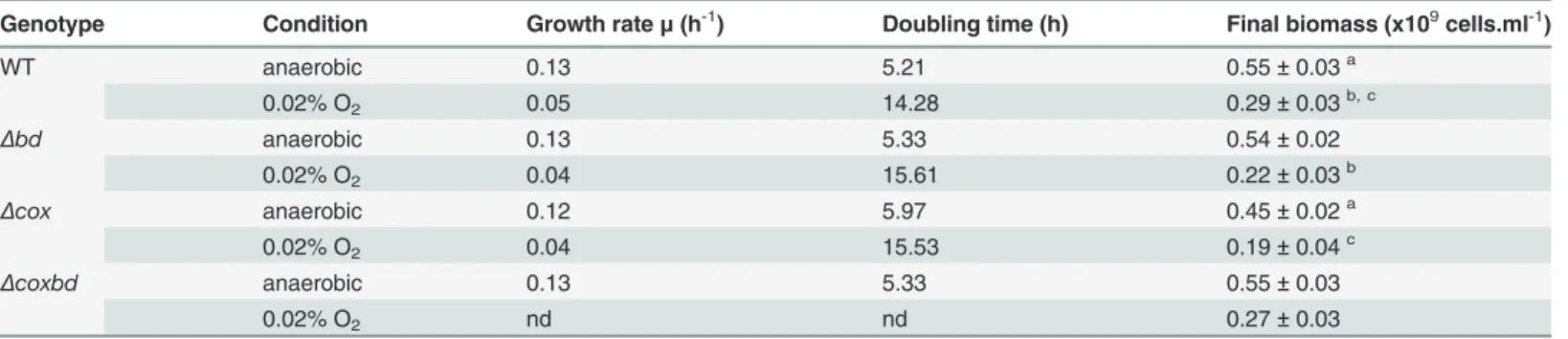
than those of the single deletion mutants and similar to that of the wild-type strain (Fig 5). Table 1. Growth parameters for the Desulfovibrio vulgaris Hildenborough strains.
Genotype Condition Growth rateμ (h-1) Doubling time (h) Final biomass (x109cells.ml-1)
WT anaerobic 0.13 5.21 0.55± 0.03a 0.02% O2 0.05 14.28 0.29± 0.03b, c Δbd anaerobic 0.13 5.33 0.54± 0.02 0.02% O2 0.04 15.61 0.22± 0.03b Δcox anaerobic 0.12 5.97 0.45± 0.02a 0.02% O2 0.04 15.53 0.19± 0.04c Δcoxbd anaerobic 0.13 5.33 0.55± 0.03 0.02% O2 nd nd 0.27± 0.03
DvH strains were grown in lactate/sulfate medium under anaerobic conditions or under constant 0.02% O2gas mixture sparging. Data are mean values of
five independent experiments. Statistical analysis using t-test was used to compare means.
a, b, c: Significant differences (p<0.05). nd: not determined.
Quantification of number of cells able to divide after oxygen exposure
When cells were exposed to a constant 0.02% O2sparging, they stopped to grow (Fig 1). In order
to quantify the number of cells that were able to divide again when conditions were then switched to anaerobic, time-lapse microscopy on both the wild-type and deletion mutant strains was performed.Table 2shows that in the case of the wild-type and theΔcoxbd strains, 100% of cells were able to divide when conditions were switched to anaerobic, while 1% and 2.4% ofΔbd andΔcox cells, respectively, were unable to restart division. The oxidative conditions induced by the constant 0.02% O2sparging did not thus have any significant effect on the capabilities of the
wild-type and theΔcoxbd strains to resume division when conditions were switched to anaerobic Fig 2. Variability in the cell length of the various DvH strains. Comparison of the distribution of the cell length in cultures under anaerobic conditions (black bars) and with constant 0.02% O2gas mixture sparging (striped bars). The average cell length was determined from two independent cultures (>200
cells were counted in each experiment). Statistical analysis using One-way ANOVA were performed to reveal significant differences between distributions (p<0.05), which were mentioned by an asterisk.
and had only a weak effect on theΔbd and Δcox deletion mutant strains for which a low percent-age of cells were found unable to resume division in anaerobic conditions.
The average values of the first division recovery time (time to recover a complete division event), calculated on at least 200 cells, were 6 hours for theΔbd mutant and about 5.3 hours for the three others strains (Table 2). When the same experimental protocol was performed on cells cultured under anaerobic conditions only (without any exposure to oxygen), the time to recover a complete division event was about 3 h for all strains (data not shown). The second doubling time (time to finish the second division event) was about 2.5 hours in average for the two single deletion mutants (Δbd and Δcox) and shorter for the Δcoxbd (2.1 hours) and the Fig 3. Lactate consumption and acetate production in the DvH strains. Quantification of consumed lactate (A) and produced acetate (B) after 40h growth under anaerobic conditions (black bars) or with constant 0.02% O2gas mixture sparging (striped bars) in medium C (with 37mM of lactate as initial
concentration).
doi:10.1371/journal.pone.0123455.g003
Fig 4. Biomass formation under various culturing conditions. Final biomass for the four DvH strains cultured under anaerobic conditions only (black bars), with constant 0.02% O2gas mixture sparging (striped
bars) and after growth resumption (open bars). Data are mean values of five independent experiments + SD. Statistical analysis using t-test was used to compare means. Significant differences (p< 0.05) are mentioned by an asterisk.
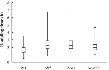
wild-type (1.9 hours) (Table 2). The distribution pattern showed that second doubling times for the single deletion mutantΔbd and Δcox strains were more scattered than that of the wild-type, with a significant proportion of cells exhibiting higher median, third quartile and maxi-mum values. The distribution pattern of double deletion mutantΔcoxbd appeared closer to that of the wild-type strain, with less scattered values (Fig 6). These data show that even if cells were able to divide again when conditions were switched to anaerobic, it took longer to com-plete a division event when cells were previously exposed to constant 0.02% O2sparging, the
Δbd strain being the most affected. In addition the absence of either one of the membrane-bound oxygen reductase had a pronounced effect on the following division event, which was longer than for the wild-type strain.
PFOR activity on the various strains
The above data point out that when DvH was cultured with constant 0.02% O2gas mixture
sparging, it was able to divide more than two times, then stopped growing and, when condi-tions were switched to anaerobic, resumed dividing. The pyruvate-ferredoxin oxidoreductase (PFOR) has been shown to be able to reversibly switch from a fully active oxidative-sensitive Fig 5. Molar growth yield for the four strains under various growth conditions. Molar growth yield on lactate for the wild-type and deletion mutant strains cultured under anaerobic conditions (black bars) and with constant 0.02% O2gas mixture sparging (striped bars). Molar growth yields are expressed in 1012cells/mole
of substrate.
doi:10.1371/journal.pone.0123455.g005
Table 2. Quantification of cells able to divide and average division parameters of the wild-type and deletion mutant strains after oxygen exposure. Genotype Cells unable to divide (%) First division recovery time (h) Second doubling time (h)
WT 0 5.29± 0.25a 1.86± 0.12b, c
Δbd 1 6± 0.43a 2.49± 0.47b
Δcox 2.4 5.27± 0.91 2.42± 0.19c
Δcoxbd 0 5.26± 0.97 2.06± 0.03
Strains were cultured with constant 0.02% O2gas mixture sparging followed by a switch to anaerobic condition. Data are mean values of three
independent experiments± SD. Statistical analysis using t-test was used to compared means.
a, b, c: Significant differences.
state (active state) to an inactive oxidative-insensitive state (protected state) when cells encoun-ter oxidative conditions by means of an autoprotective thiol-redox mechanism [24,50]. Because this enzyme plays a crucial role in the lactate metabolism, effect of constant 0.02% O2sparging
on the PFOR state was studied. Quantification of the PFOR activity in cells without and with addition of a chemical reducing agent (i. e. dithioerythritol) stands for the active versus pro-tected states of the enzyme [24]. The protected PFOR rate was determined for each strain when cells were cultured under both anaerobic conditions and with constant 0.02% O2
sparg-ing (Fig 7). While under anaerobic conditions, the protected PFOR rate was found similar in all strains (the rate being arbitrary set to 1 for the wild-type strain), when cells were cultured Fig 6. Variability of the second doubling time for the four strains after oxygen exposure. Box and whiskers plot representing the variability in the distribution of the second doubling time of the various DvH strains cultured with constant 0.02% O2gas mixture sparging followed by a switch to anaerobic condition.
These distribution patterns arose from at least 2 independent cultures (> 200 cells each). doi:10.1371/journal.pone.0123455.g006
Fig 7. Protected PFOR rate in the four strains. Quantification of the rate of protected PFOR during the exponential growth phase under anaerobic (black bars) and with constant 0.02% O2gas mixture sparging
(striped bars). Statistical analysis using Mann and Whitney test was used to compare means. Significant differences (p< 0.05) are indicated by an asterisk. All values are related to the PFOR protected rate in the wild-type strain under anaerobic conditions (value arbitrary set to 1).
with constant 0.02% O2sparging, this rate was found slightly higher in the wild-type strain
than under anaerobic conditions and much higher for the two single deletion mutant strains, theΔbd strain exhibiting the largest one. On the contrary, the double deletion mutant exhibited a similar rate as the wild-type strain. It should be noted that no variation of the amount of por transcripts induced by the oxidative conditions caused by the constant 0.02% O2sparging was
observed in any strain (data not shown). These data show that under oxidative conditions, the PFOR protected rate depended on the presence of the membrane bound oxygen reductases, their absences, and specially that of the bd oxidase, leading to a larger part of the PFOR in the protected form. In the case of the double mutant, one or several other mechanisms, still un-known, would balance the absence of the two oxygen reductases.
Discussion
To our knowledge, only one study [32] has reported the growth of Desulfovibrio species with constant oxygen sparging with more than a doubling of the initial population. In this report, we successfully grew DvH in Hungate tubes with lactate as carbon and energy sources and sul-fate as electron acceptor, with constant 0.02% O2gas mixture sparging, corresponding to a
con-stant dissolved oxygen concentration of 0.23μM. It should be noted that no formation of bacterial aggregates which would permit cells to create anaerobic micro-niches as in Desulfovi-brio oxyclinae [34] was observed. Under these conditions, DvH was able to divide more than two times but growth parameters were affected, i.e. lower growth rate and final biomass than under anaerobic conditions. An important point is that cells stopped to grow despite lactate was not completely consumed. This growth arrest was not due to cells death as time-lapse mi-croscopy revealed that all cells were able to re-start division when the conditions were switched to anaerobic. This high survival rate under oxidative conditions is in agreement with the high aerotolerance of DvH that was previously pointed out [26,44,52]. Interestingly, growth re-sumed if conditions were switched to anaerobic, with the recovery of lactate consumption until exhaustion but the final biomass did not reach the same value as when cells were grown under completely anaerobic conditions. The molar growth yield on lactate was found lower when cells were cultured with constant 0.02% O2gas mixture sparging than under anaerobic
condi-tions, indicating that a part of the energy gained from the substrate oxidation is derived toward cells protection/repairing against oxidative conditions rather than biosynthesis. A trade-off should exist between growth and defense investment, permitting bacteria to redirect energy for growth to repairing or detoxifying systems.
These observations directed our attention to the potential involvement of the two mem-brane-bound oxygen reductases, the bd-quinol oxidase and the cytochrome cc(o/b)o3oxidase,
in the growth under the continuous presence of low oxygen concentration. The study of the single deletion mutant strains of the genes encoding these oxygen reductases revealed that the absence of either of the two membrane-bound oxygen reductases led to both lower growth rates and lower final biomasses than those observed for the wild-type strain when cultured with constant 0.02% O2sparging. As for the wild-type strain, the molar growth yields on lactate
of the deletion mutant strains are lower with constant 0.02% O2sparging, compared to
anaero-bic conditions, with a larger difference for the singleΔbd, and Δcox deletion mutant strains. It suggests that the energy diverted to repairing/detoxifying systems is higher in the single dele-tionΔbd and, to a lesser extent, Δcox mutant strains, indicating that the membrane-bound oxy-gen reductases, and especially the bd-quinol oxidase, play an important role in maintaining cells healthy under oxidative conditions. They could be involved through their capability to both detoxify oxygen by reducing it to water and contribute to the generation of a proton mo-tive force inherent in their mechanistic activities [53–54]. The prevailing role of the bd-quinol
oxidase is in agreement with its function in aerobic organisms where it significantly contributes to respiration of O2under microaerobic conditions, when oxygen availability is limited [55],
which is compatible with its high affinity for oxygen [56–57]. In addition, it has to be related with our previous results that have shown a higher affinity for oxygen of the bd-quinol oxidase (Km 300 nM) than that of the cytochrome cc(o/b)o3oxidase (Km 620 nM) as well as a greater
abundance of the former enzyme in DvH cells [26]. Contrary to expectation, the double dele-tion mutant does not appear more affected than the single deledele-tion mutants when cultured under constant 0.02% O2sparging, and even, on several aspects (final biomass, Ylactate, second
division time, PFOR protected rate), has a behaviour quite similar as that of the wild type. While the viability of double deletion mutant after exposure to air or to 0.1% O2for more than
8 hours was lower than that of the single deletion mutant strains [26], the present data suggests that under these specific oxidative conditions, the absence of the two membrane-bound oxygen reductases has an beneficial effect when compared to the absence of only one. The mechanism that would balance the absence of the two membrane-bound reductases is however still un-known and studies are needed to get further insights.
The fact that DvH, when cultured with constant 0.02% O2gas mixture sparging, is able to
grow for more than two generations, then stops growing but keeps its ability to divide and to resume growing when conditions are switched to anaerobic, suggests that it is able of active dormancy as originally proposed by Le Gall and Xavier [58]. The idea was that when exposed to oxygen, cells were not killed but remained dormant with the maintenance of low-level cell activities [58–59]. Ability of anaerobes to enter in active dormancy should be considered as an adaptation strategy used to cope with temporary exposure to O2. The maintenance of cells
ac-tivity under oxidative conditions enables them to rapidly resume growth on re-entering an an-aerobic habitat. Culturing DvH with 0.02% O2sparging would induce the cells to enter active
dormancy state after several hours. The longer time to recover a complete division event when conditions are then switched to anaerobic than when DvH is continuously cultured under an-aerobic conditions (about 5.3 h versus 3 h) as observed by microscopy, could be linked to the need of a metabolic reprogramming to get out active dormancy and to resume dividing. In the case of theΔbd deletion mutant strain, the absence of the bd-quinol oxidase could have in-duced more oxidative damages that have to be fixed before the cells can divide again, which re-sult in a longer time to recover a division event (6 h versus 5.3 h).
The mechanism by which cells are able to enter active dormancy is still unknown but the Pyruvate-Ferredoxin OxidoReductase (PFOR) should play an important role through its autoprotective disulfide redox-switch mechanism [24]. This mechanism allows the enzyme to reversibly switch from a fully active oxidative-sensitive state to an inactive oxidative-insensitive state when cells encounter oxidative conditions [24,50]. Protecting PFOR from oxidative dam-ages and restoring a high rate of pyruvate oxidation by switching PFOR to its fully active form without de novo synthesis could be a key stage for rapid growth resumption. The protected state of the PFOR can be thus considered as an hallmark of the active dormancy state. Our data show that when DvH is cultured under constant 0.02% O2sparging, a part of the PFOR is
switched from active to protected states, inducing a decrease of the pyruvate oxidation activity in the cells. This decrease would lead cells to enter active dormancy, resulting in a growth ar-rest. When conditions become less oxidative, PFOR is switched to active state, restoring a high pyruvate oxidation activity, allowing cells to get out active dormancy state and to resume growth. The membrane-bound oxygen reductase, and especially the bd-quinol oxidase, seem to play an important role in the protected/active states PFOR switch. In Azotobacter vinelandii, the bd-quinol oxidase has be shown to play an important role in protecting the O2labile
nitro-genase activity by maintaining very low O2concentration [60] In the same way, one can
very low oxygen concentration and thus reducing conditions that permit PFOR to stay in its active state. When O2-reduction activity is not efficient enough, increased oxidative conditions
inside the cell would trigger it to enter active dormancy.
Characterization of the active dormancy state constitutes the next challenge to get a better understanding of the aerotolerance capabilities of anaerobic microorganisms.
Supporting Information
S1 Fig. Quantification of cox and bd genes transcripts by qRT-PCR. Transcript level of the bd-quinol oxidase encoding gene (bd gene) (A) and the cytochrome c oxydase encoding gene (cox genes) (B) in WT and deletion mutants in anaerobiosis (black bars) or continuously ex-posed to 0.02% O2sparging (striped bars). Data are mean values of two independent
experi-ments +SD. (DOC)
S1 Table. Variability in the cell length of the various DvH strains. Variability in the cell length of the various DvH strains. Values of cell length (μm) in cultures under anaerobic condi-tions and with a constant 0.02% O2gas mixture sparging for 24 hours for each strain. The
quar-tiles values (Q1, Q3) indicated in the table come from two independent cultures (>200 cells were counted in each experiment).
(DOC)
Acknowledgments
The authors are grateful to Drs. A. Fievet and C Aubert for helpful discussions on the time-lapse microscopy experiments and to Prof. G. Voordouw from providing us with the single and double deletion mutant strains.
Author Contributions
Conceived and designed the experiments: AD GB LP. Performed the experiments: FR OV MLF AH. Analyzed the data: FR AD GB LP. Contributed reagents/materials/analysis tools: MLF AH FR. Wrote the paper: FR AD GB LP.
References
1. Teske A, Wawer C, Muyzer G, Ramsing NB. Distribution of sulfate-reducing bacteria in a stratified fjord (Mariager Fjord, Denmark) as evaluated by most-probable-number counts and denaturing gradient gel electrophoresis of PCR-amplified ribosomal DNA fragments. Appl Environ Microbiol. 1996; 62: 1405. PMID:8919802
2. Santegoeds CM, Ferdelman TG, Muyzer G, de Beer D. Structural and functional dynamics of sulfate-re-ducing populations in bacterial biofilms. Appl Environ Microbiol. 1998; 64: 3731–3739. PMID:9758792
3. Okabe S, Itoh T, Satoh H, Watanabe Y. Analyses of spatial distributions of sulfate-reducing bacteria and their activity in aerobic wastewater biofilms. Appl Environ Microbiol. 1999; 65: 5107–5116. PMID:
10543829
4. Canfield DE, Des Marais DJ. Aerobic sulfate reduction in microbial mats. Science. 1991; 22: 1471– 1473.
5. Krekeler D, Sigalevich P, Teske A, Cypionka H, Cohen Y. A sulfate-reducing bacterium from the oxic layer of a microbial mat from Solar Lake (Sinai), Desulfovibrio oxyclinae sp. nov. Arch Microbiol. 1997; 167: 369–375.
6. Minz D, Fishbain S, Green SJ, Muyzer G, Cohen Y, Rittmann BE, et al. Unexpected population distribu-tion in a microbial mat community: sulfate-reducing bacteria localized to the highly oxic chemocline in contrast to a eukaryotic preference for anoxia. Appl Environ Microbiol. 1999; 65: 4659–4665. PMID:
7. Visscher PT, Prins RA, Gemerden HV. Rates of sulfate reduction and thiosulfate consumption in a ma-rine microbial mat. FEMS Microbiology Ecology. 1992; 86: 283–294.
8. Risatti JB, Capnan WC, Stahl DA. Community structure of a microbial mat: The phylogenetic dimension Proc Natl Acad Sci USA. 1994; 91: 10173–10177. PMID:7937858
9. Jorgensen B B, Bak F. Pathways and Microbiology of Thiosulfate Transformations and Sulfate Reduc-tion in a Marine Sediment (Kattegat, Denmark). Appl Environ Microbiol. 1991; 57: 847–856. PMID:
16348450
10. Blaabjerg V, Finster K. Sulphate reduction associated with roots and rhizomes of the marine macro-phyte Zostera marina. Aquatic Microbial Ecology. 1998; 15: 311–314.
11. Nielsen LB, Finster K, Welsh DT, Donelly A, Herbert RA, de Wit R, et al. Sulphate reduction and nitro-gen fixation rates associated with roots, rhizomes and sediments from Zostera noltii and Spartina mari-timameadows. Environ Microbiol. 2001; 3: 63–71. PMID:11225724
12. Kuo J. Root anatomy and rhizosphere ultrastructure in tropical seagrass. Austral J Mar Fresh Res. 1993; 44: 75–84.
13. Küsel K, Pinkart H C, Drake H L, Devereux R. Acetogenic and sulfate-reducing bacteria inhabiting the rhizoplane and deep cortex cells of the sea grass Halodule wrightii. Appl Environ Microbiol. 1999; 65: 5117–5123. PMID:10543830
14. Sigalevich P, Baev MV, Teske A, Cohen Y. Sulfate reduction and possible aerobic metabolism of the sulfate-reducing bacterium Desulfovibrio oxyclinae in a chemostat coculture with Marinobacter sp. Strain MB under exposure to increasing oxygen concentrations. Appl Environ Microbiol. 2000; 66: 5013–5018. PMID:11055957
15. Fründ C, Cohen Y. Diurnal Cycles of Sulfate Reduction under Oxic Conditions in Cyanobacterial. Mats. Appl Environ Microbiol. 1992; 58: 70–77. PMID:16348641
16. Postgate JR. The sulfate-reducing bacteria. Cambridge: Cambridge University Press, 1984.
17. Fournier M, Aubert C, Dermoun Z, Durand MC, Moinier D, Dolla A. Response of the anaerobe Desulfo-vibrio vulgarisHildenborough to oxidative conditions: proteome and transcript analysis. Biochimie. 2006; 88: 85–94. PMID:16040186
18. Mukhopadhyay A, Redding AM, Joachimiak MP, Arkin AP, Borglin SE, Dehal PS, et al. Cell-wide re-sponses to low-oxygen exposure in Desulfovibrio vulgaris Hildenborough. J Bacteriol. 2007; 189: 5996–6010. PMID:17545284
19. Pereira PM, He Q, Valente FMA, Xavier AV, Zhou J, Pereira IAC, et al. Energy metabolism in Desulfovi-brio vulgarisHildenborough: insights from transcriptome analysis. Antonie van Leeuwenhoek. 2008; 93: 347–362. PMID:18060515
20. Zhang W, Culley DE, Scholten JC, Hogan M, Vitiritti L, Brockman FJ. Global transcriptomic analysis of Desulfovibrio vulgarison different electron donors. Antonie Van Leeuwenhoek. 2006; 89: 221–237. PMID:16710634
21. Figueiredo MC, Lobo SA, Carita JN, Nobre LS, Saraiva LM. Bacterioferritin protects the anaerobe Desulfovibrio vulgarisHildenborough against oxygen. Anaerobe. 2012; 18: 454–458. doi:10.1016/j. anaerobe.2012.06.001PMID:22706208
22. Dijk van C, Berkel-Arts van A, Veeger C. The effect of reoxidation on the reduced hydrogenase of Desulfovibrio vulgarisstrain Hildenborough and its oxygen stability. FEBS Letters. 1983; 156: 340–344. 23. Stams AJM, Hansen TA. Oxygen-labile l(+) lactate dehydrogenase activity in Desulfovibrio
desulfuri-cans. FEMS Microbiol Lett. 1982; 13: 389–394.
24. Vita N, Hatchikian EC, Nouailler M, Dolla A, Pieulle L. Disulfide bond-dependent mechanism of protec-tion against oxidative stress in pyruvate-ferredoxin oxidoreductase of anaerobic Desulfovibrio bacteria. Biochemistry. 2008; 47: 957–964. PMID:18161989
25. Sass H, Berchtold M, Branke J, König H, Cypionka H, Babenzien HD. Psychrotolerant sulfate-reducing bacteria from an oxic freshwater sediment, description of Desulfovibrio cuneatus sp. nov. and Desulfo-vibrio litoralissp. nov. Syst Appl Microbiol. 1998; 21: 212–219. PMID:9704109
26. Ramel F, Amrani A, Pieulle L, Lamrabet O, Voordouw G, Seddiki N, et al. Membrane-bound oxygen re-ductases of the anaerobic sulfate-reducing Desulfovibrio vulgaris Hildenborough: roles in oxygen de-fence and electron link with periplasmic hydrogen oxidation. Microbiology. 2013; 159: 2663–2673. doi:
10.1099/mic.0.071282-0PMID:24085836
27. Marschall C, Frenzel P, Cypionka H. Influence of oxygen on sulfate reduction and growth of sulfate-re-ducing bacteria. Arch Microbiol. 1993; 159: 168–173.
28. Cypionka H, Widdel F, Pfennig N. Survival of sulfate-reducing bacteria after oxygen stress, and growth in sulfate-free oxygen-sulfide gradients. FEMS Microbiol Ecol. 1985; 31: 39–45.
29. Hardy J A and Hamilton W A. The oxygen tolerance of sulfate reducing bacteria isolated from North Sea waters. Current Microbiology.1981; 6: 259–262.
30. Krekeler D, Teske A, Cypionka H. Strategies of sulfate-reducing bacteria to escape oxygen stress in a cyanobacterial mat. FEMS Microbiol Ecol. 1998; 25: 89–96.
31. Saas H, Cypionka H, Babenzien HD. Vertical distribution of sulfate-reducing bacteria at the oxic-anoxic interface in sediments of the oligotrophic Lake Stechlin. FEMS Microbiol Ecol. 1997; 22: 245–255. 32. Johnson MS, Zhulin IB, Gapuzan MER, Taylor BL. Oxygen-dependent growth of the obligate anaerobe
Desulfovibrio vulgarisHildenborough. Journal of Bacteriology. 1997; 179: 5598–5601. PMID:9287020
33. Eschemann A, Kühl M, Cypionka H. Aerotaxis in Desulfovibrio. Environ Microbiol. 1999; 1: 489–494. PMID:11207770
34. Sigalevich P, Cohen Y. Oxygen-dependent growth of the sulfate-reducing bacterium Desulfovibrio oxy-clinaein coculture with Marinobacter sp. strain MP in an aerated sulfate-depleted chemostat. Appl Envi-ron Microbiol. 2000; 66: 5019–5023. PMID:11055958
35. Dilling W, Cypionka H. Aerobic respiration in sulphate-reducing bacteria. FEMS Microbiol Lett. 1990; 71: 123–128.
36. Dannenberg S, Kroder M, Dilling W, Cypionka H. Oxidation of H2, organic compounds and inorganic
sulfur compounds coupled to reduction of O2or nitrate by sulfate-reducing bacteria. Arch Microbiol.
1992; 158: 93–99.
37. Cypionka H. Oxygen respiration by desulfovibrio species. Annu Rev Microbiol. 2000; 54: 827–848. PMID:11018146
38. Kuhnigk T, Branke J, Krekeler D, Cypionka H, König H. A feasible role of sulfate-reducing bacteria in the termite gut. Syst Appl Microbiol. 1996; 19: 139–149.
39. Baumgarten A, Redenius I, Kranczoch J, Cypionka H. Periplasmic oxygen reduction by Desulfovibrio species. Arch Microbiol. 2001; 176: 306–309. PMID:11685376
40. Fournier M, Dermoun Z, Durand MC, Dolla A. A new function of the Desulfovibrio vulgaris Hildenbor-ough [Fe] hydrogenase in the protection against oxidative stress. J Biol Chem. 2004;16; 279: 1787– 1793. PMID:14594815
41. Chen L, Liu MY, LeGall J, Fareleira P, Santos H, Xavier AV. Rubredoxin oxidase, a new flavo-hemo-protein, is the site of oxygen reduction to water by the "strict anaerobe" Desulfovibrio gigas. Biochem Biophys Res Commun. 1993;28; 193: 100–105. PMID:8503894
42. Frazão C, Silva G, Gomes CM, Matias P, Coelho R, Sieker L, et al. Structure of a dioxygen reduction enzyme from Desulfovibrio gigas. Nat Struct Biol. 2000; 7: 1041–1045. PMID:11062560
43. Silaghi-Dumitrescu R, Kurtz DM Jr, Ljungdahl LG, Lanzilotta WN. X-ray crystal structures of Moorella thermoaceticaFprA. Novel diiron site structure and mechanistic insights into a scavenging nitric oxide reductase. Biochemistry. 2005; 44: 6492–64501. PMID:15850383
44. Wildschut JD, Lang RM, Voordouw JK, Voordouw G. Rubredoxin:oxygen oxidoreductase enhances survival of Desulfovibrio vulgaris hildenborough under microaerophilic conditions. J Bacteriol. 2006; 188: 6253–6260. PMID:16923892
45. Santana M. Presence and expression of terminal oxygen reductases in strictly anaerobic sulfate-reduc-ing bacteria isolated from salt-marsh sediments. Anaerobe. 2008; 14:145–156. doi:10.1016/j. anaerobe.2008.03.001PMID:18457966
46. Lemos RS, Gomes CM, Santana M, LeGall J, Xavier AV, Teixeira M. The 'strict' anaerobe Desulfovibrio gigascontains a membrane-bound oxygen-reducing respiratory chain. FEBS Lett. 2001; 496: 40–43. PMID:11343703
47. Machado P, Félix R, Rodrigues R, Oliveira S, Rodrigues-Pousada C. Characterization and expression analysis of the cytochrome bd oxidase operon from Desulfovibrio gigas. Curr Microbiol. 2006; 52: 274– 281. PMID:16550467
48. Lamrabet O, Pieulle L, Aubert C, Mouhamar F, Stocker P, Dolla A, et al. Oxygen reduction in the strict anaerobe Desulfovibrio vulgaris Hildenborough: characterization of two membrane-bound oxygen re-ductases. Microbiology. 2011; 157: 2720–2732. doi:10.1099/mic.0.049171-0PMID:21737501
49. Pieulle L, Guigliarelli B, Asso M, Dole F, Bernadac A, Hatchikian EC. Isolation and characterization of the pyruvate-ferredoxin oxidoreductase from the sulfate-reducing bacterium Desulfovibrio africanus. Biochim Biophys Acta. 1995; 1250: 49–59. PMID:7612653
50. Pieulle L, Stocker P, Vinay M, Nouailler M, Vita N, Brasseur G, et al. Study of the thiol/disulfide redox systems of the anaerobe Desulfovibrio vulgaris points out pyruvate:ferredoxin oxidoreductase as a new target for thioredoxin 1. J Biol Chem. 2011; 286: 7812–7821. doi:10.1074/jbc.M110.197988PMID:
51. Schindelin J, Arganda-Carreras I, Frise E, Kaynig V, Longair M, Pietzsch T, et al. Fiji: an open-source platform for biological-image analysis. Nat Methods. 2012; 9: 676–682. doi:10.1038/nmeth.2019
PMID:22743772
52. Yurkiw MA, Voordouw J, Voordouw G. Contribution of rubredoxin:oxygen oxidoreductases and hybrid cluster proteins of Desulfovibrio vulgaris Hildenborough to survival under oxygen and nitrite stress. En-viron Microbiol. 2012; 14: 2711–2725. doi:10.1111/j.1462-2920.2012.02859.xPMID:22947039
53. Jünemann S. Cytochrome bd terminal oxidase. Biochim Biophys Acta. 1997; 1321: 107–127. PMID:
9332500
54. Brzezinski P, Gennis RB. Cytochrome c oxidase: exciting progress and remaining mysteries. J Bioe-nerg Biomembr. 2008; 40: 521–531. doi:10.1007/s10863-008-9181-7PMID:18975062
55. Zhou G, Yin J, Chen H, Hua Y, Sun L, Gao H. Combined effect of loss of the caa3 oxidase and Crp reg-ulation drives Shewanella to thrive in redox-stratified environments. ISME J. 2013; 7: 1752–1763. doi:
10.1038/ismej.2013.62PMID:23575370
56. Borisov VB, Gennis RB, Hemp J, Verkhovsky MI. The cytochrome bd respiratory oxygen reductases. Biochim Biophys Acta. 2011; 1807: 1398–1413. doi:10.1016/j.bbabio.2011.06.016PMID:21756872
57. Belevich I, Borisov VB, Bloch DA, Konstantinov AA, Verkhovsky MI. Cytochrome bd from Azotobacter vinelandii: evidence for high-affinity oxygen binding. Biochemistry. 2007; 46: 11177–11184. PMID:
17784736
58. Le Gall J, Xavier AV. Anaerobes response to oxygen: the sulfate-reducing bacteria. Anaerobe. 1996; 2: 1–9. PMID:16887549
59. Postgate JR. The sulfate reducing bacteria. London: Cambridge University Press, 1979.
60. Poole RK, Hill S. Respiratory protection of nitrogenase activity in Azotobacter vinelandii—roles of the terminal oxidases. Biosci Rep. 1997; 17: 303–317. PMID:9337485