HAL Id: hal-02634392
https://hal.inrae.fr/hal-02634392
Submitted on 27 May 2020
HAL is a multi-disciplinary open access archive for the deposit and dissemination of sci-entific research documents, whether they are pub-lished or not. The documents may come from teaching and research institutions in France or abroad, or from public or private research centers.
L’archive ouverte pluridisciplinaire HAL, est destinée au dépôt et à la diffusion de documents scientifiques de niveau recherche, publiés ou non, émanant des établissements d’enseignement et de recherche français ou étrangers, des laboratoires publics ou privés.
highlights its hierarchical position in different
transcription regulatory networks
Mathilde de Taffin, Yannick Carrier, Laurence Dubois, Laetitia Bataillé, Anaïs
Painset, Stéphanie Le Gras, Bernard Jost, Michèle Crozatier, Alain Vincent
To cite this version:
Mathilde de Taffin, Yannick Carrier, Laurence Dubois, Laetitia Bataillé, Anaïs Painset, et al.. Genome-wide mapping of collier In vivo binding sites highlights its hierarchical position in different transcription regulatory networks. PLoS ONE, Public Library of Science, 2015, 10 (7), �10.1371/jour-nal.pone.0133387�. �hal-02634392�
Genome-Wide Mapping of Collier
In Vivo
Binding Sites Highlights Its Hierarchical
Position in Different Transcription Regulatory
Networks
Mathilde de Taffin1☯, Yannick Carrier1☯, Laurence Dubois1, Laetitia Bataillé1,
Anaïs Painset1,2, Stéphanie Le Gras3, Bernard Jost3, Michèle Crozatier1, Alain Vincent1*
1 Centre de Biologie du Développement, UMR 5547 CNRS Université de Toulouse 3, 118 route de Narbonne, F-31062, Toulouse cedex 09, France, 2 Plate-forme bio-informatique Genotoul/MIA-T, INRA, Borde Rouge, 31326, Castanet-Tolosan, France, 3 Institut de Génétique et de Biologie Moléculaire et Cellulaire, CNRS/INSERM/Université de Strasbourg, 67404, Illkirch, France
☯ These authors contributed equally to this work. *alain.vincent@univ-tlse3.fr
Abstract
Collier, the single Drosophila COE (Collier/EBF/Olf-1) transcription factor, is required in sev-eral developmental processes, including head patterning and specification of muscle and neuron identity during embryogenesis. To identify direct Collier (Col) targets in different cell types, we used ChIP-seq to map Col binding sites throughout the genome, at mid-embryo-genesis. In vivo Col binding peaks were associated to 415 potential direct target genes. Gene Ontology analysis revealed a strong enrichment in proteins with DNA binding and/or transcription-regulatory properties. Characterization of a selection of candidates, using transgenic CRM-reporter assays, identified direct Col targets in dorso-lateral somatic mus-cles and specific neuron types in the central nervous system. These data brought new evi-dence that Col direct control of the expression of the transcription regulators apterous and eyes-absent(eya) is critical to specifying neuronal identities. They also showed that cross-regulation between coland eya in muscle progenitor cells is required for specification of muscle identity, revealing a new parallel between the myogenic regulatory networks operat-ing in Drosophila and vertebrates. Col regulation of eya, both in specific muscle and neuro-nal lineages, may illustrate one mechanism behind the evolutionary diversification of Col biological roles.
Introduction
Differential gene expression underlying animal development and cell differentiation is medi-ated at the transcriptional level by Cis-Regulatory Modules (CRMs), which contain short DNA motifs acting as binding sites for sequence-specific transcription factors (TFs) [1]; [2]. Increas-ing organismal complexity throughout metazoan evolution has been paralleled by the
OPEN ACCESS
Citation: de Taffin M, Carrier Y, Dubois L, Bataillé L, Painset A, Le Gras S, et al. (2015) Genome-Wide Mapping of CollierIn Vivo Binding Sites Highlights Its Hierarchical Position in Different Transcription Regulatory Networks. PLoS ONE 10(7): e0133387. doi:10.1371/journal.pone.0133387
Editor: Esther Marianna Verheyen, Simon Fraser University, CANADA
Received: May 8, 2015
Accepted: June 25, 2015
Published: July 23, 2015
Copyright: © 2015 de Taffin et al. This is an open access article distributed under the terms of the
Creative Commons Attribution License, which permits unrestricted use, distribution, and reproduction in any medium, provided the original author and source are credited.
Data Availability Statement: Our ChIP-seq data have been deposited in GEO, Submission Number: GSE67805.
Funding: This work was supported by Fondation de la Recherche Médicale (FRM) grant
DEQ20090515429,http://www.frm.org/; Agence Nationale de la Recherche grant 13-BSVE2-0010-0,
http://www.agence-nationale-recherche.fr/; and Association Française contre lesMyopathies,http:// www.afm-telethon.fr/association. The funders had no role in study design, data collection and analysis, decision to publish, or preparation of the manuscript.
expansion of TF families, allowing sub-specialization of each family member, via changes in either expression pattern or/and biochemical properties. One peculiar situation is the COE (Collier/EBF/ Olf-1) family of sequence-specific TFs, which display a HLH dimerization motif associated with a specific DNA-binding domain [3]; [4]; [5]. The COE family does comprise a single member in all invertebrates, from sponges to ascidians [6]; [7]; [8], and 4 members (Early B Cell Factor; EBF1-4) in vertebrates [9]; [10], indicating that coe gene duplications only occurred at the origin of vertebrates [7]. Pioneering studies showed that EBF binds DNA in vitro as dimer, to a consensus palindromic sequence ATTCCCNNGGGAAT [11]; [12]. The high degree of primary sequence conservation and lack of expansion of COE proteins contrast with their diversity of functions, as revealed by analyses of mutants, both in vertebrates [13]; [14], [15]; [16]; [17]; [18], nematodes [19]; [20]; [21] and Drosophila ([22] and references in the text below).
Drosophila Collier (Col) (Flybase; Knot (Kn)) in involved in multiple developmental pro-grams in embryos: early head patterning; specification of muscle progenitor cells (PCs) and founder cells (FCs) at the origin of dorso-lateral somatic muscles; specification of lymph gland (LG) cells, the larval hematopoietic organ; control of neuron identities in both the peripheral and central nervous system [5]; [23]; [24]; [25]; [26]; [27]; [28]; [29]; [30]; [31]; [32]. Yet, despite a wealth of genetic and developmental studies, only two direct Col targets, hh and col itself, have been characterized so far [33]; [34].
To get deeper insight into Col regulatory roles in different developmental processes, we sought to identify direct Col target genes. Here, we used ChIP-seq to perform a genome-wide analysis of Col binding to chromatin at mid-embryogenesis, (stages 13–14), a time frame when Col is expressed in several cell types in the mesoderm and nervous system. This analysis identi-fied 415 potential direct Col target genes. Among those, 64 encode transcription regulators, including several sequence-specific TFs previously shown genetically to act downstream of Col in the head, specific somatic muscles and neuronal lineages, thereby validating our approach. More detailed analysis of a selection of targets, and corresponding CRMs, showed that Col directly regulates the expression of apterous (ap), eyes absent (eya), nerfin-1 and, very likely, even-skipped (eve), in specific neuronal lineages, thus contributing, both directly and via the direct regulation of other TFs, to transcriptional codes specifying different neuron identities. It also revealed that cross-regulation between eya and col, in somatic muscle progenitors, is required for specification of muscle identity. Col binding peaks in numerous other TFs offers as many new entries to investigate the combinatorial control of cell identity.
Materials and Methods
Chromatin immunoprecipitation and sequencing
ChIP experiments were performed according to [35], using stage 13–14 col 2.6_0.9ColCONS -lacZ embryos, with a mix of three different monoclonal antibodies recognizing separate epitopes of the Col protein. The mock was monoclonal HA antibody (HA.11 Clone 16B13-Covance, Dedham, Massachusetts, USA). To improve the purification yield, we used a compet-itive elution with purified recombinant Col protein (Kn-RB isoform;http://flybase.org); this step contaminated the ChIP-DNA with plasmid DNA, requiring bio-informatics elimination of the contaminating sequences. The precipitated DNA was quantified using Qubit dsDNA H Assay Kits (invitrogen#Q32851). Real time quantitative PCR was performed on a MyiQ single color real time PCR detection system (Biorad). CT values were collected and analysis was per-formed using the 2(-Delta Delta C(T)) method [36], using cg11964 to normalize calculations of relative expression. For comparison, we substituted the consensus EBF/Col binding motif established in vitro (TCCCNNGGGA; [11] for the endogenous TGTCNNGGGA site in the
Competing Interests: The authors have declared that no competing interests exist.
reporter construct col 2.6_0.9ColCONS-lacZ. Primers sequences for col and col 2.6_0.9ColCONS are available on request. All qRT-PCR data are representative of three independent experi-ments. DNA from two independent Col and Mock iPs was pooled before ChiP fragment ampli-fication and High-throughput sequencing (Genome analyzer IIx, Illumina; Microarray and sequencing platform, IGBMC, Illkirch). Reads were aligned to the D. melanogaster genome (BDGP5) using Bowtie v0.12.7 [37]. Data are available at Gene expression Omnibus, Accession number GSE67805.
Identification of Col binding peaks
Col peaks were searched using SISSRs v1.4 [38]; [39], run with default parameters except for the following options: pValue threshold = 0.1, eValue threshold = 1500, one read per genomic coordinate, average fragment length = 191. 559 Col binding peaks retrieved by SISSRs were fur-ther analysed.
de novo
motif discovery
MEME-ChIP v4.10.0 [40] was used to search 200 bp of DNA, centred on the summit of each of the 559 Col peaks. Motif discovery was performed by scanning both DNA strands for 4 to 25 nucleotides long motifs, with a distribution probability of zero or one occurrence per sequence and a 4-order Markov model as background reference. Results from SISSRS and MEME analy-ses are provided asS1 Fileand dataset GSE 67805.
Genome annotation and GO enrichment of Col binding peaks
Peak Analyzer [41] was used to associate each Col ChiP-Seq peaks with one gene in the Dro-sophila genome (BDGP 5.74). Peaks were associated with the overlapping gene when in introns or the nearest gene transcription start. Enrichment of Col target genes for GO biological pro-cesses was using GeneCodis (http://genecodis.cnb.csic.es/)
Reporter constructs and transgenic lines
pDEST-moeGFP and pDEST-moeRFP were made by replacing Gal4 sequences from pbGUw (addgene plasmid #17575) by moeGFP and moeRFP sequences, respectively. For candidate Col targets (Table 1), 1kb long DNA fragments centered on the Col binding site were cloned upstream of moeGFP by Gateway recombination (invitrogen–life technologies) into pDEST-moeGFP. Site-directed mutagenesis of the Col binding site(s) (S2 Table) was done by PCR. The ap_Colmutand eya_Colmut.1et eya_Colmut.2were made in pDEST-moeRFP. All moe-GFP/RFP reporters were inserted at position 68A4 on the third chromosome by injection into nos-phiC31-NLS and made homozygous. Other fly stocks are available at the Bloomington Stock Center.
Immunohistochemistry and in situ hybridization
Immunostaining and in situ hybridization procedures were as in [33]. The following primary antibodies were used: mouse β-galactosidase (Promega) 1/800; Col 1/50; rabbit anti-GFP (Torrey) 1/500; anti-RFP (Rockland 1/500); anti-Nau 1/100 (B. Paterson, Bethesda, USA), anti-β3-Tubulin 1/5000 (R. Renkawitz-Pohl, Marburg, Germany). Secondary antibodies were Alexa Fluor -488, -647, -555 conjugated antibodies (1:300; Molecular Probes). Mounting sam-ples for confocal microscopy (Leica SP2, SP5 and SPE microscopes, Wetzlar, Germany; 20x and 63x objectives) was in Vectashield medium (Vector Laboratories). The Vectastain ABC Kit PK-401, from Vector, was used for DAB (3, 3’-diaminobenzidine) immunostaining;
phosphatase-conjugated antibodies for BCIP/NBT detection of ISH transcripts were from Roche. DAB immunostaining experiments were repeated at least 3 times and NBT/BCIP ISH at least twice with large collections of embryos. At least 10 randomly selected embryos at one given developmental stage were recorded for each experiment, using a 20x objective. Effect of mutating the Col binding site(s) was considered as significant when>80% of embryos showed a change in expression pattern between the intact and mutated reporter constructs, except when otherwise indicated in the text. Images shown in figures and supporting figures are repre-sentative examples. Colocalisation of signals from 2 different fluorochromes used Image J colo-calization highlighter plugin (Pierre Bourdoncle, Institut Cochin, Paris, France).
Results
Genome-wide mapping of Col binding sites to chromatin in stage 13
–14
embryos
In order to identify Col direct targets, we used chromatin from 10 to 12h old Drosophila embryos (stages 13–14). During stages 11–13, Col is expressed the muscle PCs and FCs at the origin of the dorso-lateral (DL)—DA3, DO3, DO4, DO5, LL1 and DT1- somatic muscles, and starts to be expressed in the ventral nerve cord (VNC); it is also expressed in the hypopharyn-geal lobe (HL) (Fig 1A). At stage 14 and later, it is expressed in the differentiating DA3 muscle, about 50 VNC neurons and 2 or 3 multidendritic (md) neurons per hemisegment, and the developing LG [5]; [24]; [25]; [31] (Fig 1A). To immuno-precipitate Col-bound chromatin fragments, we opted for a mix of monoclonal anti-Col antibodies. As an internal control for IP specificity, we used a reporter transgene carrying a modified col CRM, col 2.6_0.9ColCONS, whose activity in the DA3 muscle depends upon direct Col binding [33]. Quantitative real time PCR [36] of DNA fragments covering the endogenous and transgenic col CRMs was performed
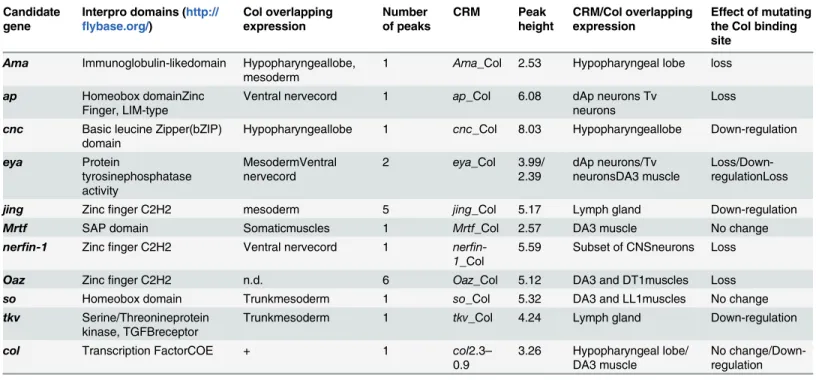
Table 1. Candidate Col direct targets and linked CRMs.
Candidate gene Interpro domains (http:// flybase.org/) Col overlapping expression Number of peaks CRM Peak height CRM/Col overlapping expression Effect of mutating the Col binding site
Ama Immunoglobulin-likedomain Hypopharyngeallobe, mesoderm
1 Ama_Col 2.53 Hypopharyngeal lobe loss
ap Homeobox domainZinc Finger, LIM-type
Ventral nervecord 1 ap_Col 6.08 dAp neurons Tv neurons
Loss
cnc Basic leucine Zipper(bZIP) domain
Hypopharyngeallobe 1 cnc_Col 8.03 Hypopharyngeallobe Down-regulation
eya Protein tyrosinephosphatase activity MesodermVentral nervecord 2 eya_Col 3.99/ 2.39 dAp neurons/Tv neuronsDA3 muscle Loss/Down-regulationLoss
jing Zincfinger C2H2 mesoderm 5 jing_Col 5.17 Lymph gland Down-regulation
Mrtf SAP domain Somaticmuscles 1 Mrtf_Col 2.57 DA3 muscle No change
nerfin-1 Zincfinger C2H2 Ventral nervecord 1 ner fin-1_Col
5.59 Subset of CNSneurons Loss
Oaz Zincfinger C2H2 n.d. 6 Oaz_Col 5.12 DA3 and DT1muscles Loss
so Homeobox domain Trunkmesoderm 1 so_Col 5.32 DA3 and LL1muscles No change
tkv Serine/Threonineprotein kinase, TGFBreceptor
Trunkmesoderm 1 tkv_Col 4.24 Lymph gland Down-regulation
col Transcription FactorCOE + 1 col2.3–
0.9 3.26 Hypopharyngeal lobe/ DA3 muscle No change/Down-regulation n.d.: not detected doi:10.1371/journal.pone.0133387.t001
on DNA samples from two independent IPs. It showed a significant enrichment of both frag-ments, of around 2 (2.08 and 2.19) and 4 (3.90 and 4.26) folds, respectively, compared to the intergenic region of cg11964, a control housekeeping gene [35]. This differential enrichment both confirmed the efficiency of Col antibodies and indicated that the nucleotide sequence of
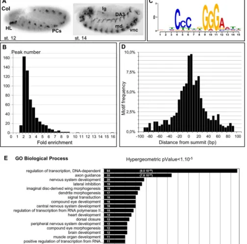
Fig 1. Genome-wide mapping of Col binding sites. (A) Col expression in stage 12 and stage 14 embryos, lateral view. In this and all subsequent figures, embryos are oriented anterior to the left. HL: hypopharyngeal lobe; PCs: dorso-lateral muscle progenitors; vnc: ventral nerve chord; lg: lymph gland; md: class IV
multidendritic neuron. (B) Fold enrichment distribution of the 559 Col ChIP peaks selected using SISSRs. (C) Single most enriched sequence motif identified by MEME analysis in the 559 Col binding peaks. (D) Graphical representation of the position of Col-binding motifs relative to the center of Col binding peaks. The axis gives the number of motifs in each cluster. (E) GO clusters enriched in putative Col direct target genes. The p-Value of the top two clusters is given in brackets.
the Col binding motif could influence the occurrence and/or stability of contextual in vivo Col binding.
Low amounts of chromatin were obtained for each IP, correlating well with the small num-ber of Col-expressing cells. We therefore pooled IP samples before sequencing. 18.3x10⁶and 14.6x10⁶immuno-precipitated fragments of 190 bp average size were sequenced for the Col-IP and mock (HA-IP) samples, respectively (dataset GSE 67805). The sequences were aligned to the D. melanogaster genome (BDGP release 5) using Bowtie v0.12.7 [37]. 16.1x10⁶and
13.1x10⁶unique reads for the Col and mock-IP, corresponding to 25 and 22 times the Drosoph-ila genome size, respectively, were kept for analysis. Peak calling using the SISSRs software [42] detected 559 Col binding peaks (pvalue threshold = 0.1, eValue threshold = 1500), with a fold enrichment ranging from 1.94 to 16.1 (Fig 1BandS1 Table). The peaks were located either in introns or intergenic regions, consistent with Col binding in vivo to cis-regulatory regions.
de novo motif discovery was then performed on the entire set of 559 Col peaks, using the MEME suite software [43]. 200 nucleotides long windows centered on each peak’s summit
were considered for this analysis (S1 File). It revealed that 97% of the peaks (542/559) contain one motif of consensus sequence CCCnnGGGA (Fig 1C). This consensus site is similar to the consensus in vivo binding site determined for mouse EBF in cultured lymphomas [44]. Signifi-cant enrichment of positions 13 and 14 for A and T nucleotides, respectively, was also consis-tent with the ATTCCCNNGGGAAT sequence of the in vitro EBF binding site defined by selex [11]. The calculated E-value: 2,1e-381 and predominant position of the CCCnnGGGA motif close to the center of the ChIP peak (Fig 1D) supported the conclusion that this motif is bound by Col in vivo. MEME analysis failed, however, to reveal other significantly enriched motifs, which could have represented binding sites for other sequence-specific TFs acting synergisti-cally with Col in the different cell types where Col is expressed.
Direct Col-target genes are enriched in transcription factors
PeakAnalyzer [41] associated the 559 Col ChIP-seq peaks to 415 genes (dm3/FlyBase R5.74). Several peaks (between 2 and 9) were associated with the same gene in 95 cases (S1 Table). 150 peaks (27%) were located 5’ and 100 (18%), 3’ of the nearest transcription start site (TSS), while most other peaks (309; 55%) were located in introns. Of course, this association did not exclude that, in some cases, the Col-bound region could act as remote enhancer for other/and additional genes [45], especially for those peaks found further than 10 kb from the nearest TSS (72 peaks; 13%). In 43 cases, the automatic peak association could be manually curated, to account for gene association issued from compared expression of Vienna Tiles (VT) reporter constructs [45] or other CRMs described in REDFly3.3., with that of nearby genes. For 7 peaks, of which 4 were located far away from the nearest transcription start, this analysis modified the associated gene (S1 Table). The described embryonic expression patterns of 176 of the 415 Col targets (http://insitu.fruitfly.org/) indicated that 100 (57%) are expressed in the CNS and 45 (25%) in the mesoderm, consistent with the Col expression pattern in stage 12–14 embryos. In order to further associate Col target genes to biological processes we used GeneCodis [46]; [47]; [48] to perform a Gene Ontology (GO) analysis (Fig 1E). 397 out of 415 Col-bound genes had GO annotations for Biological Processes and were considered by GeneCodis. 156 were rep-resented in at least one cluster (>5 genes) of p-value<10−5. Highly enriched GO terms identi-fied several developmental processes, correlating with the diverse functions played by Col during mid-embryogenesis (Fig 1E). GO terms further revealed a statistically significant enrichment in the category“regulation of transcription”, with 64 of 397 (16%, P-value 9x10-30) annotated Col targets present in this category. This could, in part, be due to larger than average regulatory regions of developmental control genes [49]. Yet, several of these TF genes were
already shown to act genetically downstream of Col in different cell types, thus validating our dataset. One example was cap n’ collar (cnc), which acts downstream of Col in head patterning [5]; [26]. In addition to cnc, the dataset included pox-meso (poxm), slouch/S59 and col itself which are regulated by Col in specific muscle lineages [33]; [32], and apterous (ap), even-skipped (eve), and eyes-absent (eya) which are regulated by Col in specific subsets of VNC neu-rons [28]; [31]. eve expression depends upon Col in a specific subset of neurons, the Eve-lateral (EL) neurons [31]. Previous dissection of eve cis-regulatory elements identified a 0.7kb geno-mic region, EveEL, specifically required for eve expression in EL neurons [50]; [51]. We found that Col binding to eve precisely mapped within EveEL (S1 Fig). The congruence between col/ eve epistatic interaction, and matching genomic positions of EveEL and in vivo Col binding, led us to propose that Col binding reflected direct eve regulation in EL neurons. By extension, GO analysis suggested that Col directly regulates the transcription of various other TFs.
In summary, our ChIP-seq analysis identified 415 potential direct Col targets, among which 64 transcription regulators, suggesting that Col occupies a hierarchical position in a diversity of transcription regulatory networks. More than half of putative Col targets are“unknown” genes for which Col binding provides an entry site for studying their developmental expression and biological role.
Selection of candidate Col direct targets
Identification of Col chromatin binding sites provided an opportunity to identify genes regu-lated in different Col-expressing cells and the corresponding CRMs. Here, we focused on a small set of 10 candidate Col targets. Selection was based on a number of criteria, including a range of peak heights (from 2.39 to 8.03); genes displaying from 1 to 6 Col peaks; documented expression, especially in the nervous system and/or mesoderm (http://insitu.fruitfly.org/); GO analysis (Fig 1E) and previous literature on Col embryonic functions. It included 8 TFs or co-factors—ap, eya, jing, Myocardin-related transcription factor (Mrtf), nervous fingers-1 (nerfin-1), O/E-associated zinc finger protein (Oaz), and sine oculis (so)—and two transmembrane pro-teins, Amalgam (Ama), and thickveins (tkv) (Table 1). Among TFs, ap and eya were chosen because of prior evidence for their regulation by Col in specific neurons, with no evidence that this control was direct [28]; [52]. eya encodes a protein with tyrosine phosphatase activity which is a partner of SIX homeodomain TFs (D-Six4, Optix and So in Drosophila) [53,54]. so is the only SIX gene bound by Col in vivo. Oaz is the Drosophila ortholog of vertebrate Oaz/ Zfp243, the only TF reported to physically interact with EBF [55]; [56].
For each gene in our selection, we cloned 1 kb fragments centered on the Col peak summit, upstream of a moe-GFP reporter construct and followed GFP expression in transgenic Dro-sophila lines. Each CRM construct was given the name of the gene, followed by _Col (Table 1
andS2 Table). For jing and Oaz which display 5 and 4 Col peaks, respectively, we analyzed one peak per gene, where the nucleotide sequence of the predicted Col recognition site was well conserved between 12 different Drosophila species ([57]; [58]; Dataset GSE 67805). For eya, which displays 2 nearby peaks, they were analysed together as well as separately. To precisely determine the role of Col binding for CRM activity, we made a parallel series of constructs in which the predicted Col binding site(s) was mutated at 3 nucleotide positions by substituting CCCNNCCC for CCCNNGGG (S2 Table). This substitution was shown to abolish EBF bind-ing in vitro [11]. As an in vivo control, we mutated the Col binding site in the col2.3–0.9 CRM (col2.3–0.9mut). col-2.3–0.9mutactivation in the head was not modified, while expression was severely reduced in the DA3 muscle where col2.3–0.9 is subject to autoregulation [33], showing that our reporter strategy could identify context-dependent direct regulation by Col (S2 Fig).
Analysis of the 10 candidate CRMs revealed overlap with Col expression in various tissues (Table 1), including the head hypopharyngeal lobe (HL) (Ama, cnc;Fig 2), the LG (jing and tkv,S3 Fig), specific VNC neurons (ap, eya, nerfin-1; Figs3and4andS4 Fig), body wall muscles (eya, Mrtf, oaz, so,Fig 5andS5–S7Figs). tkv_Col and jing_Col were expressed in numerous cell types, including the LG. Since LG development is a dynamic process [27], we compared tkv_Col with tkv_Colmut, and jing_Col with jing_Colmutexpression by ISH at two developmental stages, in order to detect subtle transcriptional regulation potentially hindered by the time lag between moeGFP transcription and protein accumulation (S3 Fig). Upon muta-tion of the Col binding site, expression of both reporters was specifically decreased in the LG, indicating that Col positively regulates tkv_Col and jing_Col activity in this tissue. However, since the decreased expression was only indubitable in about half of the embryos, these targets were not investigated in more detail.
Col targets in the head
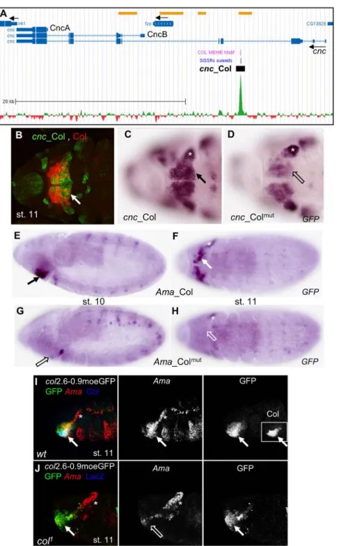
Col is required for head patterning in the embryo, downstream of gap genes [23], more specifi-cally in parasegment 0 (posterior intercalary and anterior mandibular segment; also described as HL from stage 11), where it regulates expression of the segment polarity gene hh and the seg-ment identity gene cnc. Other than col itself [33], hh was the only direct Col target previously characterized [34]. Col ChIP-seq failed to detect binding to hh, perhaps because it is regulated at earlier developmental stages than used for our ChIP-seq [23]; [34]. Concerning cnc, cnc_Col expression reproduced cnc expression in the HL, i.e. partly overlapping that of Col (Fig 2B) [26]. Since rapid morphogenetic movements occur in the head between embryonic stages 8 and 12, we compared cnc_Col and cnc_Colmutexpression by ISH. Lower cnc_Colmutexpression was observed in the HL, compared to cnc_Col, suggesting that Col regulation of cnc via cnc_Col contributes to robustness of cnc expression (Fig 2C and 2D). Ama_Col expression also over-laped that of Col in the HL (Fig 2E and 2F). Ama_Colmutexpression was not detected in the HL, suggesting a direct control of Ama activation by Col (Fig 2G and 2H). We then examined endogenous Ama transcription and found that it was specifically lost in the HL, in col mutant embryos, confirming that this expression is under direct Col control (Fig 2I and 2J). Together, analysis of Ama_Col and cnc_Col indicated that Ama and cnc expression is under direct Col regulation. Beyond, it showed that Col binding to embryonic chromatin could identify new genes involved in head development.
Col targets in neurons
Col is expressed in various subsets of interneurons displaying diverse molecular identities and neurotransmitter phenotypes [31]. nerfin-1, a TF essential for the expression of a subset of axon guidance genes in nascent neurons and expressed in most neuroblasts and nascent neu-rons [60], was found among Col targets. We found that nerfin-1_Col overlaps with the neuro-nal nerfin-1-6 enhancer [61] and is widely active in the VNC at stages 14–15, raising the question of a modular regulation in Col expressing-neurons. We noticed, however, that, beyond stage 15, nerfin-1_Colmutexpression was lost in a few lateral neurons (S4 Fig), which we identified as EL neurons by Eve antibody staining (data not shown). As mentionned above, Col controls eve expression in EL neurons [31], likely directly (S1 Fig). Whether nerfin-1 regu-lation by Col is only direct or also contributed by Eve in a feed-forward process is an open question. Several Col peaks were found to overlap with DNA fragments driving reporters active in EL neurons in stage 16 embryos (http://www.janelia.org/gal4-gen1) [62]. It will be interest-ing to see in the future which of the associated genes are direcly regulated by Col in these neurons.
Fig 2. Col control of cnc and Ama expression in the head. (A) Annotation of the Col peak in cnc, adapted from Gene Browser (GEO submission GSE67805). 39,5 kb of cnc genomic region are shown (Chr3R: 19.009.000–19.048.500) with the Flybase gene annotation indicated by bars (transcribed regions) and intervening blue lines (introns). Black arrows indicate the direction of transcription of cnc and fuzzy onions (fzo), inwardly rectifying potassium channel 1 (Irk1). The cnc transcripts coding for the protein isoforms CncA and CncB are indicated. ChIP-seq data for Col (green) substracted from HA (mock) data (red) are shown on the bottom. The Col Dam-ID binding regions [59] are indicated by yellow bars, top line. The summit of the ChIP-Col peak identified by SISSRs and position of the Col binding site(s) identified by MEME are indicated by blue and violet lines, respectively; the position of cnc_Col is represented by a black box; scale is indicated. (B-D) Ventral anterior views of stage 11 embryos. (B) Overlap between cnc_Col (GFP, green) and Col (red)
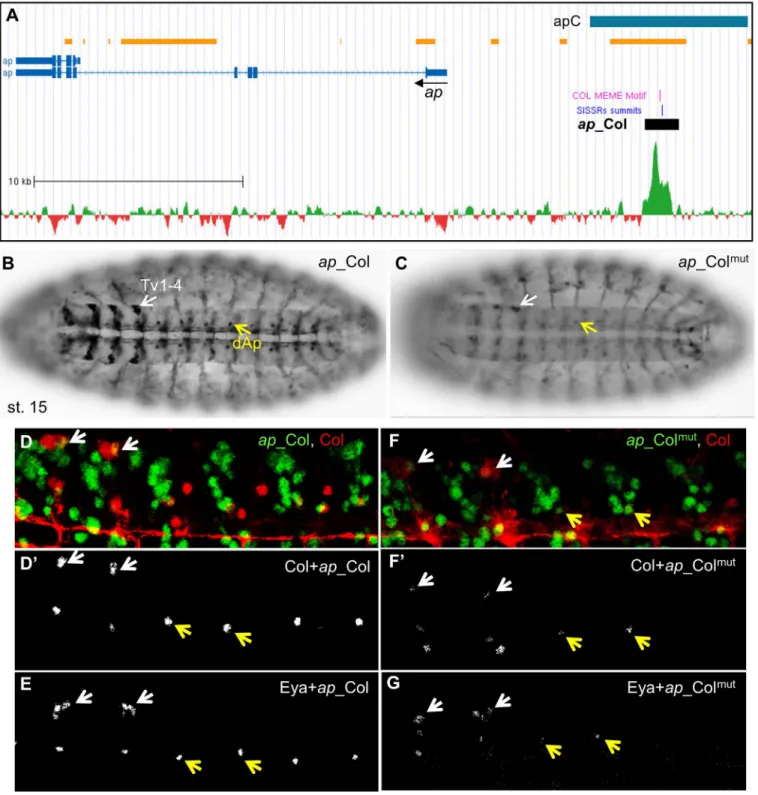
Col directly regulates ap and eya in peptidergic Ap-neurons. Studies of the transcrip-tional regulatory network controlling the peptidergic identity of Ap-expressing neurons showed that Col regulates both Ap and Eya expression in the segmental dorsal AP (dAP) neu-rons and the Tv1-4 neuneu-rons which form only in thoracic T1-T3 segments, and dimmed (dimm, FBgn0023091; a bHLH transcription Factor), expressed in the Tv1 (Tvb) neuron, from stage 16 [28]. Col, Ap, Eya, and Dimm act together to activate Dopamine D1 Receptor (DopR) and Neuropeptide-like precursor 1 (Nplp1) specifically in Tv1 and, possibly the dAp neuron, thereby specifying their peptidergic identity. Although this cascade represents one of the best-studied neuron subtype transcription networks [28]; [52], direct transcriptional regulations remained to be established. We did not detect Col peaks in either dimm, DopR or nplp1, sug-gesting that regulation by Col was indirect or occurred later than stages used for ChIP-seq. However, the presence of Col peaks in ap and eya suggested a direct regulation, especially since the ap Col peak mapped within apC, an upstream region driving neuronal ap expression [63]; [28] (Fig 3A). ap_Col reproduced ap expression in the dAp and 4 Tv neurons in stage 15 embryos (Fig 3B). Immunostaining for ap_Col and either Col or Eya at stage 16 confirmed that only Tv1 and dAp maintain Col expression [28]; [52] (Fig 3D and 3E), confirming transient Col activity in the other Tv neurons. ap_Col encompasses two close Col binding sites which were both mutated in ap_Colmut(S2 Table). ap_Colmutexpression was drastically reduced, when detected, in Tv and dAP neurons (Fig 3C, 3F and 3G), indicating that regulation of ap by Col in these neurons is direct.
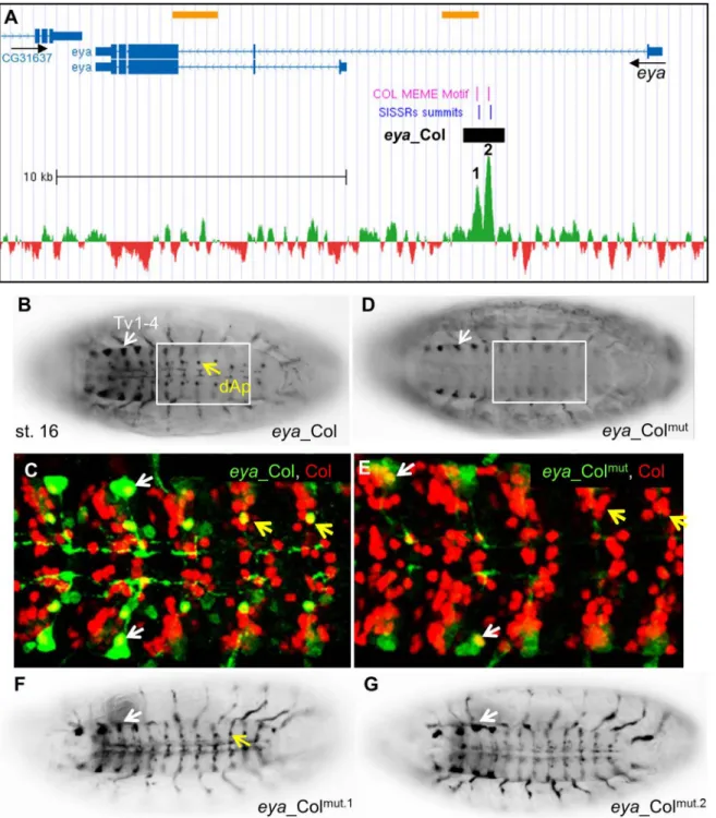
Concerning eya, two nearby Col peaks were identified in the first intron, each centered on a Col consensus recognition site (Fig 4AandS2 Table). eya_Col was designed to encompass both sites. It reproduced eya expression in the dAp and Tv neurons at stage 16, i.e., at a later developmental stage than ap_Col (Fig 4B and 4C). When both Col sites were mutated, eya_-Colmutexpression was completely lost in dAp but only reduced in the Tv1-4 cluster (Fig 4D and 4E), indicating that Col is required for eya activation in dAp neurons, and up-regulation in Tv neurons. We then mutated separately each predicted Col site. eya_Colmut.2showed no expression in dAP neurons while eya_Colmut.1was still expressed, although more weakly than eya_Col (Fig 4F and 4G). We thus conclude that Col site 2 is strictly required for eya transcrip-tion in dAP neurons, while Col site 1 could confer robustness to regulatranscrip-tion by Col. In conclu-sion, Col binding to chromatin showed that it directly controls ap and eya expression in specific subsets of neurons and identified the related CRMs.
Col targets in dorso-lateral muscles
Col is transiently expressed in muscle PCs at the origin of DL muscles, before being uniquely maintained in the elongating DA3 muscle. Time-series datasets for Mef2 and RNA Pol II bind-ing to chromatin and marks of open chromatin and/or active enhancers, H3K79me3 and H3K27ac [64]; [65]; [66] were suggestive of eya_Col, Mrtf_Col, Oaz_Col and so_Col activity in the mesoderm (S2 Table). We found that eya_Col was stochastically expressed in the DA3 muscle, mainly in thoracic segments (see below,Fig 5A). so_Col and Oaz_Col were expressed in the DA3 and other DL muscles. The expression pattern of so_Colmutdid not, however,
expression in the HL (white arrow). (C) cnc_Col and (D) cnc_ColmutmRNA expression, showing
down-regulation of cnc_Colmutin the mandibular segment (open arrow). (E,F) Ama_Col, (G,H) Ama_Colmut expression in stage 10 (E,G) and 11 (F,H) embryos. HL Ama_Col expression (arrow) is lost in Ama_Colmut (open arrow). (I, J) Overlap between Ama (red), Col (blue), and col2.6–0.9moeGFP (green) expression in the HL (white arrow) in stage 11 wt (I), and col1mutant embryos (J). Separate signals for Ama, GFP (I, J) and Col
(inset in I, left panel) are shown in black and white. Ama expression is specifically lost in the HL in col1 mutants. The asterisk in C, D, F, H, J indicates Ama and cnc expression independent on Col. doi:10.1371/journal.pone.0133387.g002
Fig 3. Col direct control of ap expression in Ap neurons. (A) Annotation of the Col peak in ap, same representation as inFig 2A; 35.8 kb of the ap genomic region are shown (Chr2R: 1.593.000–1.628.800); the previously described apC enhancer is represented by a blue box. (B) ap_Col (GFP) expression in the dAp (yellow arrow) and Tv1-Tv4 neurons (white arrow) in stage 15 embryos, ventral view. (C) ap_Colmutexpression is severely reduced in
dAP neurons and Tv neurons. (D,D’) Close up view of 4 segments of stage 16 embryos, showing the specific overlap between Col (red) and ap_Col (green) in the Tv1 and dAp neurons. (E) all Tv neurons express ap_Col and Eya. (F,G) ap_Colmutexpression is lost in dAp and strongly reduced in Tv neurons.
significantly differ from that of so_Col (S5 Fig) while Oaz_Colmutexpression was lost (S6 Fig). Lastly, Mrtf_Col expression was observed in virtually all muscles, but not altered upon
Fig 4. Col direct control of eya expression in Ap neurons. (A) Annotation of the Col peaks in eya, same representation as inFig 2A; 24 kb of the eya genomic region are shown (Chr2L: 6.524.500–6.548.500); the summits of the two ChIP-Col peaks are numbered 1 and 2. (B, C) eya_Col (GFP) expression in the dAp (yellow arrow) and Tv1-Tv4 neurons (white arrow) in stage 16 embryos, ventral view. (C) Close up view of abdominal segments, showing the specific overlap between Col (red) and eya_Col (green) in the dAp (yellow arrow) and Tv1 neurons (white arrow). (D, E) eya_Colmutexpression is lost in dAP neurons
and reduced in TV neurons. (F, G) Mutation of the Col binding site 2 (G), but not site 1 (F) eliminates eya_Col (RFP) expression in dAp neurons (yellow arrow).
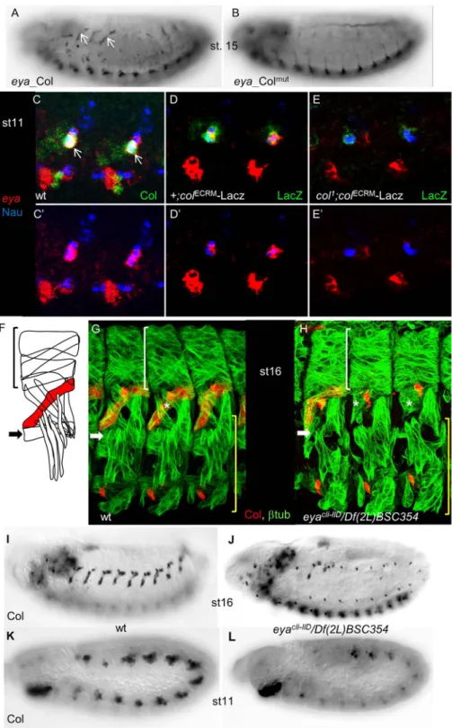
Fig 5. Cross-regulation between eya and col is required to specify dorso-lateral muscles. (A) eya_Col (GFP) expression in the DA3 muscle (arrows) in stage 15 embryos, is not detected for eya_Colmut(B). (C-E)
Triple staining of st11 wt (C), +;coleCRM-lacZ(D) and col1;coleCRM-lacZ(E) embryos for Nau (blue), eya transcripts (red), and either Col (C) orβ-galactosidase (D,E) (green), shows co-expression of Col, cole-CRM -LacZ, Nau and eya in DL muscle PCs (white arrow). (C’-E’) only Nau and eya stainings are shown. eya transcription is specifically lost in DL PCs in col1mutant embryos (E,E
’); dorso-lateral view of the T2 and T3 segments is shown. (F) Schematic drawing of the dorsal, dorso-lateral and lateral transverse muscles in a stage 16 wt embryo, with the DA3 muscle in red and the LL1 muscle indicated by an arrow. (G, H) Staining of
mutation of the Col binding site (S7 Fig). In two cases, eya_Col and Oaz_Col, their expression was dependent upon the predicted Col binding site. In 2 other cases, so_Col and Mrtf_Col, a direct regulatory role for Col binding was left uncertain.
Cross-regulation between col and eya reveals new complexity in the
muscle specification process
eya_Col expression was also observed in the DA3 muscle. Furthermore, this expression was lost with eya_Colmut(Fig 5A and 5B) and down-regulated by mutating either Col site 1 or 2 (data not shown). eya was previously reported to be broadly expressed in the somatic meso-derm at stage 9, and assigned a rather general function in modulating somatic myogenesis [67], while col was only required in DL muscles [32]. The eya and col mutant phenotypes and eya_-Col expression evoked the possibility of eya/col cross-regulation in DL muscle lineages. Immu-nostaining stage 11 embryos for both Col and Nautilus (Nau), to identify muscle PCs and FCs [68], together with eya ISH, revealed that eya and Col are co-expressed in DL PCs, including the PC giving rise to the DA3 muscle (Fig 5C and 5C’). We therefore examined eya expression in col1mutants. To circumvent the lack of detectable Col protein in these mutants, we used the early mesodermal col CRM (colECRM-LacZ; [69] to follow col-expressing myoblasts (Fig 5D and 5D’). Triple staining forβ-gal, Nau and eya showed that eya expression was strongly decreased, when not lost, in col mutant DL PCs, showing that Col regulates eya transcription at that stage (Fig 5E and 5E’). Since a DA3 phenotype has not been previously noted, we re-exam-ined the eya DL muscle phenotype [67]. Loss of LL1, and malformation of DA3 muscles was observed with high frequency in eyacli-IID/Df(2L)BSC354 embryos completely lacking eya func-tion (Fig 5F–5H). This was correlated with the loss of muscle Col expression (Fig 5I and 5J), which could be traced back to stage 11, during the process of selection of PCs from the Col-expressing promuscular cluster (Fig 5K and 5L). These results indicated that eya activity was required for high level col expression at that step. Together, the reciprocal changes of eya and col expression in col and eya mutants and related muscle defects, led us to conclude that regula-tion of eya by Col in muscle PCs is direct, and that cross-regularegula-tion between col and eya genes is critical for establishing the Drosophila muscle pattern.
Col appears to directly regulate eya in specific muscle and neuronal lineages, via binding to the same genomic sites (Fig 6). This provides the first example of diversification of Col regula-tory functions, through differential regulation of the same gene in different cell contexts.
Discussion
Col is dynamically expressed and is required in various developmental processes during Dro-sophila embryogenesis. Through genome-wide mapping of Col chromatin binding sites, we identified direct Col targets, among which transcription regulators, indicating that Col contrib-utes both directly, and through activation of other TFs, to various TF combinatorial codes defining cell identities. Examples are the direct regulation of ap and eya in specific Ap neurons and the cross-regulation brought to light here, between eya and col, during specification of dorso-lateral muscle identity.
stage 16 wt (G) and (H), eyacIi-IID/Df(2L)BSC354(null) mutant embryos for Col (red) and
β3-tubuliin (green). Lateral view of 3 segments. In absence of eya, Col expression is lost in most segments, the LL1 muscle is missing (white arrow) and the DA3 muscle (asterisk) malformed. White brackets indicate dorsal, unaffected muscles while the lateral transverse and ventral muscles (yellow brackets) are moderately affected. (I-J) Col immunostaining of st.16 wt (I), and (J) eya mutant embryos, showing the loss of Col muscle expression in most segments (see also G, H), in eya mutants. (K, L) promuscular Col expression, early stage 11, is reduced in eya mutant embryos (L), compared to wt (K).
Col direct gene targets
ChIP-Seq analysis of chromatin from 10–12h old embryos identified 559 Col binding peaks, corresponding to 415 potential Col direct target genes. Studies of 10 targets revealed regulation by Col in different cell-types, confirming the versatility of Col regulatory functions. Of note, our selection of Col targets included genes with known expression profiles and was therefore biased towards positive regulation. It does not exclude that Col could repress some of its tar-gets. 792 regions binding Col/Kn were independently identified in 0 to 12h embryos by the modENCODE cis-regulatory annotation project [70]. The modENCODE genomic regions did not significantly overlap with the positions of Col peak summits mapped here, however, and we could not detect an enrichment of the EBF/Col motif in the modENCODE peak collection. One possibility to explain this lack of overlap is the different source of antibodies used for ChIP. Another genome-wide search for Col/Kn chromatin binding sites was performed in 3rd instar larvae by the DamID method [59]. 99 genes were found to be both bound and regulated by Col in class IV md neurons, with a broad range of molecular functions possibly contributing to dendritic arbor formation [59]. 15 were found among our 415 genes set (S1 Table), suggest-ing that they are bound by Col already in embryos. Another set of 34“neuronal” genes bound by Col in larvae, though not regulated by Col in md neurons, is bound by Col in embryos. (S1 Table) [59]. 9 of them code for TFs, including nerfin-1, for which we obtained evidence of direct regulation by Col in EL neurons. It will be interesting to determine in which neuron sub-types, TFs bound by Col in embryos and larvae are expressed, as potential entry sites to new networks controlling neuron identity from embryos to larvae. A study in the nematode C. ele-gans concluded that the COE factor UNC-3 was a master regulator of « cholinergic genes » in motor neurons [21]. Our ChIP-seq data failed to detect Col binding to either the Acetylcholine esterase, or Acetylcholine receptor genes, or the choline acetyl transferase upstream region driv-ing expression in cholinergic neurons [71], correlating with the observation that Col is expressed in one cholinergic interneuron per hemisegment, but not in embryonic motor neu-rons. Among the Col-expressing neurons, the best characterized are Tv1 and dAp. CRM activ-ity demonstrated that both ap and eya expression is under independent, direct Col control in
Fig 6. Scheme for Col direct regulation of ap and eya in specific neuron and muscle lineages. Col directly controls ap_Col and eya_Col CRM activity in the Tv and dAP neurons and eya_Col and col_Col CRM in the DA3 muscle lineage. The two Col binding sites in eya_Col CRM are not functionally equivalent (double arrow). eya and col cross-regulate each other in the DA3 PC. Control of DopR and Nplp1 expression in Tv and dAP neurons (Baumgardt et al., 2007) could be indirect.
these neurons, with ap_Col being active earlier than eya_Col. Col requirement for ap and eya expression in the Tv1 neuron persists to the adult stage [52]. It will be interesting to see whether the same CRMs remain active from embryo to adult.
Context-dependent Col binding to chromatin and gene regulation
Over-expression experiments showed that Col ability to auto-regulate or activate target genes is highly cell-context dependent, indicating a need to co-operate with other TFs [23]; [33]; [25]. The consensus CCCnnGGGA motif was found close to the center of a large majority of Col ChIP-seq peaks, showing that Col binds in vivo preferentially, if not exclusively, to this motif in the chromatin of different cell types. No other significantly enriched motif was found by MEME analysis of the whole set of 559 Col peaks. This can be related to the diversity of expression profiles and therefore cis-regulatory signatures of Col-regulated genes. Mutating the Col binding site resulted into either loss (e.g., ap, Ama, eya) or decrease (e.g., cnc) of CRM activity, suggesting that Col either activates or up-regulates/maintains gene transcription, depending upon the gene and cell type/developmental process. In other cases, such as so and Mrtf, CRM activity failed to reveal a requirement for Col binding, raising the possibility that Col could prime some genes in the chromatin for their later regulation by other lineage-specific transcription factors, as proposed for EBF [44]; [72]. The genome distribution of Col peaks shows cases of several peaks associated with the same gene (S1 Table), for example eya. Inde-pendent mutation of each Col binding site in eya_Col showed that one site is strictly required for CRM activity in dAp neurons, while the other provides robustness to this activity. Both sites contribute to CRM activity in Tv neurons and the DA3 muscle. The presence of several Col peaks can therefore reflect redundance of cis-elements required for robustness/precision of Col regulation in a given cell, or/and differential regulation in different cell types, with possible evolutionary implications.
New loops in transcriptional regulatory networks controlling skeletal
muscle identity
eya_Col activity revealed that eya is a direct target of Col in DL muscle PCs. Previously, eya was assigned a rather general role in modulating somatic myogenesis, downstream of the myo-genic factor Tinman (Tin)/Nkx2.5 [67], while col was shown to be negatively regulated by Tin and Tail-up/Islet1 in dorsal muscles and cooperate with Nautilus/D-MyoD in DL muscles [68]; [32]. Our discovery of col/eya cross regulation in DL muscle PCs therefore brings to light a new layer of intricacy in the transcriptional control of muscle identity, with evolutionary implica-tion. Indeed, cooperation between Eya1/2, Six1/4 and MyoD (Mrf), and Ebfs and MyoD has been shown to operate during vertebrate myogenesis [73]; [74]; [75]; [76]; [18]. Future investi-gation of epistatic interactions between muscle iTFs such as Col and myogenic regulators such as eya, Six, and Mrf(s) in patterning the Drosophila somatic musculature, should provide deeper insight into which aspects of the myogenic regulatory network have been conserved and/or diversified during evolution. The question of ancestral functions of COE proteins, which appeared with metazoans [7], is highly speculative, in view of COE pleiotropic functions in extant phyla ([8]; [31]; [77], and references in the above text). Col direct regulation of eya, both in specific muscle and neuronal lineages, may illustrate one mechanism behind the evolu-tionary diversification of Col biological roles.
Supporting Information
S1 Fig. The Col peak in eve.Annotation of the Col peak in eve, adapted from Gene Browser (GEO submission GSE67805). 17.5 kb of the eve genomic region are shown (Chr2R:
5.859.800–5.877.300) with the Flybase gene annotation indicated by blue bars (transcribed regions) and intervening lines (introns). Black arrows indicate the direction of transcription of cnc and cg12134. The Col Dam-ID binding regions [59] are indicated by yellow bars, top line. The summit of the ChIP-Col peak identified by SISSRs and position of the Col binding site(s) identified by MEME are indicated by blue and violet lines, respectively. The position of EveEL is indicated by a blue box. Scale is indicated.
(TIF)
S2 Fig. col direct autoregulation.(A,B) Col expression in A, stage 11 and B, stage 16 embryos. (C,D) col2.3–0.9 expression. (E,F) col2.3–0.9mutexpression. A,C,E: ventral view; B,D,F: lateral view. Mutation of the Col binding site specifically affects col2.3–0.9mutexpression in the DA3 muscle (arrow).
(TIF)
S3 Fig. Col regulates tkv_Col and jing_Col expression in the developing lymph gland. (A-H) In situ hybridization to GFP transcripts in stage 14 (A,B,E,F) and 16 (C,D,G,H) tkv_Col (A,C), tkv_Colmut(B,D), jing_Col (E,G) and jing_Colmut(F,H) embryos. Col regulates both tkv_Col (A-D) and jing_Col (E-H) activity specifically in the developing LG (white arrow). Dorsal views.
(TIF)
S4 Fig. Col regulates nerfin-1-_Col expression in EL neurons.(A) Annotation of the Col peak in nerfin-1, with the same representation as inFig 2A; 10.6 kb of the nerfin-1 genomic region are shown (Chr3L: 903.800–914.400). The position of the previously characterized ner-fin-1-6 enhancer is indicated by a blue box. (B, C) Staining of stage 15 nerfin-1_Col (B) and nerfin-1_Colmut(C) embryos for Col (red), and GFP (green). Only Col and GFP stainings are shown in white in B’,C’ and B”,C”, respectively. A close up view of the squared area (3 seg-ments) is shown below in each panel. The white arrow points to the nerfin-1_Col site of expres-sion lost in nerfin-1_Colmutembryos.
(TIF)
S5 Fig. so_Col expression in developing DL muscles.(A-D) Staining for moeGFP expression of stage 14 (A,C) and stage 16 (B,D) so_Col (A,B) and so_Colmut(C,D) embryos. The brackets in A,C indicate the position of the DL muscles. The DA3, DO5 and LL1 muscles are indicated in some segments in B,D, by an asterisk, a vertical arrow and a double asterisk, respectively. Lateral views.
(TIF)
S6 Fig. Col regulates Oaz_Col expression in the DA3 muscle.(A,B) View of one abdominal segment of stage 13 (A) and stage 15 (B) Oaz_Col embryo stained for Col (DA3 muscle, red) and moeGFP (green); only moeGFP staining is shown on the right, in white. Oaz_Col is expressed in the DA3 and other DL muscles. Staining of stage 14 (C,E) and 16 (D,F) Oaz_Col (C,D) and Oaz_Colmut(E,F) embryos. The bracket in C indicates the position of DL muscle precursors expressing Oaz_Col. Oaz_Colmutexpression is not detected in DL muscles. The DA3 muscle is indicated by an asterisk in D.
(TIF)
S7 Fig. Mrtf_Col expression in somatic muscles.(A-D) Staining of stage 17 Mrtf_Col (A,B) and Mrtf_Colmut(C,D) embryos for Col (red) and moeGFP (green); only moeGFP staining is shown in B,D. The DA3 muscle is indicated in some segments by an asterisk. Lateral views. (TIF)
S1 File. Analyses of Col-ChIP seq data. ChIP-seq summits and MEME sites.DATASET: GEO accession number: GSE 67805.
(TXT)
S1 Table. 415 genes bound by Col in vivo.(A) Gene Name. (B) Number of in vivo Col peaks. (C) Chromosomal position and height of each peak. (D) Peaks genomic coordinates. (E) Anno-tation symbol. (F) Flybase ID number.
(PDF)
S2 Table. CRM_Col constructs and mutated Col binding sites. (PDF)
Acknowledgments
We thank the Bloomington Stock Center for Drosophila strains. We acknowledge Muriel Boube and Alice Davy for critical reading of the manuscript, and the help of Brice Ronsin, Tou-louse RIO Imaging platform, for confocal microscopy and Julien Favier for establishing the transgenic lines and maintenance of fly stocks.
Author Contributions
Conceived and designed the experiments: MdT YC AV. Performed the experiments: MdT YC LB LD SLG. Analyzed the data: MdT YC AP SLG BJ AV. Contributed reagents/materials/anal-ysis tools: MC. Wrote the paper: AV.
References
1. Levine M, Tjian R (2003) Transcription regulation and animal diversity. Nature 424: 147–151. PMID:
12853946
2. Wilczynski B, Furlong EE (2010) Challenges for modeling global gene regulatory networks during development: insights from Drosophila. Dev Biol 340: 161–169. doi:10.1016/j.ydbio.2009.10.032
PMID:19874814
3. Hagman J, Belanger C, Travis A, Turck CW, Grosschedl R (1993) Cloning and functional characteriza-tion of early B-cell factor, a regulator of lymphocyte-specific gene expression. Genes Dev 7: 760–773. PMID:8491377
4. Wang MM, Reed RR (1993) Molecular cloning of the olfactory neuronal transcription factor Olf-1 by genetic selection in yeast. Nature 364: 121–126. PMID:8321284
5. Crozatier M, Valle D, Dubois L, Ibnsouda S, Vincent A (1996) Collier, a novel regulator of Drosophila head development, is expressed in a single mitotic domain. Curr Biol 6: 707–718. PMID:8793297
6. Simionato E, Ledent V, Richards G, Thomas-Chollier M, Kerner P, Coornaert D, et al. (2007) Origin and diversification of the basic helix-loop-helix gene family in metazoans: insights from comparative geno-mics. BMC Evol Biol 7: 33. PMID:17335570
7. Daburon V, Mella S, Plouhinec JL, Mazan S, Crozatier M, Vincent A (2008) The metazoan history of the COE transcription factors. Selection of a variant HLH motif by mandatory inclusion of a duplicated exon in vertebrates. BMC Evol Biol 8: 131. doi:10.1186/1471-2148-8-131PMID:18454855
8. Jackson DJ, Meyer NP, Seaver E, Pang K, McDougall C, Moy VN, et al. (2010) Developmental expres-sion of COE across the Metazoa supports a conserved role in neuronal cell-type specification and mesodermal development. Dev Genes Evol 220: 221–234. doi:10.1007/s00427-010-0343-3PMID:
21069538
9. Garel S, Marin F, Mattei MG, Vesque C, Vincent A, Charnay P (1997) Family of Ebf/Olf-1-related genes potentially involved in neuronal differentiation and regional specification in the central nervous system. Dev Dyn 210: 191–205. PMID:9389446
10. Wang SS, Betz AG, Reed RR (2002) Cloning of a novel Olf-1/EBF-like gene, O/E-4, by degenerate oligo-based direct selection. Mol Cell Neurosci 20: 404–414. PMID:12139918
11. Travis A, Hagman J, Hwang L, Grosschedl R (1993) Purification of early-B-cell factor and characteriza-tion of its DNA-binding specificity. Mol Cell Biol 13: 3392–3400. PMID:8497258
12. Hagman J, Gutch MJ, Lin H, Grosschedl R (1995) EBF contains a novel zinc coordination motif and multiple dimerization and transcriptional activation domains. EMBO J 14: 2907–2916. PMID:7796816
13. Lin H, Grosschedl R (1995) Failure of B-cell differentiation in mice lacking the transcription factor EBF. Nature 376: 263–267. PMID:7542362
14. Garel S, Marin F, Grosschedl R, Charnay P (1999) Ebf1 controls early cell differentiation in the embry-onic striatum. Development 126: 5285–5294. PMID:10556054
15. Wang SS, Lewcock JW, Feinstein P, Mombaerts P, Reed RR (2004) Genetic disruptions of O/E2 and O/E3 genes reveal involvement in olfactory receptor neuron projection. Development 131: 1377–1388. PMID:14993187
16. Kieslinger M, Hiechinger S, Dobreva G, Consalez GG, Grosschedl R (2010) Early B cell factor 2 regu-lates hematopoietic stem cell homeostasis in a cell-nonautonomous manner. Cell Stem Cell 7: 496– 507. doi:10.1016/j.stem.2010.07.015PMID:20887955
17. Gyory I, Boller S, Nechanitzky R, Mandel E, Pott S, Liu E, et al. (2012) Transcription factor Ebf1 regu-lates differentiation stage-specific signaling, proliferation, and survival of B cells. Genes Dev 26: 668– 682. doi:10.1101/gad.187328.112PMID:22431510
18. Jin S, Kim J, Willert T, Klein-Rodewald T, Garcia-Dominguez M, Mosqueira M, et al. (2014) Ebf factors and MyoD cooperate to regulate muscle relaxation via Atp2a1. Nat Commun 5: 3793. doi:10.1038/ ncomms4793PMID:24786561
19. Prasad BC, Ye B, Zackhary R, Schrader K, Seydoux G, Reed RR (1998) unc-3, a gene required for axonal guidance in Caenorhabditis elegans, encodes a member of the O/E family of transcription fac-tors. Development 125: 1561–1568. PMID:9502737
20. Richard JP, Zuryn S, Fischer N, Pavet V, Vaucamps N, Jarriault S (2011) Direct in vivo cellular repro-gramming involves transition through discrete, non-pluripotent steps. Development 138: 1483–1492. doi:10.1242/dev.063115PMID:21389048
21. Kratsios P, Stolfi A, Levine M, Hobert O (2012) Coordinated regulation of cholinergic motor neuron traits through a conserved terminal selector gene. Nat Neurosci 15: 205–214.
22. Dubois L, Vincent A (2001) The COE—Collier/Olf1/EBF—transcription factors: structural conservation and diversity of developmental functions. Mech Dev 108: 3–12. PMID:11578857
23. Crozatier M, Valle D, Dubois L, Ibnsouda S, Vincent A (1999) Head versus trunk patterning in the Dro-sophila embryo; collier requirement for formation of the intercalary segment. Development 126: 4385– 4394. PMID:10477305
24. Crozatier M, Vincent A (1999) Requirement for the Drosophila COE transcription factor Collier in forma-tion of an embryonic muscle: transcripforma-tional response to notch signalling. Development 126: 1495– 1504. PMID:10068642
25. Crozatier M, Vincent A (2008) Control of multidendritic neuron differentiation in Drosophila: the role of Collier. Dev Biol 315: 232–242. doi:10.1016/j.ydbio.2007.12.030PMID:18234173
26. Seecoomar M, Agarwal S, Vani K, Yang G, Mohler J (2000) knot is required for the hypopharyngeal lobe and its derivatives in the Drosophila embryo. Mech Dev 91: 209–215. PMID:10704845
27. Crozatier M, Ubeda JM, Vincent A, Meister M (2004) Cellular immune response to parasitization in Dro-sophila requires the EBF orthologue collier. PLoS Biol 2: E196. PMID:15314643
28. Baumgardt M, Miguel-Aliaga I, Karlsson D, Ekman H, Thor S (2007) Specification of neuronal identities by feedforward combinatorial coding. PLoS Biol 5: e37. PMID:17298176
29. Hattori Y, Sugimura K, Uemura T (2007) Selective expression of Knot/Collier, a transcriptional regulator of the EBF/Olf-1 family, endows the Drosophila sensory system with neuronal class-specific elaborated dendritic patterns. Genes Cells 12: 1011–1022. PMID:17825045
30. Jinushi-Nakao S, Arvind R, Amikura R, Kinameri E, Liu AW, Moore AW (2007) Knot/Collier and cut con-trol different aspects of dendrite cytoskeleton and synergize to define final arbor shape. Neuron 56: 963–978. PMID:18093520
31. Demilly A, Simionato E, Ohayon D, Kerner P, Garces A, Vervoort M (2011) Coe genes are expressed in differentiating neurons in the central nervous system of protostomes. PLoS One 6: e21213. doi:10. 1371/journal.pone.0021213PMID:21695052
32. Enriquez J, de Taffin M, Crozatier M, Vincent A, Dubois L (2012) Combinatorial coding of Drosophila muscle shape by Collier and Nautilus. Dev Biol 363: 27–39. doi:10.1016/j.ydbio.2011.12.018PMID:
22200594
33. Dubois L, Enriquez J, Daburon V, Crozet F, Lebreton G, Crozatier M, et al. (2007) Collier transcription in a single Drosophila muscle lineage: the combinatorial control of muscle identity. Development 134: 4347–4355. PMID:18003742
34. Ntini E, Wimmer EA (2011) Second order regulator Collier directly controls intercalary-specific segment polarity gene expression. Dev Biol 360: 403–414. doi:10.1016/j.ydbio.2011.09.035PMID:22005665
35. Sandmann T, Jakobsen JS, Furlong EE (2006) ChIP-on-chip protocol for genome-wide analysis of tran-scription factor binding in Drosophila melanogaster embryos. Nat Protoc 1: 2839–2855. PMID:
17406543
36. Livak KJ, Schmittgen TD (2001) Analysis of relative gene expression data using real-time quantitative PCR and the 2(-Delta Delta C(T)) Method. Methods 25: 402–408. PMID:11846609
37. Langmead B, Trapnell C, Pop M, Salzberg SL (2009) Ultrafast and memory-efficient alignment of short DNA sequences to the human genome. Genome Biol 10: R25. doi:10.1186/gb-2009-10-3-r25PMID:
19261174
38. Jothi R, Cuddapah S, Barski A, Cui K, Zhao K (2008) Genome-wide identification of in vivo protein-DNA binding sites from ChIP-Seq data. Nucleic Acids Res 36: 5221–5231. doi:10.1093/nar/gkn488
PMID:18684996
39. Narlikar L, Jothi R (2012) ChIP-Seq data analysis: identification of protein-DNA binding sites with SISSRs peak-finder. Methods Mol Biol 802: 305–322. doi:10.1007/978-1-61779-400-1_20PMID:
22130889
40. Machanick P, Bailey TL (2011) MEME-ChIP: motif analysis of large DNA datasets. Bioinformatics 27: 1696–1697. doi:10.1093/bioinformatics/btr189PMID:21486936
41. Salmon-Divon M, Dvinge H, Tammoja K, Bertone P (2010) PeakAnalyzer: genome-wide annotation of chromatin binding and modification loci. BMC Bioinformatics 11: 415. doi:10.1186/1471-2105-11-415
PMID:20691053
42. Pepke S, Wold B, Mortazavi A (2009) Computation for ChIP-seq and RNA-seq studies. Nat Methods 6: S22–32. doi:10.1038/nmeth.1371PMID:19844228
43. Bailey TL, Boden M, Buske FA, Frith M, Grant CE, Clementi L, et al. (2009) MEME SUITE: tools for motif discovery and searching. Nucleic Acids Res 37: W202–208. doi:10.1093/nar/gkp335PMID:
19458158
44. Treiber T, Mandel EM, Pott S, Gyory I, Firner S, Liu ET, et al. (2010) Early B cell factor 1 regulates B cell gene networks by activation, repression, and transcription- independent poising of chromatin. Immunity 32: 714–725. doi:10.1016/j.immuni.2010.04.013PMID:20451411
45. Kvon EZ, Kazmar T, Stampfel G, Yanez-Cuna JO, Pagani M, Schernhuber K, et al. (2014) Genome-scale functional characterization of Drosophila developmental enhancers in vivo. Nature 512: 91–95. doi:10.1038/nature13395PMID:24896182
46. Tabas-Madrid D, Nogales-Cadenas R, Pascual-Montano A (2012) GeneCodis3: a non-redundant and modular enrichment analysis tool for functional genomics. Nucleic Acids Res 40: W478–483. doi:10. 1093/nar/gks402PMID:22573175
47. Nogales-Cadenas R, Carmona-Saez P, Vazquez M, Vicente C, Yang X, Tirado F, et al. (2009) Gene-Codis: interpreting gene lists through enrichment analysis and integration of diverse biological informa-tion. Nucleic Acids Res 37: W317–322. doi:10.1093/nar/gkp416PMID:19465387
48. Carmona-Saez P, Chagoyen M, Tirado F, Carazo JM, Pascual-Montano A (2007) GENECODIS: a web-based tool for finding significant concurrent annotations in gene lists. Genome Biol 8: R3. PMID:
17204154
49. Zeitlinger J, Stark A (2010) Developmental gene regulation in the era of genomics. Dev Biol 339: 230– 239. doi:10.1016/j.ydbio.2009.12.039PMID:20045679
50. Fujioka M, Emi-Sarker Y, Yusibova GL, Goto T, Jaynes JB (1999) Analysis of an even-skipped rescue transgene reveals both composite and discrete neuronal and early blastoderm enhancers, and multi-stripe positioning by gap gene repressor gradients. Development 126: 2527–2538. PMID:10226011
51. Fujioka M, Yusibova GL, Patel NH, Brown SJ, Jaynes JB (2002) The repressor activity of Even-skipped is highly conserved, and is sufficient to activate engrailed and to regulate both the spacing and stability of parasegment boundaries. Development 129: 4411–4421. PMID:12223400
52. Eade KT, Fancher HA, Ridyard MS, Allan DW (2012) Developmental transcriptional networks are required to maintain neuronal subtype identity in the mature nervous system. PLoS Genet 8: e1002501. doi:10.1371/journal.pgen.1002501PMID:22383890
53. Pignoni F, Hu B, Zavitz KH, Xiao J, Garrity PA, Zipursky SL (1997) The eye-specification proteins So and Eya form a complex and regulate multiple steps in Drosophila eye development. Cell 91: 881–891. PMID:9428512
54. Ohto H, Kamada S, Tago K, Tominaga SI, Ozaki H, Sato S et al. (1999) Cooperation of six and eya in activation of their target genes through nuclear translocation of Eya. Mol Cell Biol 19: 6815–6824. PMID:10490620
55. Tsai RY, Reed RR (1997) Cloning and functional characterization of Roaz, a zinc finger protein that interacts with O/E-1 to regulate gene expression: implications for olfactory neuronal development. J Neurosci 17: 4159–4169. PMID:9151733
56. Formstecher E, Aresta S, Collura V, Hamburger A, Meil A, Trehin A, et al. (2005) Protein interaction mapping: a Drosophila case study. Genome Res 15: 376–384. PMID:15710747
57. Blanchette M, Schwikowski B, Tompa M (2002) Algorithms for phylogenetic footprinting. J Comput Biol 9: 211–223. PMID:12015878
58. Odenwald WF, Rasband W, Kuzin A, Brody T (2005) EVOPRINTER, a multigenomic comparative tool for rapid identification of functionally important DNA. Proc Natl Acad Sci U S A 102: 14700–14705. PMID:16203978
59. Hattori Y, Usui T, Satoh D, Moriyama S, Shimono K, Itoh T, et al. (2013) Sensory-neuron subtype-spe-cific transcriptional programs controlling dendrite morphogenesis: genome-wide analysis of Abrupt and Knot/Collier. Dev Cell 27: 530–544. doi:10.1016/j.devcel.2013.10.024PMID:24290980
60. Kuzin A, Brody T, Moore AW, Odenwald WF (2005) Nerfin-1 is required for early axon guidance deci-sions in the developing Drosophila CNS. Dev Biol 277: 347–365. PMID:15617679
61. Kuzin II, Bagaeva L, Young FM, Bottaro A (2008) Requirement for enhancer specificity in immunoglob-ulin heavy chain locus regulation. J Immunol 180: 7443–7450. PMID:18490744
62. Manning L, Heckscher ES, Purice MD, Roberts J, Bennett AL, Kroll JR, et al. (2012) A resource for manipulating gene expression and analyzing cis-regulatory modules in the Drosophila CNS. Cell Rep 2: 1002–1013. doi:10.1016/j.celrep.2012.09.009PMID:23063363
63. Lundgren SE, Callahan CA, Thor S, Thomas JB (1995) Control of neuronal pathway selection by the Drosophila LIM homeodomain gene apterous. Development 121: 1769–1773. PMID:7600992
64. Sandmann T, Jensen LJ, Jakobsen JS, Karzynski MM, Eichenlaub MP, Bork P, et al. (2006) A temporal map of transcription factor activity: mef2 directly regulates target genes at all stages of muscle develop-ment. Dev Cell 10: 797–807. PMID:16740481
65. Sandmann T, Girardot C, Brehme M, Tongprasit W, Stolc V, Furlong EE (2007) A core transcriptional network for early mesoderm development in Drosophila melanogaster. Genes Dev 21: 436–449. PMID:17322403
66. Bonn S, Zinzen RP, Girardot C, Gustafson EH, Perez-Gonzalez A, Delhomme N, et al. (2012) Tissue-specific analysis of chromatin state identifies temporal signatures of enhancer activity during embryonic development. Nat Genet 44: 148–156. doi:10.1038/ng.1064PMID:22231485
67. Liu YH, Jakobsen JS, Valentin G, Amarantos I, Gilmour DT, Furlong EE (2009) A systematic analysis of Tinman function reveals Eya and JAK-STAT signaling as essential regulators of muscle develop-ment. Dev Cell 16: 280–291. doi:10.1016/j.devcel.2009.01.006PMID:19217429
68. Boukhatmi H, Frendo JL, Enriquez J, Crozatier M, Dubois L, Vincent A (2012) Tup/Islet1 integrates time and position to specify muscle identity in Drosophila. Development 139: 3572–3582. doi:10.1242/ dev.083410PMID:22949613
69. Enriquez J, Boukhatmi H, Dubois L, Philippakis AA, Bulyk ML, Michelson AM, et al. (2010) Multi-step control of muscle diversity by Hox proteins in the Drosophila embryo. Development 137: 457–466. doi:
10.1242/dev.045286PMID:20056681
70. Negre N, Brown CD, Ma L, Bristow CA, Miller SW, Wagner U, et al. (2011) A cis-regulatory map of the Drosophila genome. Nature 471: 527–531. doi:10.1038/nature09990PMID:21430782
71. Kitamoto T, Ikeda K, Salvaterra PM (1992) Analysis of cis-regulatory elements in the 5' flanking region of the Drosophila melanogaster choline acetyltransferase gene. J Neurosci 12: 1628–1639. PMID:
1374460
72. Rajakumari S, Wu J, Ishibashi J, Lim HW, Giang AH, Won KJ, et al. (2013) EBF2 determines and main-tains brown adipocyte identity. Cell Metab 17: 562–574. doi:10.1016/j.cmet.2013.01.015PMID:
23499423
73. Heanue TA, Reshef R, Davis RJ, Mardon G, Oliver G, Tomarev S, et al. (1999) Synergistic regulation of vertebrate muscle development by Dach2, Eya2, and Six1, homologs of genes required for Drosophila eye formation. Genes Dev 13: 3231–3243. PMID:10617572
74. Grifone R, Demignon J, Houbron C, Souil E, Niro C, Seller MJ, et al. (2005) Six1 and Six4 homeopro-teins are required for Pax3 and Mrf expression during myogenesis in the mouse embryo. Development 132: 2235–2249. PMID:15788460
75. Green YS, Vetter ML (2011) EBF proteins participate in transcriptional regulation of Xenopus muscle development. Dev Biol 358: 240–250. doi:10.1016/j.ydbio.2011.07.034PMID:21839736
76. Relaix F, Demignon J, Laclef C, Pujol J, Santolini M, Niro C, et al. (2013) Six homeoproteins directly activate Myod expression in the gene regulatory networks that control early myogenesis. PLoS Genet 9: e1003425. doi:10.1371/journal.pgen.1003425PMID:23637613
77. Razy-Krajka F, Lam K, Wang W, Stolfi A, Joly M, Bonneau R, et al. (2014) Collier/OLF/EBF-dependent transcriptional dynamics control pharyngeal muscle specification from primed cardiopharyngeal pro-genitors. Dev Cell 29: 263–276. doi:10.1016/j.devcel.2014.04.001PMID:24794633