HAL Id: hal-01064954
https://hal.archives-ouvertes.fr/hal-01064954
Submitted on 17 Sep 2014
HAL is a multi-disciplinary open access
archive for the deposit and dissemination of
sci-entific research documents, whether they are
pub-lished or not. The documents may come from
teaching and research institutions in France or
abroad, or from public or private research centers.
L’archive ouverte pluridisciplinaire HAL, est
destinée au dépôt et à la diffusion de documents
scientifiques de niveau recherche, publiés ou non,
émanant des établissements d’enseignement et de
recherche français ou étrangers, des laboratoires
publics ou privés.
4DGVF segmentation of vector-valued images
Vincent Jaouen, Paulo Gonzalez, Sylvie Chalon, Denis Guilloteau, Irène
Buvat, Clovis Tauber
To cite this version:
Vincent Jaouen, Paulo Gonzalez, Sylvie Chalon, Denis Guilloteau, Irène Buvat, et al.. 4DGVF
seg-mentation of vector-valued images. IEEE International Conference in Image Processing, Oct 2014,
Paris, France. pp.1. �hal-01064954�
4DGVF SEGMENTATION OF VECTOR-VALUED IMAGES
V. Jaouen
1P. González
1,2S. Chalon
1D. Guilloteau
1I. Buvat
3C. Tauber
11
UMR INSERM U930 «Imagerie et cerveau», Université François-Rabelais de Tours
2 bd Tonnelé, 37044 Tours Cedex, France
2
Universidad Católica del Maule, Talca, Chile
3SHFJ/CEA, Orsay, France
ABSTRACT
In this paper, we extend the gradient vector flow field to the vector-valued case for robust variational segmentation of 4D images with active surfaces. Instead of only exploiting scalar edge strength in order to identify vector edges, we propagate both directions and amplitudes of vector gradients computed from the analysis of a structure tensor of the vector-valued image. To reduce contributions from noise in the calculation of the structure tensor, image channels are weighted accor-ding to a blind estimator of contrast that take profit of the deformable models framework. The proposed 4DGVF vector field is validated on synthetic image datasets and applied to biological volume delineation in dynamic PET imaging.
Index Terms— Deformable models, Image segmenta-tion, Gradient vector flow, Vector-valued images, Positron emission tomography
1. INTRODUCTION
Active surface models attempt to recover a region of in-terest by conforming to its boundary an evolving surface su-perimposed onto the image domain [1, 2] . The evolution of the surface toward equilibrium can be seen as a force-balance relation between forces acting on the surface : inter-nal forces, which control the smoothness of the model, and external forces, derived from image information. In this pa-per, we focus on edge-based parametric deformable models. Among external force fields used for parametric models, gra-dient vector flow (GVF) fields [4, 5] aroused great interest due to their reduced sensitivity to noise and their ability to progress into concave regions in the image. However, their extension to the vector-valued case is not straightforward.
It is generally not satisfying to average the gradient orien-tations of the different channels in order to express the gra-dient of a vector-valued image, or vector gragra-dient [6]. A more relevant approach was proposed by Di Zenzo who considered the image as a vector field [7]. In this context, the gradient
The research leading to these results has received funding from the Euro-pean Union’s Seventh Framework Programme (FP7/2007-2013) under grant agreement HEALTH-F2-2011-278850 (INMiND).
direction and amplitude are associated with the maximization of a quadratic form of the total differential of the image, a problem which can be expressed algebraically using the local structure tensor(LST) formalism [8]. Previous segmentation approaches that rely on this paradigm use the amplitude of the vector gradient to identify vector edges [9, 10, 11, 12]. Howe-ver, relevant directional information can also be drawn from the analysis of the LST in order to further enhance gradient identification, and hence segmentation results. Moreover, the different channels generally participate equally in the calcu-lus of the gradient, even those where the feature of interest is badly represented.
The aim of this work is to propose a new external force field for parametric deformable surfaces evolving in vector-valued images. We introduce a new gradient vector flow field, the 4DGVF field (Four Dimensional Gradient Vector Flow) that takes advantage of both direction and amplitude of vector gradients to propagate them throughout the image. The vector gradients are drawn from the analysis of a LST of the image that combines gradient information available in the different channels. We focus our study on imaging modalities where detection is hampered by varying contrast and noise over the channels, but where physical edges locations do not vary. In such situations, while image edges may be ill-defined on in-dividual channels, the combination of information along the different channels may improve edge location. By weighting the structure tensor dynamically, we control the influence of the different channels in order to favor the ones where the features can be better detected, hence reducing sensitivity to noise.
We assess the quality of our model using synthetic images, realistic Monte Carlo simulations of dynamic Po-sitron Emission Tomography (dPET) images and apply it to real dPET images. dPET images consist of subsequent ac-quisitions of the same field of view at different time intervals and can therefore be considered as vector-valued images. We compare our results to single-channel approaches based on Generalized GVF [5] and Vector Field Convolution (VFC) [13], and to the Color GVF model of Yang et al. for vector-valued images [11].
2. PROPOSED METHOD 2.1. Vector geometry
In the continuous domain, we denote by I a3D vector-valued image constituted ofM channels :
I(x, c) : (Ωs⊗ Ωc) ∈ R3⊗ N → R,
whereΩsis the3D spatial domain of the image and Ωc the
channel dimension. x= (x1, x2, x3) ∈ Ωsis the spatial
po-sition of the voxel. We denote byIk thekthchannel of the
image. A parametric active surface Sτat timeτ of its defor-mation is represented as a mapping of a bivariate parameter (m, n) on a regular grid Ω, superimposed on the spatial image domain : (m, n) → Sτ(m, n) = [x 1(m, n), x2(m, n), x3(m, n)] T . (1) The vector gradient is associated with the direction that maximizes the quadratic form of the total differentialdI of I [7]. In the3D case, I is a 3D→M D vector field and a weigh-ted quadratic form, or first fundamental form ofdI is expres-sed as :
kdIk2ω= dxTGωdx, (2) withGωa regularized, weighted LST [14] :
Gω= Kσ∗ M
X
k=1
ωk(∇Ik⊗ ∇IkT), (3)
whereKσis a Gaussian kernel of scaleσ, ∗ is the
convolu-tion operaconvolu-tion,⊗ is the tensor product, and ωkis a weighting
factor for channel Ik. The weighting factorsωk,k ∈ {1..M }
allow to characterize the reliability of the different channels. Without a priori knowledge, the extension of Di Zenzo’s ap-proach to3D consists in weighting all channels equally by choosingωk = 1/M, ∀k. With such weights, contributions
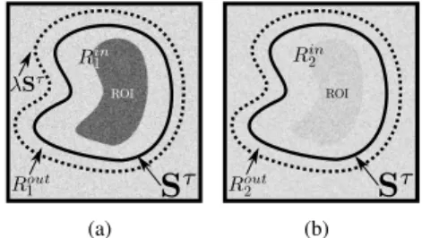
from noise in channels where the object is poorly represen-ted might hamper edge detection. Taking profit of the defor-mable model framework, we propose a blind estimation of the weights based on the object representativeness in the chan-nels. For each channel Ik, letRink be the set of voxels located
inside St, and letRout
k be the set of voxels located outside
Stand insideλSt, a homothetic transformation of Stof pa-rameterλ. The limitation of Rout
k toλSt prevents possible
influence from further regions. We compare the intensities of voxels located inside the region to voxels of its outer neigh-borhood in order to define a contrast measure. We define the weighting factor for channel Ik as follows :
ωk := ¯Iink − ¯Ioutk γ PM j=1 ¯Iinj − ¯Ioutj γ ! , (4)
where summation is over theM channels. ¯Iin
k and ¯Ioutk are
the average intensities inRin
k andRoutk respectively. γ is a
parameter that controls the linearity of the influence of the channels. Figure 1 contains an illustration of the weighting scheme on a conceptual 2D example. Figure 1a displays a high contrast channel where average intensitiesRin1 andR
out 1
are significantly different and for which the corresponding weight would be large. On the contrary, Figure 1b shows a low contrast channel where the corresponding weight would be low.
(a) (b)
Fig. 1. Illustration of the weighting method on a2D repre-sentation of the active surface Staround a region of interest (ROI). (a) High contrast channel. (b) Low contrast channel.
2.2. 4DGVF-based active surface 2.2.1. 4DGVF external force field
The eigenvaluesλ+
> λ−1 > λ −
2 of the LSTGωgive the
scalar rates of change of the first fundamental form in a local basis of extremal variations. Depending on the applications, these eigenvalues can be combined to define different norms of the LST [15, 16, 17, 18]. In our study, we choose a cohe-rence norm that measures the amount of local anisotropy [18], a generalization to the3D case of the norm presented by Sa-piro in [16]. This measure exhibits oriented gradient patterns in the image : kdIk2ω= q (λ+− λ− 1) 2 + (λ+− λ− 2) 2 + (λ−1 − λ − 2)2. (5) We define the gradient amplitudeNωas :Nω = kdIkω.
The directions of the eigenvectors ofGω give the directions
of local extrema of the quadratic form (2). The eigenvector ~
θ+ associated with the maximum eigenvalue λ+ gives the
gradient direction, and the other two define orthogonal "iso-phote" directions. We propose here, rather than only exploi-ting the eigenvalues of the LST [9, 11, 12], to also take advan-tage of the directional information carried by ~θ+. We define
a vectorial edge map ~V, a vector field collinear to the local dominant eigenvectors ofGω, but oriented toward the nearest
vector edge : ~
V= ~θ+sign < ~θ+, ∇Nω>, (6)
The 4DGVF external force field is the result of nonlinear diffusion of the vectorial edge map ~V throughout the image. The 4DGVF field is defined as the steady-state solution of the following vector partial differential equation :
∂~Fext
∂t = g(Nω)∆~Fext− h(Nω)(~Fext− ~V), (7) whereg(s) = e−|∇s|/κ andh = 1 − g are two functions
that control the tradeoff between the first and second terms through parameterκ [5], and ∆ is the vector Laplace opera-tor. In the vicinity of vector edges, as measured byNω, the
directions of ~Fextare constrained by ~V, while isotropic dif-fusion of ~Fext prevails in homogeneous regions. We obtain a regularized vector field oriented locally toward the nearest vector edge.
2.2.2. Model deformation
At each iterationτ of the deformation, the surface Sτ
un-dergoes locally the external force field ~Fext. To avoid conver-gence issues and ensure a smooth deformation, the deforma-tion force field is projected on the normal direcdeforma-tion to Sτ. The surface is iteratively moved according to the following Euler-Lagrange equation :
∂Sτ
∂τ = α∆S
τ− β∆2Sτ+ < ~F
ext, ~n >, (8)
where elasticity terms are weighted byα and rigidity terms by β. ~n denotes the normal direction to the local surface element dSτ. The LST is computed according to the proposed weigh-ting scheme, and at each timestepτ , weights are recomputed to construct a more accurate external force field for the next iteration. Internal energy parameters were set for all methods to typical values found in the literatureα = 0.2 and β = 1.0.
2.3. Initialization
In general, the energy landscape associated with a varia-tional segmentation problem is not convex, requiring the ini-tial model to be close to the desired optimum [19]. To this end, we propose to initialize the 4DGVF model with the Poisson Inverse Gradient algorithm [20] adapted here to the vector-valued case. We build an initialization field ~F0
extbased upon
the 4DGVF framework for which we use equal weighting of all channels, as finer weights such as proposed in eq. (4) can only be derived after an initial surface is defined. Once ~F0
ext
is computed, we estimate the scalar potentialEextby solving
the Poisson equation :
∆Eext= −∇ · ~F0ext, (9)
This equation is solved numerically by matrix inversion for which Dirichlet boundary conditions are applied on the boundary of the image domain. We scaleEext in the range
[0, −1], and perform P energy isosurface reconstructions Ep = (E1, ..., EP), Ep ∈ [0, −1], using a marching cubes
algorithm [21]. We then select the closed model with mi-nimal total external energy. This shape is then used for the computation of the initial weights prior to the deformation.
3. RESULTS
The algorithm was implemented using MATLAB using a finite difference scheme. We assessed the proposed 4DGVF active surface model by comparing it with three other models of the literature :
1) Generalized Gradient Vector Flow (GGVF) active sur-face model [5]. The diffusion of the gradient vectors was per-formed on a channel-by-channel basis, where the edge map of each channelIkwas defined asfk = Kσ∗ |∇Ik| ;
2) Vector Field Convolution (VFC) active surface model [13]. A convolution was performed on a channel-by-channel basis betweenfkand a vector field kernel ~C in which all
vec-tors point toward the kernel’s center : ~Fext= ~C ∗ fk. As per-formances of the two above models depended on the channel studied, we retained the result produced in the channel with best Jaccard index value.
3) Color Gradient Vector Flow (CGVF) active surface mo-del [11]. This vector-valued momo-del computes a single edge map based on Di Zenzo’s vector gradient amplitude, wherein all channels are considered equally for the calculation of the LST. On the contrary of GGVF and VFC, a single active sur-face result was thus obtained for each dynamic image with both CGVF and 4DGVF approaches.
For all methods, the parameters were established so as to maximize the Jaccard similarity criterion [22] between the segmentation result and the ground truth. All models were ini-tialized using the same surface model to prevent results from being affected by this step.
3.1. Synthetic images
Fig. 2. Top :2D slices of the 10 channels of a synthetic 4D image that exhibits varying contrast with background. Bot-tom : corresponding 4DGVF weights obtained for this image.
A set of images of70 × 50 × 40 × 10 voxels was genera-ted for which the contrast of the object with the background varied along the channels. The different channels of one of
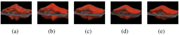
(a) (b) (c)
Fig. 3. Surface plots of the gradient amplitude of a synthe-tic 4D image : (a) fk (best contrasted channel), (b) with
equal weighting of all channels, (c) with proposed weighting scheme
these synthetic 4D images are displayed in the top row of fi-gure 2, where 2D slices are shown for the sake of readabi-lity. The bottom row of figure 2 displays the corresponding weights obtained with the proposed 4DGVF approach, which are consistent with the observed variations of contrast.
In order to study the influence of weighting the LST, we compared the gradient amplitudefk obtained in the best
contrasted channelIk (Fig. 3a) to the vector-gradient
ampli-tude obtained with equal weighting of all channels (Fig. 3b) and to the proposed weighted vector gradient amplitudeNω
(Fig. 3c). In this image, the proposed weighting scheme led to an enhancement of the edge signal while, in flat regions, spu-rious variations due to noise were kept at lower levels, leading to better edge detection. Figure 4 shows segmentation results obtained on this image for the different tested models. The 4DGVF segmentation result (fig. 4e) is the closest to ground truth result (fig. 4a). These observations were confirmed by the Jaccard similarity scores displayed in the middle column of table 1.
(a) (b) (c) (d) (e)
Fig. 4. Segmentation results for a synthetic 4D image. (a) ground truth (b) GGVF (c) VFC (d) CGVF (e) 4DGVF.
Table 1. Average Jaccard similarity scores Image 4D synthetic images 4D PET simulations GGVF 0.89±0.12 0.80±0.26 VFC 0.91±0.11 0.82±0.20 CGVF 0.81±0.15 0.81±0.12 4DGVF 0.95±0.14 0.85±0.11
3.2. Monte Carlo simulations
We assessed the 4DGVF approach with quantitative re-sults on realistic Monte Carlo simulations of dPET images
ge-(a) (b) (c) (d) (e)
Fig. 5. Cerebellum segmentation in a4D PET simulation. (a) ground truth (b) GGVF (c) VFC (d) CGVF (e) 4DGVF
nerated using GATE, an highly realistic PET image simulator based on the CERN’s GEANT4 particles interaction platform [23]. Two4D images based on the Zubal head phantom [24] were created, which necessitated 90 days of parallel compu-tations on a 12 cores 48 GB RAM computer. For each of the reconstructed images, we studied the segmentation of the ce-rebellum. Representative segmentation results are shown in Fig. 5. The average quantitative results obtained for the two simulations are displayed in the right column of table 1. The 4DGVF-based model obtained the best Jaccard similarity in-dex (0.85).
3.3. Real images
Fig. 6. Left : segmentation of a quinolinic acid lesion in the striatum region of a rat brain. Middle, right : result (in red), superimposed onto the 4DGVF field (in white).
To illustrate the behavior of the 4DGVF method in a real pre clinical context, we performed a dPET acquisition of a rat brain using [18F]-DPA-714, a radiotracer specific to the trans-locator protein (TSPO). This protein is over expressed under pathologic neuroinflammatory conditions. The inflammation was produced by performing unilateral quinolinic acid lesions in the right striatum of the rat. Figure 6 shows a representa-tive segmentation result for one rat. The shape produced was consistent with the morphology and location of the injured region.
4. CONCLUSION
We have proposed a novel external force field in order to perform robust active surface segmentation of 3D volumes in vector-valued images. The proposed method exploits the whole spatio-spectral information available in order to in-crease its accuracy and reduce its sensitivity to noise. Re-sults on synthetic images as well as real dPET images have confirmed the potentiality of the proposed method for the segmentation of vector-valued images.
5. REFERENCES
[1] M. Kass, A. Witkin, and D. Terzopoulos, “Snakes : Ac-tive contour models,” International journal of computer vision, vol. 1, no. 4, pp. 321–331, 1988.
[2] A. Dufour, R. Thibeaux, E. Labruyère N. Guillen and J.-C. Olivo-Marin “3-D Active Meshes : Fast Discrete De-formable Models for Cell Tracking in 3-D Time-Lapse Microscopy” Image Processing, IEEE Transactions on, vol. 20, no. 7, pp. 1925–1937, 2011.
[3] S. Osher and J.A. Sethian, “Fronts propagating with curvature-dependent speed : algorithms based on hamilton-jacobi formulations,” Journal of computatio-nal physics, vol. 79, no. 1, pp. 12–49, 1988.
[4] C. Xu and J.L. Prince, “Snakes, shapes, and gradient vector flow,” Image Processing, IEEE Transactions on, vol. 7, no. 3, pp. 359–369, 1998.
[5] C. Xu and J.L. Prince, “Generalized gradient vector flow external forces for active contours,” Signal Processing, vol. 71, no. 2, pp. 131–139, 1998.
[6] Thomas Brox, Joachim Weickert, Bernhard Burgeth, and Pavel Mrázek, “Nonlinear structure tensors,” Image and Vision Computing, vol. 24, no. 1, pp. 41–55, 2006. [7] S. Di Zenzo, “A note on the gradient of a multi-image,”
Computer Vision, Graphics, and Image Processing, vol. 33, no. 1, pp. 116–125, 1986.
[8] H.C. Lee and D.R. Cok, “Detecting boundaries in a vec-tor field,” Signal Processing, IEEE Transactions on, vol. 39, no. 5, pp. 1181–1194, 1991.
[9] G. Sapiro, “Vector (self) snakes : A geometric frame-work for color, texture, and multiscale image segmenta-tion,” in Image Processing, 1996. Proceedings., Interna-tional Conference on. IEEE, 1996, vol. 1, pp. 817–820. [10] X. Xie and M. Mirmehdi, “RAGS : Region-aided
geo-metric snake,” Image Processing, IEEE Transactions on, vol. 13, no. 5, pp. 640–652, 2004.
[11] L. Yang, P. Meer, and D.J. Foran, “Unsupervised seg-mentation based on robust estimation and color active contour models,” Information Technology in Biomedi-cine, IEEE Transactions on, vol. 9, no. 3, pp. 475–486, 2005.
[12] V. Jaouen, P. González, S. Stute, D. Guilloteau, I. Bu-vat, and C. Tauber, “Vector-based active surfaces for segmentation of dynamic PET images,” in Biomedical Imaging (ISBI), 2013 IEEE 10th International Sympo-sium on. IEEE, 2013, pp. 61–64.
[13] B. Li and S.T. Acton, “Active contour external force using vector field convolution for image segmentation,” Image Processing, IEEE Transactions on, vol. 16, no. 8, pp. 2096–2106, 2007.
[14] J. Weickert, Anisotropic diffusion in image processing, vol. 1, Teubner Stuttgart, 1998.
[15] A. Cumani, “Edge detection in multispectral images,” CVGIP : Graphical models and image processing, vol. 53, no. 1, pp. 40–51, 1991.
[16] G. Sapiro and D.L. Ringach, “Anisotropic diffusion of multivalued images with applications to color filtering,” Image Processing, IEEE Transactions on, vol. 5, no. 11, pp. 1582–1586, 1996.
[17] P. Blomgren and T.F. Chan, “Color TV : total varia-tion methods for restoravaria-tion of vector-valued images,” Image Processing, IEEE Transactions on, vol. 7, no. 3, pp. 304–309, 1998.
[18] J. Weickert, “Coherence-enhancing diffusion filtering,” International Journal of Computer Vision, vol. 31, no. 2-3, pp. 111–127, 1999.
[19] C. Tauber, H. Batatia, and A. Ayache, “Quasi-automatic initialization for parametric active contours,” Pattern Recognition Letters, vol. 31, no. 1, pp. 83–90, 2010. [20] B. Li and S.T. Acton, “Automatic active model
initiali-zation via Poisson inverse gradient,” Image Processing, IEEE Transactions on, vol. 17, no. 8, pp. 1406–1420, 2008.
[21] W.E. Lorensen and H.E. Cline, “Marching cubes : A high resolution 3D surface construction algorithm,” in ACM Siggraph Computer Graphics. ACM, 1987, vol. 21, pp. 163–169.
[22] P. Jaccard, “Distribution de la flore alpine dans le bassin des Dranses et dans quelques régions voisines,” Bulletin de la Société Vaudoise des Sciences Naturelles, vol. 37, pp. 241–272, 1901.
[23] S. Jan et al., “GATE v6 : a major enhancement of the gate simulation platform enabling modelling of CT and radiotherapy,” Physics in medicine and biology, vol. 56, no. 4, pp. 881, 2011.
[24] G. Zubal et al., “Computerized three-dimensional seg-mented human anatomy,” Medical Physics-New York-Institute of Physics, vol. 21, no. 2, pp. 299–302, 1994.