HAL Id: inserm-00000108
https://www.hal.inserm.fr/inserm-00000108
Submitted on 28 Oct 2008HAL is a multi-disciplinary open access archive for the deposit and dissemination of sci-entific research documents, whether they are pub-lished or not. The documents may come from teaching and research institutions in France or abroad, or from public or private research centers.
L’archive ouverte pluridisciplinaire HAL, est destinée au dépôt et à la diffusion de documents scientifiques de niveau recherche, publiés ou non, émanant des établissements d’enseignement et de recherche français ou étrangers, des laboratoires publics ou privés.
The envelope protein of a human endogenous
retrovirus-W family activates innate immunity through
CD14/TLR4 and promotes Th1-like responses.
Alexandre Rolland, Evelyne Jouvin-Marche, Christophe Viret, Mathias Faure,
Hervé Perron, Patrice Marche
To cite this version:
Alexandre Rolland, Evelyne Jouvin-Marche, Christophe Viret, Mathias Faure, Hervé Perron, et al.. The envelope protein of a human endogenous retrovirus-W family activates innate immunity through CD14/TLR4 and promotes Th1-like responses.: The HERV family MSRV retrovirus activates in-nate immunity. Journal of Immunology, Publisher : Baltimore : Williams & Wilkins, c1950-. Lat-est Publisher : Bethesda, MD : American Association of Immunologists, 2006, 176 (12), pp.7636-44. �inserm-00000108�
The Envelope Protein of a HERV-W family Human Retrovirus Activates Innate
Immunity Through CD14/TLR4 and Promotes Th1 Like Responses.
Alexandre Rolland, Evelyne Jouvin-Marche, Christophe Viret, Mathias Faure†,
Hervé Perron‡,§ and Patrice N. Marche§
Laboratoire d’Immunochimie, Institut National de la Santé et de la Recherche Médicale U548; Commissariat à l’Energie Atomique; Université J. Fourier, Grenoble, France.
†
INSERM U503; Université Lyon 1; IFR128-Biosciences Gerland, Lyon, France. ‡
bioMérieux, R&D, Marcy l’Etoile, France.
Running title: The HERV family MSRV retrovirus activates innate immunity Keywords: Inflammation, Monocytes/Macrophages, Dendritic Cells, Viral
Abstract
MSRV is a retroviral element the sequence of which served to define the W family of
human endogenous retroviruses (HERV). MSRV viral particles display pro-inflammatory
activities both in vitro in human mononuclear cell cultures and in vivo in a humanized SCID
mice model. To understand the molecular basis of such properties, we have investigated the
inflammatory potential of the surface unit of the MSRV envelope protein (ENV-SU), the
fraction that is poised to naturally interact with host cells. We report here that MSRV
ENV-SU induces, in a specific manner, human monocytes to produce major pro-inflammatory
cytokines through engagement of CD14 and Toll Like Receptor 4 (TLR4) that are pattern
recognition receptors of primary importance in innate immunity. ENV-SU could also trigger a
maturation process in human dendritic cells. Finally, ENV-SU endowed dendritic cells with
the capacity to support a Th1-like type of T helper cell differentiation. The data are discussed
Introduction
HERV (Human Endogenous RetroViruses) represent about 8% of the human genome
and result from integration of exogenous retroviruses that have infected the germline of their
host during primate evolution (1). Although most HERV elements are partly or completely
deleted following integration, the human genome does contain HERV sequences with open
reading frames encoding functional proteins (2). For instance, the so called W family of
HERV possesses a copy on chromosome 7 well known to express an envelope protein called
syncytin that plays an important role in the placenta physiology through syncytiotrophoblast
formation (3-5). Our knowledge of the possible influence of HERV-encoded components in
human disorders remains rather limited. In particular, little is known about the effects of
HERV proteins on the innate immune system which represents the first line of defense against
viruses and operates largely through detection of invariant microbial molecular patterns by
pattern recognition receptors (PRR) expressed on antigen presenting cells (APC) such as
monocytes/macrophages and dendritic cells as well as other cell types. Examples of PRR are
the trans-membrane Toll-Like Receptors (TLR) that can sense distinct microbial products and
are central in innate immune response to various classes of pathogens (6-9).
MSRV is an enveloped virus with reverse transcriptase activity (10) that represents the
prototype genome which defined the HERV-W family in human DNA (3, 11) and was
initially isolated in cell cultures from patients affected by the severe inflammatory
demyelinating disorder of the central nervous system (CNS) multiple sclerosis (12, 13). The
origin of MSRV particles is still unclear. They could originate from a modified endogenous
HERV-W provirus or from a transmissible exogenous member of the same family (11, 14,
15). It was previously observed that MSRV virions trigger the secretion of IL-6 and TNF-α pro-inflammatory cytokines by human peripheral blood mononuclear cells in culture (16). In
blood cells, intra-peritoneally-injected MSRV virion caused an over-expression of TNF-α leading to death by brain hemorrhages within few days (17). Thus, MSRV particles exert
potent pro-inflammatory effects both in mononuclear cells in culture and in vivo in a
humanized-SCID mice model.
In the present study, we have investigated the mechanisms of the pro-inflammatory
properties of the surface unit (ENV-SU) of MSRV envelope protein. This fraction contains
the binding site to the cellular receptor and allows the virus to naturally interact with host
cells. We report that ENV-SU is able to specifically activate cells of the innate immune
system, such as monocytes, through pattern recognition receptors CD14 and TLR4. This
activation is associated with the production of major pro-inflammatory cytokines such as
IL-1β, IL-6 or TNF-α. Moreover, we also show that ENV-SU can activate dendritic cells and promote the development of Th1 like responses.
Materials and methods
Proteins and toxins.
ENV-SU is a 33 KDa and 293 amino acids fraction of the full length MSRV (Multiple
Sclerosis-associated Retro-Viral element) envelope protein (ENV pV14; genbank AF331500).
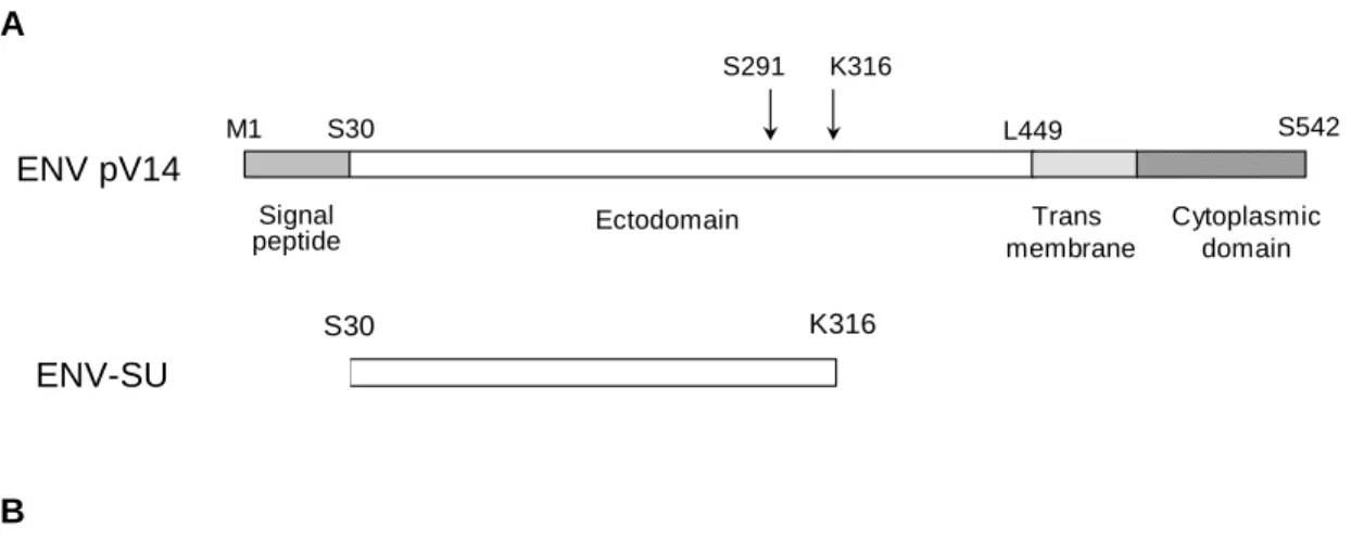
ENV-SU and ENV pV14 organizations are presented in Figure 1A and amino acid sequence
of ENV-SU is presented in Fig. 1B. Recombinant ENV-SU protein was produced and purified
by Protein’eXpert (Grenoble, France) by using the pET-15b expression vector (Novagen,
Merck Biosciences, UK) and AD494 (DE3) E. Coli cells. Briefly,
isopropyl-beta-D-thiogalactopyranoside (IPTG)-treated cultures grown at 37°C were centrifuged and the pellet
was washed. After centrifugation, the pellet was resuspended in wash buffer in the presence
of a cocktail of protease inhibitors (Roche-Diagnostics) and subjected to sonication. The cell
lysate was then centrifuged and the pellet, containing the inclusion bodies, was resuspended in
buffer plus protease inhibitors. After centrifugation, the pellet was washed in the presence of
2% TritonX100, then washed again to remove detergent. Inclusion bodies were solubilized by
resuspension in the presence of 8 M Urea prior to centrifugation. The supernatant was diluted
in Tris HCl pH 8, 1 mM β-mercaptoethanol and loaded onto a Chelating Sepharose Fast Flow column (Amersham Biosciences) for purification under denaturing conditions. ENV-SU was
refolded by dialysis against Tris 50 mM pH 7, NaCl 300 mM, β-mercaptoethanol 1 mM, Sucrose 2%, Glycerol 2%, 2 M urea. Under these conditions, the protein remained soluble.
Amino acid sequence determination of purified ENV-SU based on Edman degradation was in
accordance with the known sequence. Fifty µg/ml aliquots were flash frozen into liquid
nitrogen and stored at –80°C. SDS-PAGE and mass spectrometry (MALDI-TOF) analysis
profiles of recombinant ENV-SU (Protein’eXpert) are shown in Fig. 1C-D. A mock protein,
casein kinase, was used as negative control in experiments and was synthesized and purified
endotoxins by a Lymulus Amebocyte Lysate (LAL) test performed by CleanCells (Bouffere,
France) and all fractions were below the detection level of 5 UI/ml. Staphylococcal
enterotoxin B (SEB) was obtained from Toxin Technology (Sarasota, FL) and was 95 % pure.
Lipopolysaccharide (LPS) from E. coli strain 026:B6 was obtained from Sigma (St Louis,
Mi).
Cell isolation and preparation.
Human peripheral blood mononuclear cells (PBMC) were isolated from healthy donor buffy
coats by density gradient centrifugation over Ficoll Paque. Unlabeled monocytes were
isolated from PBMCs by depletion of T cells, B cells, dendritic cells, basophils and NK cells
by using a Monocyte Isolation Kit II from Miltenyi Biotec (Bergisch Gladbach, Germany).
Briefly, cells were incubated with a cocktail of biotinylated antibodies against CD3, CD7,
CD16, CD19, CD56, CD123 and CD235a prior to addition of Anti-Biotin MicroBeads. The
cell suspension was applied onto a MACS Column placed in the magnetic field of a MACS
Separator and the effluent was collected along with fractions corresponding to three washes.
The purity of monocytes was assessed by immuno-staining (anti-CD14 MOP9 mAb) and flow
cytometry and ranged from 96 to 98%. For the generation of monocyte-derived dendritic cells
(MDDC), purified monocytes were cultured for 5 days in the presence of recombinant IL-4
(25 ng/ml) and GM-CSF (100 ng/ml) (both from R&D systems, Minneapolis, MN). Cells
were fed at day three of culture with full amount of cytokines. As assessed by morphology
and flow cytometry analysis the resulting cell preparation contained more than 90 % of CD1a
positive dendritic cells. Similar results were obtained in experiments when MDDC culture
were used directly or after depletion of possible residual CD14+ cells by immuno-magnetic
cell enrichment cocktail (StemCell Technologies, Vancouver, CA). The recovered CD4+ T
cell populations were always more than 90 % pure.
Cell stimulation.
Cells (PBMC, monocytes or MDDC) were plated in 24-well plates at a concentration of 1 x
106 per well in 1 ml of medium consisting in RPMI 1640 (Gibco, Rockville, MD)
supplemented with 1 % L-glutamine, 1 % penicillin/streptomycin, 1 % sodium-pyruvate, 1 %
non-essential amino acids (all from Sigma), and 10 % heat inactivated FCS (BioWest,
Nouaille, France). After stimulation with ENV-SU, LPS, SEB or Mock, cells were incubated
at 37°C in 5 % CO2 in humidified atmosphere for various periods of time. Where indicated,
proteins and toxins were pre-treated for 30 minutes at 37°C with 25 μg/ml of polymyxin B (PB) (Sigma) prior to stimulation. In some cases, cells were pre-incubated before stimulation
with either 20 μg/ml of anti-CD14 (Polyclonal sheep IgG; R&D systems), anti-TLR4 (Monoclonal mouse IgG2a; clone HTA125, eBioscience, San Diego, CA)(18, 19) or
anti-TLR2 antibodies (Monoclonal mouse IgG2a; clone TL2.1, eBioscience). In some
experiments, ENV-SU, Mock, LPS and SEB were boiled for 30 min before cell stimulation.
To determine the specificity of our results, prior to cell stimulation, 1 μg of ENV-SU, Mock and LPS were pre-incubated for 45 min at 4°C with 30 μg/ml of monoclonal antibodies (mAbs) directed against either ENV-SU (Monoclonal mouse IgG1; clone 13H5A5 and
3B2H4; bioMérieux, Marcy l’Etoile, France) or GAG (Monoclonal mouse IgG1; clone
3H1H6; BioMérieux) recombinant proteins. 13H5A5, 3B2H4 and 3H1H6 mAbs were
obtained by immunization of mice with respectively ENV-SU and GAG recombinant
proteins. The specificity of the antibodies was controlled by ELISA and only 13H5A5 and
Immunofluorescence staining and flow cytometry.
Cells were harvested, washed in PBS and immuno-stained for surface expression of distinct
markers. The following mAbs were obtained from Becton-Dickinson (San Jose, CA):
anti-CD1a allophycocyanin (HI149-APC), anti-CD14 fluorescein isothiocyanate (MOP9-FITC),
CD40 phycoerythrin (5C3-PE), CD80 phycoerythrin (L307.4-PE), CD86 phycoerythrin
(IT2.2-PE) and HLA-DR peridin chlorophyll (L243-PerCP). Briefly, direct
immunofluorescence staining of cells was performed in ice cold PBS supplemented with 2 %
of FCS in the presence of mAbs at concentrations recommended by the manufacturers. After
30 min at 4°C, cells were washed and analyzed using a FACS Calibur and the software
CellQuest (Becton-Dickinson).
Cytokine production and T cell polarization assays.
Culture supernatants were harvested and preserved at -20°C before evaluation of cytokine
production by ELISA. OptEIA ELISA kits from PharMingen (San Diego, CA) for detection
of human IL-1β, IL-6, IL-12p40, IL-12p70 and TNF-α, were used according to the manufacturer’s instructions.
For T cell polarization assay, stimulated MDDC were used as stimulators. Responder cells
were purified allogeneic CD4+CD45RA+ T cells used at 1 x 10⁵ per well (96-well round
bottom microtiter plates). Stimulatory cells were added to T cells in graded doses and cultures
were set up in triplicates in a final volume of 200 μl of medium supplemented with 10% of human AB serum (Sigma). After 5 days of incubation at 37°C, cell supernatants were
Results
ENV-SU stimulates the production of pro-inflammatory cytokines in human PBMC
cultures. We first studied ENV-SU abilities to stimulate cytokine production in PBMC
cultures from healthy controls. Human PBMC were incubated with graded doses of ENV-SU
for 24 hours and secretions of TNF-α, IL-1β and IL-6 were analyzed by ELISA. We observed that ENV-SU induced the production of all three cytokines in a dose/dependent manner even
at doses as low as 10 ng/ml (Fig. 2A). In parallel, a mock control protein (Mock), produced
and purified under the same conditions as ENV-SU, was also tested and no cytokine
production was observed (not shown). We then studied the kinetics of cytokine secretion
induced by ENV-SU in PBMC cultures and compared it with Mock, SEB (a well
characterized bacterial SAg) and LPS. All proteins and toxins were used at a concentration of
1 μg/ml, found to be the optimal concentration for pro-inflammatory cytokine production. As shown in figure 2B, ENV-SU-mediated kinetics of cytokine secretion was more similar to that
of LPS rather than SEB. Both ENV-SU and LPS induced the secretion of high amounts of
TNF-α, IL-6 and IL-1β already after 24 hours. TNF-α and IL-1β reached their peak of production at this time point and then decreased while IL-6 levels constantly increased
following stimulation. SEB did not induce any IL-1β or IL-6, but a constant TNF-α secretion was observed. Mock-induced cytokines remained below the detection threshold. Finally,
separate experiments have revealed that secretion of IFN-γ and IL-10 are marginal in PBMCs cultures treated for 24 hrs with solubleENV-SU (20). Thus, ENV-SU induces the secretion of
major pro-inflammatory cytokines in human PBMC cultures from healthy donors.
ENV-SU-mediated pro-inflammatory properties are specifically inhibited by
monoclonal antibodies and are not due to endotoxin contaminations. The possibility that
ruled out by successive analysis. As a first control, the protein preparations were tested in a
FDA-approved LAL assay (see methods) and were negative. Indeed, it was unlikely that trace
amounts (<5 UI/ml) of endotoxin could cause a higher release of IL-6 by PBMC than the
secretion observed in the presence of 1 μg/ml of pure LPS (Fig. 2B) and doses of LPS corresponding to the detection threshold of the LAL assay effectively did not cause such
effects (not shown). In order to establish the specificity of ENV-SU pro-inflammatory
properties, we studied the effects of anti-ENV-SU monoclonal antibodies (mAb) on the
cytokine production previously described. PBMC were incubated for 24 hours with Mock,
ENV-SU or LPS pre-incubated with antibodies directed against either MSRV ENV-SU or
MSRV GAG. As shown in figure 3A, two of the anti-ENV-SU monoclonal antibodies
(13H5A5 and 3B2H4) tested specifically blocked ENV-SU-mediated TNF-α secretion but not that induced by LPS. In contrast, cytokine secretion was not affected in either case by
treatment with anti-GAG mAb. Since the 13H5A5 and 3B2H4 mAbs had no effect on
LPS-induced TNF-α secretion, the data make the additional point that the inhibition of ENV-SU pro-inflammatory activity cannot be explained by a hypothetic toxic effect of these antibodies
on PBMC. These results definitely demonstrate that ENV-SU can specifically cause PBMC
activation and cytokine release.
Nevertheless, we performed complementary control experiments either by boiling or
treating the proteins and toxins with the cationic antibiotic polymyxin B (PB) that neutralizes
LPS activity, prior to their addition to the culture. Figure 3B shows that while PB treatment
completely abolished LPS-induced TNF-α secretion, it only marginally affected ENV-SU-induced TNF-α production. Conversely, boiled ENV-SU lost most of its stimulatory capacity whereas LPS stimulatory potential remained unaffected by boiling. Taken together, the results
indicate that residual endotoxin contamination cannot account for the pro-inflammatory
ENV-SU directly activates purified human monocytes.
While analysing total PBMC cultures exposed to ENV-SU for 24 and 48 hours, we
observed that the frequency of both CD25+ and CD69+ T lymphocytes remained at the
background level. By intracellular staining, CD3+ mononuclear cells were also devoid of
TNF-α, IFN-γ and IL-4 production (data not shown). Thus, the data failed to reveal any signs of T cell activation in response to soluble ENV-SU in short term cultures. In addition, the
profile of cytokine production induced by ENV-SU was comparable to that induced by LPS
and is reminiscent of the pattern observed upon monocyte activation. We therefore examined
whether ENV-SU was able to directly activate monocytes. Unmanipulated CD14+
mononuclear cells isolated by negative immuno-magnetic separation (>96 % purity) were
stimulated with Mock, ENV-SU or LPS for 24 hours and the expression of the activation
marker CD80 was evaluated by flow cytometry. When compared to Mock, ENV-SU induced
the up-regulation of CD80 expression at a level similar to that induced by LPS (Fig. 4A).
Several other markers such as CD40, CD86 and HLA-DR were also tested but no differences
were observed at this time point (not shown). We then studied the profile of cytokine
secretion induced by ENV-SU in purified monocytes and observed that high amounts of
TNF-α, IL-1β, IL-6 and IL-12p40 were produced in response to ENV-SU (Fig. 4B) while only marginal levels of IL-12p70 were obtained (not shown). Similar observations were made
when using heat-inactivated MSRV particles indicating that the pro-inflammatory effects of
recombinant ENV-SU on purified monocytes is recapitulated by viral particles (Fig. 5). Taken
together these observations indicate that ENV-SU induces a rapid and direct monocyte
CD14 and TLR4 pattern recognition receptors are involved in the pro-inflammatory
response to ENV-SU.
The detection of pathogen-associated molecular patterns by antigen presenting cells
relies largely on a group of trans-membrane PRRs named Toll-Like Receptors (TLR) (6, 7, 9).
For instance, TLR4 engagement mediates monocytes/macrophages activation in response to
Gram-negative bacteria through LPS detection (21). Findings supporting a role for TLR in
virus detection include the sensing of double stranded viral RNA by TLR3 (22), a role for
TLR2 in IL-6 secretion by macrophages in response to the measles virus (23) and the
identification of TLR4 as an essential component of the response to respiratory syncytial virus
(RSV) fusion protein (24) and possibly Mouse Mammary Tumor Virus (MMTV) (25). To
determine whether ENV-SU is susceptible to engage activation pathways similar to those
triggered by LPS or RSV fusion protein, (namely TLR4 and the accessory glycosyl
phosphadityl inositol anchored protein CD14), we pre-treated purified monocytes with
anti-CD14, anti-TLR4 and TLR2 neutralizing antibodies prior to activation with ENV-SU, LPS or
PMA which activates monocytes in a TLR4/CD14 independent manner. The levels of TNF-α secreted were then measured (Fig. 6). No significant inhibition of TNF-α production was observed with any blocking antibody tested when monocytes were stimulated with PMA. In
contrast, when tested on ENV-SU- or LPS-stimulated monocytes, both CD14 and TLR4
blocking antibodies caused substantial inhibition of TNF-α secretion. The inhibition of ENV-SU-induced cytokine production by anti-CD14 antibodies was consistently stronger than that
observed in the presence of the weak affinity anti-TLR4 HTA125 antibody. Such inhibitory
effects were found to be dose sensitive (not shown). TLR2 blocking antibody, which was used
as an isotype control did not induce any inhibition under any conditions tested. In addition,
TLR4, but not TLR2, blocking antibody was also shown to interfere with cytokine production
and CD14 pattern recognition receptors are involved in the pro-inflammatory effects of
ENV-SU on human monocytes.
ENV-SU directly activates Monocyte Derived Dendritic Cells and confer them the
potential to support the development of Th1 like responses.
Dendritic cells (DC) are professional antigen presenting cells that express a variety of
TLRs. TLR engagement induces DC to up regulate antigen presenting and costimulatory
molecules expression as well as to secrete pro-inflammatory cytokines. These maturation
events render them highly potent at activating naïve T cells and promoting their
differentiation in effector cells (26, 27).We therefore examined whether ENV-SU was able to
directly activate monocyte-derived DC (MDDC) and whether these MDDC would show any
capacity to polarize naïve T cell responses. MDDC were generated in vitro from highly
purified human monocytes by using IL-4 and GMCSF and stimulated during 24 hours with
Mock, ENV-SU or LPS. In contrast to the differentiation marker CD1a whose expression
level remained unaltered, we observed that ENV-SU induced a substantial increase in the
surface expression level of CD80, CD86, CD40 and HLA-DR molecules, indicating that
ENV-SU triggers a phenotypic maturation process in MDDC (Fig. 8A). Regarding the
secretion of pro-inflammatory cytokines, IL-6, TNF-α, IL-12p40 and IL-12p70 were produced at rather high levels only in the presence of ENV-SU or LPS (Fig. 8B).
IL-12 is a major cytokine for committing T cells to Th1 lineage differentiation (28). Th1 and
Th2 subsets can develop upon interaction of mature DC with the same T cell precursor, which
is a naïve CD4 T cell. Thus, in order to evaluate the ability of stimulated MDDC to polarize
naïve T lymphocytes, MDDC were co-cultured with purified allogeneic CD4+ CD45RA+ T
cells and cytokine production was measured. MDDC pulsed with ENV-SU were much more
cytokines respectively). High amounts of IFN-γ (Fig. 8C, left panel) were indeed produced even at responder/stimulator ratios as low as 1/100 while the amount of IL-4 (right panel)
obtained when MDDC were stimulated with ENV-SU was consistently below the background
level of the Mock control. By performing inhibition experiments, we found that anti-CD14
antibodies could detectably interfere with the phenotypic activation of immature MDDC
induced by ENV-SU (Fig. 9A). Inhibition by the anti-TLR4 HTA125 mAb was marginal (not
shown). Nevertheless, we consistently noticed that HTA125 was able to reinforce the
inhibitory effect of the anti-CD14 antibodies on ENV-SU-induced cytokine secretion by
MDDCs. This effect was indeed reminiscent of that observed in experiments performed with
purified monocytes (see Figure 6). Altogether, the data indicate that ENV-SU is able to
induce phenotypic and functional maturation of dendritic cells and confer them the potential
Discussion
The present study provides evidence that the surface unit of the envelope protein
(ENV-SU) of MSRV, a retroviral element of the HERV-W family is capable of activating
innate immunity through pattern recognition receptors TLR4/CD14. This conclusion arises
from the observation that ENV-SU induced human monocytes to produce major
pro-inflammatory cytokines in a CD14 and TLR4 dependent fashion. The specificity of such
phenomenon was unequivocally shown by the inhibitory effect of two anti-MSRV-ENV
monoclonal antibodies. ENV-SU also induced dendritic cell maturation and conferred them
the capacity to support a T helper 1 type of T cell differentiation.
CD14 and TLR4 are evolutionary conserved receptors with critical roles for the
activation of antigen presenting cells and pro-inflammatory cytokine production in response
to lipopolysacharide (21, 29, 30). In addition, Respiratory Syncytial Virus (RSV) induce
cytokine secretion by human monocytes via CD14 and TLR4 engagement and TLR4 deficient
mice infected with RSV showed deficient IL-12 production and NK cell response as well as
an increased titer of viral particles in the lung relative to control mice (31). Thus, CD14 and
TLR4 are PRR that cooperate for the activation of innate immunity in response to both
bacteria and viruses. Our observations that CD14 and TLR4 are involved in mediating the
pro-inflammatory effect of the MSRV envelope protein identify proteins of HERV-W family
as a putative new class of viral ligand for these PRR. As dendritic cells do not express CD14
at their surface, their capacity to react to MSRV ENV-SU most certainly relies on the
recruitment of soluble CD14 that is present in the serum as it is known to be the case for the
response of dendritic cell to LPS stimulation (32, 33).
The activation of the innate immune system might contribute to the development of
neurodegenerative diseases and a possible role for CD14 and TLR4 in
naturally present in plasma was elevated in diseases such as multiple sclerosis (34),
rheumatoid arthritis (35) or systemic lupus erythematosus (36). Moreover, specific activation
of CNS innate immunity through TLR4 can lead to neuro-degenerative phenomena (37) via
activation of brain-resident macrophages (microgliocytes) which are the only glial cells with
TLR4 expression. Finally, TLR4 is necessary for LPS-induced oligodendrocyte injury in the
CNS (38). These results indicate that the engagement of TLR4 expressed on CNS resident,
and/or peri-vascular, macrophages by its ligands might contribute to oligodendrocytes damage
and neuro-degeneration.
Since MSRV was first isolated in choroïd plexus/leptomeningeal cell cultures from
multiple sclerosis patients (12, 13), and virion-associated MSRV RNA can be detected in sera
and/or cerebrospinal fluids of such patients (39, 40), it is plausible that its envelope protein
can exert its CD14/TLR4 dependent pro-inflammatory effect within the CNS and therefore
initiates, and/or substantially exacerbates the disorder. This idea is in line with the findings
that (i) HERV-W ENV and GAG glycoproteins are expressed in the white matter of patients
with multiple sclerosis (41-43), and that (ii) the virus load detected in the CSF increases with
MS progression and thus may have prognosis value (38). HERV-W 7q ENV expression is
enhanced relative to tissues from healthy controls or patients with other neurological disorders
and has the potential to cause inflammation and oligodendrocyte death through the induction
of redox reactants in astrocytes (41).
A role for TLR has also been argued in initiation of autoimmunity. One possible way
by which MSRV-mediated activation of the TLR4 signalling pathway could contribute to the
development of autoimmunity is by interference with the immune suppression naturally
mediated by a subclass of CD4+ T lymphocytes named regulatory T cells (Treg). For
instance, Treg can readily suppress the development of auto-immune responses in
(44). A recent report documented that IL-6 produced following TLR-mediated recognition of
microbial products renders naive CD4+ T cells insensitive to the suppressive activity of CD4+
CD25+ Treg (45). We have here demonstrated that via ENV-SU, MSRV could induce the
production of massive amounts of IL-6 in human monocyte and MDDC cultures. Thus,
through the TLR4 dependent secretion of IL-6, ENV-SU could interfere with the suppressive
activity of Treg cells and therefore, facilitate the priming of auto-reactive T cells.
DC are antigen-presenting cells, which upon activation, have the unique ability to
induce primary specific immune responses. Various pathogen products can induce the
maturation of dendritic cells through TLR signalling. In the present study, MDDC pulsed with
ENV-SU displayed all features of a mature phenotype. Interestingly, in the context of multiple
sclerosis, some DC were shown to express the maturation marker CD83 in active lesions (46)
and to secrete pro-inflammatory cytokines such as IL-6 and TNF-α in peripheral blood (47). Depending on the maturation stimuli, MDDC can induce the activation/polarization of naive
T cells towards either Th1 or Th2 lineage. MDDC stimulated with ENV-SU produced large
amounts of pro-inflammatory cytokines including IL-12p70, and were able to promote the
development of naive CD4+ CD45RA+ T cells into IFN-γ secreting Th1 like cells. Induction of Th1 cells insufficiently counterbalanced by Th2 cells has indeed been proposed in the
pathogenesis of demyelinating disorders (48).
Altogether, our observations support the notion that ENV-SU of the HERV-W family
MSRV element, can activate the innate immune system through a TLR4/CD14 dependent
pathway and is susceptible to promote the development of a Th1 type of immune responses
upon DC activation. The data are compatible with the idea that, through the pro-inflammatory
properties of its surface envelope protein, MSRV could be involved in the
immuno-pathological cascades associated with chronic inflammatory and/or neurodegenerative
Acknowledgements
We thank the virology laboratory of the Centre Hospitalier Régional Universitaire in
Grenoble for providing us with blood samples from healthy donors.
References
1. Belshaw, R., V. Pereira, A. Katzourakis, G. Talbot, J. Paces, A. Burt, and M. Tristem. 2004. Long-term reinfection of the human genome by endogenous retroviruses. Proc
Natl Acad Sci U S A 101:4894-4899.
2. Lower, R., J. Lower, and R. Kurth. 1996. The viruses in all of us: characteristics and biological significance of human endogenous retrovirus sequences. Proc Natl Acad Sci
U S A 93:5177-5184.
3. Blond, J. L., F. Beseme, L. Duret, O. Bouton, F. Bedin, H. Perron, B. Mandrand, and F. Mallet. 1999. Molecular characterization and placental expression of HERV-W, a new human endogenous retrovirus family. J Virol 73:1175-1185.
4. Blond, J. L., D. Lavillette, V. Cheynet, O. Bouton, G. Oriol, S. Chapel-Fernandes, B. Mandrand, F. Mallet, and F. L. Cosset. 2000. An envelope glycoprotein of the human endogenous retrovirus HERV-W is expressed in the human placenta and fuses cells expressing the type D mammalian retrovirus receptor. J Virol 74:3321-3329.
5. Mi, S., X. Lee, X. Li, G. M. Veldman, H. Finnerty, L. Racie, E. LaVallie, X. Y. Tang, P. Edouard, S. Howes, J. C. Keith, Jr., and J. M. McCoy. 2000. Syncytin is a captive retroviral envelope protein involved in human placental morphogenesis. Nature
403:785-789.
6. Janeway, C. A., Jr., and R. Medzhitov. 2002. Innate immune recognition. Annu Rev
Immunol 20:197-216.
7. Medzhitov, R. 2001. Toll-like receptors and innate immunity. Nat Rev Immunol
1:135-145.
8. Janeway, C. A., Jr. 1989. Approaching the asymptote? Evolution and revolution in immunology. Cold Spring Harb Symp Quant Biol 54 Pt 1:1-13.
9. Akira, S. 2003. Mammalian Toll-like receptors. Curr Opin Immunol 15:238. 10. Perron, H., J. A. Garson, F. Bedin, F. Beseme, G. Paranhos-Baccala, F.
Komurian-Pradel, F. Mallet, P. W. Tuke, C. Voisset, J. L. Blond, B. Lalande, J. M. Seigneurin, and B. Mandrand. 1997. Molecular identification of a novel retrovirus repeatedly isolated from patients with multiple sclerosis. The Collaborative Research Group on Multiple Sclerosis. In Proc Natl Acad Sci USA, Vol. 94, p. 7583-7588.
11. Komurian-Pradel, F., G. Paranhos-Baccala, F. Bedin, A. Ounanian-Paraz, M. Sodoyer, C. Ott, A. Rajoharison, E. Garcia, F. Mallet, B. Mandrand, and H. Perron. 1999. Molecular cloning and characterization of MSRV-related sequences associated with retrovirus-like particles. Virology 260:1-9.
12. Perron, H., C. Geny, A. Laurent, C. Mouriquand, J. Pellat, J. Perret, and J. M. Seigneurin. 1989. Leptomeningeal cell line from multiple sclerosis with reverse transcriptase activity and viral particles. Res Virol 140:551-561.
13. Perron, H., B. Lalande, B. Gratacap, A. Laurent, O. Genoulaz, C. Geny, M. Mallaret, E. Schuller, P. Stoebner, and J. M. Seigneurin. 1991. Isolation of retrovirus from patients with multiple sclerosis. Lancet 337:862-863.
14. Perron, H., J. P. Perin, F. Rieger, and P. M. Alliel. 2000. Particle-associated retroviral RNA and tandem RGH/HERV-W copies on human chromosome 7q: possible
components of a 'chain-reaction' triggered by infectious agents in multiple sclerosis? J
Neurovirol 6 Suppl 2:S67-75.
15. Perron, H., B. Gratacap, B. Lalande, O. Genoulaz, A. Laurent, C. Geny, M. Mallaret, P. Innocenti, E. Schuller, P. Stoebner, and et al. 1992. In vitro transmission and antigenicity of a retrovirus isolated from a multiple sclerosis patient. Res Virol
16. Perron, H., E. Jouvin-Marche, M. Michel, A. Ounanian-Paraz, S. Camelo, A. Dumon, C. Jolivet-Reynaud, F. Marcel, Y. Souillet, E. Borel, L. Gebuhrer, L. Santoro, S. Marcel, J. M. Seigneurin, P. N. Marche, and M. Lafon. 2001. Multiple sclerosis
retrovirus particles and recombinant envelope trigger an abnormal immune response in vitro, by inducing polyclonal Vbeta16 T-lymphocyte activation. Virology
287:321-332.
17. Firouzi, R., A. Rolland, M. Michel, E. Jouvin-Marche, J. J. Hauw, C.
Malcus-Vocanson, F. Lazarini, L. Gebuhrer, J. M. Seigneurin, J. L. Touraine, K. Sanhadji, P. N. Marche, and H. Perron. 2003. Multiple sclerosis-associated retrovirus particles cause T lymphocyte-dependent death with brain hemorrhage in humanized SCID mice model. J Neurovirol 9:79-93.
18. Shimazu, R., S. Akashi, H. Ogata, Y. Nagai, K. Fukudome, K. Miyake, and M. Kimoto. 1999. MD-2, a molecule that confers lipopolysaccharide responsiveness on Toll-like receptor 4. J Exp Med 189:1777-1782.
19. Wang, R., J. Stephens, and M. J. Lacy. 2003. Characterization of monoclonal antibody HTA125 with specificity for human TLR4. Hybrid Hybridomics 22:357-365.
20. Rolland, A., E. Jouvin-Marche, M. Saresella, P. Ferrante, R. Cavaretta, A. Creange, P. Marche, and H. Perron. 2005. Correlation between disease severity and in vitro
cytokine production mediated by MSRV (Multiple Sclerosis associated RetroViral element) envelope protein in patients with multiple sclerosis. J Neuroimmunol
160:195-203.
21. Poltorak, A., X. He, I. Smirnova, M. Y. Liu, C. Van Huffel, X. Du, D. Birdwell, E. Alejos, M. Silva, C. Galanos, M. Freudenberg, P. Ricciardi-Castagnoli, B. Layton, and B. Beutler. 1998. Defective LPS signaling in C3H/HeJ and C57BL/10ScCr mice: mutations in Tlr4 gene. Science 282:2085-2088.
22. Alexopoulou, L., A. C. Holt, R. Medzhitov, and R. A. Flavell. 2001. Recognition of double-stranded RNA and activation of NF-kappaB by Toll-like receptor 3. Nature
413:732-738.
23. Bieback, K., E. Lien, I. M. Klagge, E. Avota, J. Schneider-Schaulies, W. P. Duprex, H. Wagner, C. J. Kirschning, V. Ter Meulen, and S. Schneider-Schaulies. 2002. Hemagglutinin protein of wild-type measles virus activates toll-like receptor 2 signaling. J Virol 76:8729-8736.
24. Kurt-Jones, E. A., L. Popova, L. Kwinn, L. M. Haynes, L. P. Jones, R. A. Tripp, E. E. Walsh, M. W. Freeman, D. T. Golenbock, L. J. Anderson, and R. W. Finberg. 2000. Pattern recognition receptors TLR4 and CD14 mediate response to respiratory syncytial virus. Nat Immunol 1:398-401.
25. Rassa, J. C., J. L. Meyers, Y. Zhang, R. Kudaravalli, and S. R. Ross. 2002. Murine retroviruses activate B cells via interaction with toll-like receptor 4. Proc Natl Acad
Sci U S A 99:2281-2286.
26. Banchereau, J., and R. M. Steinman. 1998. Dendritic cells and the control of immunity. Nature 392:245-252.
27. Palucka, K., and J. Banchereau. 1999. Linking innate and adaptive immunity. Nat Med
5:868-870.
28. Trinchieri, G., S. Pflanz, and R. A. Kastelein. 2003. The IL-12 family of heterodimeric cytokines: new players in the regulation of T cell responses. Immunity 19:641-644. 29. Hoshino, K., O. Takeuchi, T. Kawai, H. Sanjo, T. Ogawa, Y. Takeda, K. Takeda, and
S. Akira. 1999. Cutting edge: Toll-like receptor 4 (TLR4)-deficient mice are
hyporesponsive to lipopolysaccharide: evidence for TLR4 as the Lps gene product. J
30. Qureshi, S. T., L. Lariviere, G. Leveque, S. Clermont, K. J. Moore, P. Gros, and D. Malo. 1999. Endotoxin-tolerant mice have mutations in Toll-like receptor 4 (Tlr4). J
Exp Med 189:615-625.
31. Haynes, L. M., D. D. Moore, E. A. Kurt-Jones, R. W. Finberg, L. J. Anderson, and R. A. Tripp. 2001. Involvement of toll-like receptor 4 in innate immunity to respiratory syncytial virus. J Virol 75:10730-10737.
32. Frey, E. A., D. S. Miller, T. G. Jahr, A. Sundan, V. Bazil, T. Espevik, B. B. Finlay, and S. D. Wright. 1992. Soluble CD14 participates in the response of cells to lipopolysaccharide. J Exp Med 176:1665-1671.
33. Verhasselt, V., C. Buelens, F. Willems, D. De Groote, N. Haeffner-Cavaillon, and M. Goldman. 1997. Bacterial lipopolysaccharide stimulates the production of cytokines and the expression of costimulatory molecules by human peripheral blood dendritic cells: evidence for a soluble CD14-dependent pathway. J Immunol 158:2919-2925. 34. Brettschneider, J., D. Ecker, A. Bitsch, D. Bahner, T. Bogumil, A. Dressel, E. Elitok,
B. Kitze, S. Poser, F. Weber, and H. Tumani. 2002. The macrophage activity marker sCD14 is increased in patients with multiple sclerosis and upregulated by interferon beta-1b. J Neuroimmunol 133:193-197.
35. Yu, S., N. Nakashima, B. H. Xu, T. Matsuda, A. Izumihara, N. Sunahara, T. Nakamura, M. Tsukano, and T. Matsuyama. 1998. Pathological significance of elevated soluble CD14 production in rheumatoid arthritis: in the presence of soluble CD14, lipopolysaccharides at low concentrations activate RA synovial fibroblasts.
Rheumatol Int 17:237-243.
36. Nockher, W. A., R. Wigand, W. Schoeppe, and J. E. Scherberich. 1994. Elevated levels of soluble CD14 in serum of patients with systemic lupus erythematosus. Clin
Exp Immunol 96:15-19.
37. Lehnardt, S., L. Massillon, P. Follett, F. E. Jensen, R. Ratan, P. A. Rosenberg, J. J. Volpe, and T. Vartanian. 2003. Activation of innate immunity in the CNS triggers neurodegeneration through a Toll-like receptor 4-dependent pathway. Proc Natl Acad
Sci U S A 100:8514-8519.
38. Lehnardt, S., C. Lachance, S. Patrizi, S. Lefebvre, P. L. Follett, F. E. Jensen, P. A. Rosenberg, J. J. Volpe, and T. Vartanian. 2002. The toll-like receptor TLR4 is necessary for lipopolysaccharide-induced oligodendrocyte injury in the CNS. J
Neurosci 22:2478-2486.
39. Dolei, A., C. Serra, G. Mameli, M. Pugliatti, G. Sechi, M. C. Cirotto, G. Rosati, and S. Sotgiu. 2002. Multiple sclerosis-associated retrovirus (MSRV) in Sardinian MS
patients. Neurology 58:471-473.
40. Sotgiu, S., C. Serra, G. Mameli, M. Pugliatti, G. Rosati, G. Arru, and A. Dolei. 2002. Multiple sclerosis-associated retrovirus and MS prognosis: an observational study.
Neurology 59:1071-1073.
41. Antony, J. M., G. van Marle, W. Opii, D. A. Butterfield, F. Mallet, V. W. Yong, J. L. Wallace, R. M. Deacon, K. Warren, and C. Power. 2004. Human endogenous
retrovirus glycoprotein-mediated induction of redox reactants causes oligodendrocyte death and demyelination. Nat Neurosci 7:1088-1095.
42. Perron, H., F. Lazarini, K. Ruprecht, C. Pechoux-Longin, D. Seilhean, V. Sazdovitch, A. Creange, N. Battail-Poirot, G. Sibai, L. Santoro, M. Jolivet, J. L. Darlix, P.
Rieckmann, T. Arzberger, J. J. Hauw, and H. Lassmann. 2005. Human endogenous retrovirus (HERV)-W ENV and GAG proteins: physiological expression in human brain and pathophysiological modulation in multiple sclerosis lesions. J Neurovirol
43. Garson, J., A. Creange, A. Dolei, P. Ferrante, E. Jouvin-Marche, P. N. Marche, F. Rieger, K. Ruprecht, M. Saresella, S. Sotgiu, R. Tedder, and H. Perron. 2005. MSRV, Syncytin and the role of endogenous retroviral proteins in demyelination. Mult Scler
11:249-250.
44. Kohm, A. P., P. A. Carpentier, H. A. Anger, and S. D. Miller. 2002. Cutting edge: CD4+CD25+ regulatory T cells suppress antigen-specific autoreactive immune responses and central nervous system inflammation during active experimental autoimmune encephalomyelitis. J Immunol 169:4712-4716.
45. Pasare, C., and R. Medzhitov. 2003. Toll pathway-dependent blockade of
CD4+CD25+ T cell-mediated suppression by dendritic cells. Science 299:1033-1036. 46. Plumb, J., M. A. Armstrong, M. Duddy, M. Mirakhur, and S. McQuaid. 2003.
CD83-positive dendritic cells are present in occasional perivascular cuffs in multiple sclerosis lesions. Mult Scler 9:142-147.
47. Huang, Y. M., B. G. Xiao, V. Ozenci, M. Kouwenhoven, N. Teleshova, S. Fredrikson, and H. Link. 1999. Multiple sclerosis is associated with high levels of circulating dendritic cells secreting pro-inflammatory cytokines. J Neuroimmunol 99:82-90. 48. Charlton, B., and K. J. Lafferty. 1995. The Th1/Th2 balance in autoimmunity. Curr
Footnotes:
§
H.P. and P.N.M. equally contributed to this study. The present address for C.V. is: INSERM
U563 and Université P. Sabatier, C.H.U. Purpan, Toulouse, France. This work was supported
by institutional grants from Institut National de la Santé et de la Recherche Médicale
(INSERM), and a specific grant from the Programme Région Rhône-Alpes (#: 02.020691.0).
A. Rolland is a recipient of fellowships from bioMérieux and from the Fondation pour la
Recherche Médicale, C.V. is supported by the Centre National de la Recherche Scientifique
(CNRS) and M.F. was a recipient of a postdoctoral fellowship from the Région Rhône-Alpes.
Corresponding author: Dr. Patrice N. Marche, CEA DRDC and INSERM U548, 17 rue des
Martyrs, 38054 Grenoble cedex 9 (France) ; Tel: (33) 4 38 78 38 73; Fax: (33) 4 38 78 98 03;
Figure legends
Figure 1. ENV-pV14 and ENV-SU organizations and amino acid sequences.
(A) Organizations of pV14 (full length MSRV envelope protein) and SU.
ENV-SU is a 293 amino acids fraction, representing the extracellular surface unit cleaved at K316
of the full length ENV pV14 protein. (B) Amino acid sequence of ENV-SU (bold characters).
(C) SDS-12,5% PAGE analysis of recombinant ENV-SU after purification by affinity
chromatography under denaturing conditions (molecular mass is in kDa).(D) Mass spectrum
of the recombinant ENV-SU protein. The profile represents the transformation of the mass to
charge ratio on the real mass scale. Vertical axis: % relative intensity; horizontal axis: molecular weight.
Figure 2. ENV-SU induces the production of pro-inflammatory cytokines in PBMC
cultures.
(A) PBMC from healthy donors were stimulated for 24 hours with graded doses of ENV-SU
and culture supernatants were then analyzed by ELISA for TNF-α, IL-1β and IL-6 production. (B) PBMC were stimulated with 1 μg/ml of Mock, ENV-SU, LPS or SEB and incubated for 24, 48 and 72 hours before analysis of cytokine secretion by ELISA. Results are
presented as the mean +/- standard error of three independent experiments.
Figure 3. ENV-SU mediated TNF-α production is inhibited by specific monoclonal
antibodies and is not due to endotoxin contamination.
(A) PBMC were stimulated for 24 hours with 1 μg/ml of mock control, ENV-SU and LPS pre-incubated or not for 45 min at 4°C with mAbs (30 μg/ml) specific for ENV-SU (Clones 13H5H5 and 3B2H4) or GAG (Clone 3H1H6) proteins. Culture supernatants were harvested
pre-treated for 30 minutes at 37°C with 25 μg/ml of PB prior to PBMC stimulation. In parallel, cells were also incubated with proteins and toxins boiled for 30 min (100°C). Culture
supernatants were then harvested and tested by ELISA for TNF-α release. Results represent the mean +/- standard error of three independent experiments.
Figure 4. ENV-SU directly activates purified human monocytes.
(A) Human monocytes were purified from human PBMC (> 95% purity) and then stimulated
with Mock, ENV-SU or LPS at 1 μg/ml for 24 hours. Cells were harvested and surface expression of the activation marker CD80 was analyzed by flow cytometry. One
representative experiment of Mock, ENV-SU and LPS stimulation is presented. (B) Cytokine
production (TNF-α, IL-1β, IL-6 and IL-12p40) was analyzed by ELISA. Results represent the mean +/- standard error of three independent experiments.
Figure 5. MSRV particles directly activate purified monocytes.
(A) Purified monocytes were stimulated for 24 hours with 2 x 10³ MSRV particles or MOCK
control and the expression of several activation markers such as CD80 and CD86 were
assessed by flow cytometry. The surface expression of both CD80 and CD86 were up
regulated following MSRV treatment. MSRV particles were obtained from MSRV infected
immortalized B cell cultures after ultra-centrifugation. MSRV particles were quantified by
reverse transcriptase activity measurement. All MSRV particles preparations were
mycoplasma free. The MOCK control was prepared from MSRV negative B cell cultures.
(B) The supernatants were then tested for pro-inflammatory cytokine secretion by ELISA.
The values are the mean +/- standard error of replicate from one experiment representative of
Figure 6. CD14 and TLR4 are involved in ENV-SU mediated pro-inflammatory properties.
Purified monocytes were pre-incubated at 37°C for one hour with or without neutralizing
antibodies to CD14 (polyclonal IgG), TLR4 (HRTA125 mAb, functional grade) and TLR2
(TL2.1 mAb) (all at 20 μg/ml) and then further stimulated for 24 hours with PMA at 100 ng/ml, ENV-SU at 200 ng/ml or LPS at 50ng/ml. The effects of PB were also tested in
parallel as a control. Culture supernatants were harvested and TNF-α release was evaluated by ELISA. Isotype controls (Functional grade mouse IgG2a and polyclonal sheep IgG) were
also tested and no inhibitory effects were observed (not shown). The values represent the
mean +/- standard error of replicate from one experiment representative of three.
Figure 7. MSRV particles activate purified monocytes through TLR4.
Purified monocytes were pre-incubated at 37°C for one hour with or without (x) neutralizing
antibodies to TLR4 (α TLR4) or TLR2 (α TLR2) (both at 20 μg/ml) and then further stimulated for 24 hour with 2 x 10³ MSRV particles. Culture supernatants were harvested and
TNF-α release was evaluated by ELISA. The values correspond to the mean +/- standard error of three independent experiments.
Figure 8. MDDC activated with ENV-SU secrete IL-12 and promote the
development of Th1 like responses.
(A) MDDC were generated from purified monocytes and then stimulated with Mock,
ENV-SU (1 μg/ml) or LPS (100 ng/ml) for 24 hours. Surface staining of activation markers HLA-DR, CD80, CD86 and CD40 was made after Mock (dotted line), ENV-SU (shaded histogram)
and LPS (bold line) treatments and then analyzed by flow cytometry. One representative
were analyzed by ELISA. (C) Various concentrations of MDDC stimulated either with Mock
(dotted line), ENV-SU (bold line) or LPS were co-cultured for 5 days with purified allogeneic
CD4+ CD45RA+ T cells. Supernatants were then tested for IFN-γ (left panel) and IL-4 release (right panel). The values represent the mean +/- standard error of three (B) and two
(C) independent experiments.
Figure 9. CD14 and TLR4 receptors are involved in ENV-SU-mediated activation of
immature MDDC.
Monocytes-derived MDDC were stimulated with ENV-SU (5 ng/ml) and analyzed for
phenotypic maturation and cytokine secretion.
(A) Inhibition of ENV-SU-induced MDDC phenotypic maturation by anti-CD14 mAbs.
Surface expression of CD40, CD86, CD80, and HLA-DR were analyzed by immunostaining
and flow cytometry (vertical axis: cell count, horizontal axis: Log fluorescence intensity).
Vehicle (closed histograms), ENV-SU (open histograms, bold line), ENV-SU + 20 μg/ml anti-CD14 (open histograms, dotted line), and ENV-SU + 20 μg/ml control IgG (open histograms, regular line). The histograms shown are representative of three independent
experiments.
(B) Additive inhibitory effect of anti-TLR4 and anti-CD14 antibodies on cytokine secretion
by ENV-SU-treated immature MDDC. Cytokine productions (TNF-α, IL-6 and IL-12p40) were analyzed by ELISA after stimulation in the presence of CD14, TLR4,
anti-TLR2 or anti-CD14 + anti-TLR4. The data are representative of two independent
Figure 1 C D 4 1 0 0 0 4 1 0 0 0 32925.54 20 30 40 50 60 70 80 90 100 4 1 0 0 0 4 1 0 0 0 32925.54 4 1 0 0 0 4 1 0 0 0 32925.54 20 30 40 50 60 70 80 90 100 20 30 40 50 60 70 80 90 100
32925.54
MALPYHTFLFTVLLPPFALTAPPPCCCTTSSSPYQEFLWRTRLPGNIDAPSYRSLSKGNSTFTAHTHMPRNCY NSATLCMHANTHYWTGKMINPSCPGGLGATVCWTYFTHTSMSDGGGIQGQAREKQVKEAISQLTRGHSTPS PYKGLVLSKLHETLRTHTRLVSLFNTTLTRLHEVSAQNPTNCWMCLPLHFRPYISIPVPEQWNNFSTEINTTS LVGPLVSNLEITHTSNLTCVKFSNTIDTTSSQCIRWVTPPTRIVCLPSGIFFVCGTSAYHCLNGSSESMCFLSFL VPPMTIYTEQDLYNHVVPKPHNK A B ENV pV14 ENV-SU Signal peptide V S30 K316 Signal peptide Ectodomain Cytoplasmic domain K316 S291 S30 L449 M1 S542 Trans membrane M10 0.1 0.2 0.3 0.4 0.5 0.6 0.01 0.1 1 0 1 2 3 4 5 6 7 8 9 0 0.2 0.4 0.6 0.8 1 1.2 IL-6 IL-1β TNF-α ENV-SU (μg/ml) ng /m l A B IL-1β 0 0.2 0.4 0.6 0.8 1.0 1.2 1.4 0 24h 48h 72h ENV-SU LPS Mock SEB ng/m l IL-6 0 5 10 15 20 25 0 24h 48h 72h TNF-α 0 24h 48h 72h 0 0.1 0.2 0.3 0.4 0.5 0.6 0.7 0.8 0.9 Figure 2
0 0.2 0.4 0.6 0.8 1.0 MOCK ENV-SU LPS TN F -α (ng/m l) No treatment Anti ENV-SU (3B2H4) Anti ENV-SU (13H5A5) Anti GAG (3H1H6) A B 0 0.2 0.4 0.6 0.8 1.0 1.2 1.4 MOCK ENV-SU LPS TN F -α (ng/m l) No treatment PB (25μg/ml) Boiling (100°C 30min) Figure 3
a ENV-SU LPS MOCK CD80 Ce ll co un t Log Fluorescence B IL-6 10 20 30 40 50 60 70 IL-12p40 0.2 0.4 0.6 0.8 1 0 MOC K EN V-S U LPS TNF-α 0 0.5 1 1.5 2 2.5 IL-1β 1 2 3 4 5 6 7 8 ng/ m l A MOC K EN V-SU LPS Figure 4
0 0.1 0.2 0.3 MOCK MSRV TNF-α IL-1β IL-6 0.2 0.4 0.6 0.8 MOCK MSRV 0.2 0.4 0.6 0.8 1.0 1.2 1.4 MOCK MSRV TN F -α (ng/ m l) B A CD80 PE C e ll co unt MOCK MSRV
CD 80
MOCK MSRVCD 86
MOCK MSRV Figure 5X αCD14 α TLR4 αCD14 + TLR4 PB 0 0.2 0.4 0.6 0.8 ENV-SU LPS PMA 0 0.2 0.4 0.6 0.8 0 0.2 0.4 0.6 0.8 TN F -α (ng/m l) α TLR2 Figure 6
MSRV 0 0.1 0.2 0.3 X α TLR4 α TLR2 TN F -α (ng/m l) * C Figure 7
A B CD80 CD86 CD40 HLA-DR C IFN-γ 0 2 4 6 8 10 12 IF N -γ (ng /ml ) 1/100 1/20 1/10 T cells / MDDC ratio Moc k EN V-S U LP S IL-12p40 0 4 8 12 16 IL-12p70 0 0.2 0.4 0.6 TNF-a 0 1 2 3 IL-6 0 10 20 30 ng /m l Moc k EN V-S U LP S IL-4 0 2 4 6 8 10 12 IL -4 (ng /ml ) 1/100 1/20 1/10 T cells / MDDC ratio Ce ll c o un t Log Fluorescence Figure 8
TNF-α 0 200 400 600 800 1000 1200 1400 1600 1800 ENV SU @CD14/TLR4 ENV SU @TLR2 ENV SU @TLR4 ENV SU @CD14 ENV SU MEDIA pg/ml IL-6 0 2000 4000 6000 8000 10000 12000 14000 16000 18000 20000 ENV SU @CD14/TLR4 ENV SU @TLR2 ENV SU @TLR4 ENV SU @CD14 ENV SU MEDIA pg/ml IL-12p40 0 1000 2000 3000 4000 5000 6000 7000 8000 9000 ENV SU @CD14/TLR4 ENV SU @TLR2 ENV SU @TLR4 ENV SU @CD14 ENV SU MEDIA pg/ml