HAL Id: tel-02050010
https://tel.archives-ouvertes.fr/tel-02050010
Submitted on 26 Feb 2019
HAL is a multi-disciplinary open access
archive for the deposit and dissemination of sci-entific research documents, whether they are pub-lished or not. The documents may come from teaching and research institutions in France or abroad, or from public or private research centers.
L’archive ouverte pluridisciplinaire HAL, est destinée au dépôt et à la diffusion de documents scientifiques de niveau recherche, publiés ou non, émanant des établissements d’enseignement et de recherche français ou étrangers, des laboratoires publics ou privés.
Genetic Diversity, Phylogeny and Conservation of
Rainbowfish (Melanotaeniidae) in West Papua Indonesia
and Its Prospect for New Ornamental Fish Commodity
Media Fitri Isma Nugraha
To cite this version:
Media Fitri Isma Nugraha. Genetic Diversity, Phylogeny and Conservation of Rainbowfish (Melano-taeniidae) in West Papua Indonesia and Its Prospect for New Ornamental Fish Commodity. Genetics. Université Montpellier; Graduate School of the Bogor Agricultural University, Bogor (Indonésie), 2015. English. �NNT : 2015MONTS208�. �tel-02050010�
0
Délivré par UNIVERSITÉ DE MONTPELLIER
Préparée au sein de l’école doctorale
GAIA - Biodiversité, Agriculture, Alimentation, Environnement, Terre, Eau
UMR-ISEM
Spécialité : EERGP
Ecologie, Évolution, Ressources Génétique, Paléontologie
Présentée par
MEDIA FITRI ISMA NUGRAHA
Soutenue le 03 /11 /2015 devant le jury composé de
Jean –Christophe AVARRE, Charge de Recherche IRD (Directeur de Thèse UM) Odang CARMAN, Associate Professeur, IPB (Directeur de Thèse IPB) Ketut SUGAMA, Professeur, Centre de recherche pour l'aquaculture (Rapporteur)
Phillippe KEITH, Professeur, Muséum National d’Histoire Naturelle Paris (Rapporteur) M. Fatuchri SOEKADI, Proffeseur, Comité R & D des affaires maritimes
et de la pêche (Président) Membre invite
Laurent POUYAUD, Charge de Recherche IRD (Co-directueur de thèse UM)
Genetic Diversity, Phylogeny and Conservation of Rainbowfish
(Melanotaeniidae) in West Papua Indonesia and Its Prospect for
1
THÈSE
En vue de l’obtention duDOCTORAT de L’UNIVERSITE de
MONTPELLIER
Spécialité : Écologie, Evolution, Ressources Génétique, Paléobiologie Présentée et soutenue par: Media Fitri Isma NUGRAHA
Genetic Diversity, Phylogeny and Conservation of
Melanotaeniidae in West Papua Indonesia and Its Prospect for
New Ornamental Fish Commodity
Soutenue le 03/11/2015 devant le jury composé de
JURY
Jean –Christophe AVARRE, Charge de Recherche IRD (Directeur de These UM) Odang CARMAN, Associate Professeur, IPB (Directeur de These IPB) Ketut SUGAMA, Professeur, Research Centre for Aquaculture (Rapporteur)
Phillippe KEITH, Professeur, Museum National d’Histoire Naturelle (Rapporteur) M. Fatuchri SOEKADI, Proffeseur Comité R & D des affaires
Maritimes et de la pêche RI (President) Membre invite
2
THÈSE
En vue de l’obtention duDOCTORAT de L’UNIVERSITE de
MONTPELLIER
Spécialité : Écologie, Evolution, Ressources Génétique, Paléobiologie Présentée et soutenue par: Media Fitri Isma NUGRAHA
Genetic Diversity, Phylogeny and Conservation of
Melanotaeniidae in West Papua Indonesia and Its Prospect for
New Ornamental Fish Commodity
Soutenue le 03/11/2015 devant le jury composé de
JURY
Jean –Christophe AVARRE, Charge de Recherche IRD (Directeur de These UM) Odang CARMAN, Associate Professeur, IPB (Directeur de These IPB) Ketut SUGAMA, Professeur, Research Centre for Aquaculture (Rapporteur)
Phillippe KEITH, Professeur, Museum National d’Histoire Naturelle (Rapporteur) M. Fatuchri SOEKADI, Proffeseur Comité R & D des affaires
Maritimes et de la pêche RI (President) Membre invite
3 ACKNOWLEGMENT
Thank to My God for all blessings and loves, therefore this Disertation has done. The topic chosen is a new topic in ichthyology, aquaculture, population genetic, conservation and evolution. Many biodiversity in New Guinea Island, particularly West Papua makes many misinterpretations about flora and fauna, particularly fresh water fish in West Papua area.
My heart thanks to :
1. My Supervisors DR. Jean-Christphe Avarre and DR. Laurent Pouyaud, which fired my imagination and support me to begin endeavor in laboratory and science, and also supported me in many material and immaterial ways, ideas, spirit and other moral that are very priceless have been given from research, article writing, publication and my these. Without you all, these would not be a thesis. With the most fully of my heart Thank you so much for every thing.
2. DR. Esstelle Massaret and familly, Thank you so much for every thing. Its very beautiful i know you.
3. My supervisor In IPB: DR. Odang Carman, DR. Utut Widyastuti, Pr. DR. M. Zairin Junior, and Pr. DR. Komar Sumantadina (Alm)
4. Dr. Jacques Slembrouck and family. Thank you so much for your gatherings. 5. Mr. Michel larue and Madame Fida Larue, thank you for finding and guiding
for my step “Fish do not jump alone into the plastic bag”. Thank you so much.
6. France government, thank you for the scholarship with “Bourse Government France” and IRD Scholarship with “Bourse de These”.
7. Team CAVIAR IRD: DR. Jean Paul Toutain, DR. Marc Legendre, DR. Etienne Baras, DR. Remi Dugue, DR. Emmanuel Paradis, DR. Dominico Carrusso, DR. Jean Francois Agnese, Madame Monique Saltel, M. Christophe coche, Ibu Etny, Bapak Sumanta. Thank you for your wonderfull company and support.
8. Prof. Dr. Ir. Indrajaya, M.Sc and Prof. Dr. Ir. Dachrul Syach. M.Sc, Thank you for your help in my co-tutelle program.
4 9. Dr. Kadarusman, thank you for sharing and knowledge about Papua and rainbow fish, also the exploration and expedition therefore there are many fish revealed there.
10. All academic staffs in IPB and Université Montpellier II France.
11. Franch embassy and Campus France – Mle. Apreita Putri, M. Farid Saadoun and M. Florian Libourel, thank you for all financial and administration supports.
12. CROUS Montpellier having helped all my life aspects during my studying in Montpellier.
13. Platforme Génomique Environnmentale du Labex Centre Méditerranéen Environnement Biodiversité Plateforme Génotypage – séquencage Université Montpellier II team (Dr. Fréderique Cerqueira, Dr. Erick Desmarais, Dr. Philippe Clair)
14. Thank you to my institution Ministry Marine and Fisheries Republic Indonesia - Balai Penelitian dan Pengembangan Budidaya Ikan Hias – Pusat
Penelitian dan Pengembangan Perikanan budidaya - Badan Penelitian dan Pengembangan Kelautan dan Perikanan.
15. Thank you to all teachers and friends in BGF 2010 in Institut Francaise Indonesia.
16. My friends in PPI Montpellier and Franco-Indonesia family in Montpellier having given me much wonderful memory.
17. Finally my heart felt thanks to all my big family, my parents, my uncles, my aunties, all my siblings and all my cousins.
My hopefully this these will give many advantages particularly for Ichthyology, Aquaculture and Genetic Science.
5
A ma Mitochondrial
Ma grand-mère Nurcaya Syam
et Ma mère Asma Murni
6 LIST OF PUBLICATION ASSOCIATED WITH THE THESIS
1. Media Fitri Isma Nugraha, Laurent Pouyaud, Odang Carman, Kadarusman, Utut Widyastuti, Jean-Christophe Avarre. 2014. Development of twelve novel polymorphic microsatellite DNA markas for the Boeseman's rainbowfish (Melanotaenia boesemani) and test for their cross-utility in 21 rainbowfish species from West Papua (Indonesia). European Journal Wild Life Research. 60:941-946. doi: 10. 1007/s10344-014-0868-2.
2. Media Fitri Isma Nugraha, Kadarusman, Nicolas Hubert, Jean-Christophe Avarre, Renny Kurnia Hadiaty, Jacques Slembrouck, Odang Carman, Sudarto, Ran Ogistira and Laurent Pouyaud. 2015. Eight new species of Rainbowfishes (Melanotaeniidae) from the Birds Head Region, West Papua, Indonesia.
Cybium 39(2): 99-130.
3. Media Fitri Isma Nugraha, Laurent Pouyaud, Odang Carman, Utut Widyastuti, M. Zairin Junior, Kadarusman, Jean-Christophe Avarre. 2015. Genetic diversity of Boeseman’s rainbowfish Melanotaenia boesemani reared in Indonesian farms compared to endangered natural population. Tropical
7 TABLE OF CONTENTS
I. GENERAL INTRODUCTION 10
II. LITERATURE REVIEW 14
2.1. RAINBOWFISH DISTRIBUTION AND DIVERSITY 14
2.2. THE MELANOTAENIIDAE FAMILY 19
2.3. THREATS ON THE MELANOTAENIIDAE 23
2.4. HISTORY OF RAINBOWFISH AS WORLD AQUARIUM
PEARLS AND AQUACULTURE 24
2.5. MAJOR MOLECULAR MARKERS USED IN FISH
GENETICPOPULATION STUDIES 28
2.6. ISOLATION OF MICROSATELLITE MARKERS IN
NON MODEL ORGANISMS 35
2.7. THE APPLICATION OF MICROSATELLITES IN
AQUACULTURE 39
III. DEVELOPMENT OF NEW MICROSATELLITE MARKERS FROM M.
BOESEMANI USING NEXT GENERATION SEQUENCING
TECHNOLOGY 43
3.1. INTRODUCTION 43
3.2. MATERIAL AND METHODS 45
3.2.1. Origin of the fish 45
3.2.2. Extraction of genomic DNA 45
3.2.3. Isolation of microsatellite markers 46
3.2.4. Amplification and size analysis of microsatellite markers 46 3.2.5. Test of the Mendelian inheritance pattern of the selected
microsatellite markers 48
3.2.6. Microsatellite data analysis 49
3.3. RESULTS 50
3.3.1. Selection and test of candidate microsatellite markers on several
Melanotaenia species. 50
3.3.2. Experimantal validation of the 12 new microsatellite markers 52
3.4. DISCUSSION 58
IV. GENETIC DIVERSITY OF WILD POPULATIONS OF MELANOTAENIA
FROM WEST PAPUA 59
4.1. INTRODUCTION 59
4.2. MATERIAL AND METHODS 61
4.2.1 Origin of the investigated fish 62
4.2.2. Genotyping of all populations with the 12 nuclear DNA
Microsatellites 63
4.2.3. Phylogeny of West Papua populations based on the
mitochondrial DNA barcode 64
8
4.3. RESULTS 71
4.3.1. Analysis of all the wild populations with the microsatellite
markers 71
4.3.2. Confirmation by the microsatellite markers of valid species and
8 new Melanotaenia species from West Papua 76
4.3.3. Mitochondrial DNA barcode and tree-based delimitation of West-
Papuan species 82
4.4. DISCUSSION 87
V. GENETIC DIVERSITY AND GEOGRAPIC ORIGIN OF MELANOTAENIA BOESEMANI REARED IN INDONESIAN FARMS 89 5. 1. INTRODUCTION 89 5. 2 MATERIAL AND METHODS 91
5.2.1. Origin and sampling of the investigated fish 92
5.2.2. DNA extraction and microsatellite amplification 92 5.2.3. Genetic diversity analysis 93 5.3. RESULTS 93 5.3.1. Genetic variability and heterozygosity of M. boesemani in Aquaculture settings and in natural populations 93 5.3.2. Geographic origin of M. boesemani reared in Indonesian farms 100
5.4 DISCUSSION 101
VI. GENERAL DISCUSSION AND PERSPECTIVES 104
REFERENCES 108
9 LIST OF FIGURE
1. Melanotaenia boesemani ... 13
2. Map of the Melanotaeniidae distribution based on molecular phylogenies... 22
3. Seven endangered Melanotaenia species from Bird’s head peninsula (West papua) ... 23
4. Tree species rainbowfish, have been domesticated and cultivated in Indonesia. ... 23
5. Captive breeding of M. boesemani in Indonesian farms ... 28
6. General flow chart for the development of simple sequence repeats (SSR) markers ... 39
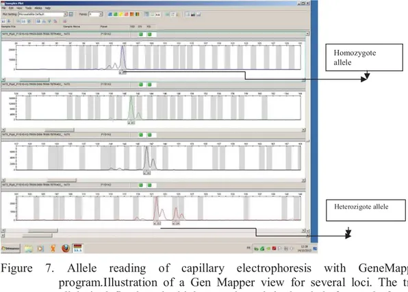
7. Allele reading of capillary electrophoresis with GeneMapper program. ... 40
8. Example of agarose gel results. ... 53
9. Factorial component analysis 5 mate-pairs of M. Boesemani ... 58
10. Type localities of the 24 valid Melanotaenia species and the 8 new species from the Bird’s Head region... 68
11. Factorial correspondence analysis of genotypes all the species belonging to the phylogenetic cluster “Central Ayamaru Plateau” and to M. arfakensis. . ... 78
12. Factorial correspondence analysis of genotypes all valid species belonging to cluster “Southern Ayamaru Plateau”, to cluster “Central Ayamaru Plateau” and to M. arfakensis. ... 79
13. Factorial correspondence analysis of genotypes from M. manibuii, M. arfakensis, M. multiradiata, M. fredericki, M. batanta, M. misoolensis, M. salawati. ... 79
14. Factorial correspondence analysis of genotypes from M. manibuii, and all valid species belonging to the cluster ‘Central Ayamaru Plateau”. ... 80
15. Factorial correspondence analysis of genotypes from the five new species belonging to COI “Northern Bird’s Head” clade. ... 80
16. Factorial correspondence analysis of genotypes from all the species belonging to cluster “Bird’s Neck”. ... 82
17. Maximum likelihood tree for Bird’s Head region Melanotaenia species based on analysis of partial COI sequences (650 bp). ... 85
18. Description and corresponding picture of the eight new species of Melanotaenia from West Papua. ... 87
19. Coloration patterns of Boeseman´s Rainbowfish Melanotaenia boesemani from Ayamaru and from Uter lake. ... 92
20. Geographic localization of Ayamaru and Uter Lakes in West Papua, Indonesia. ... 93
21. Melanotaenia boesemani with a malformation morfologie and coloration. ... 94
22. Factorial Component Analysis based on the multilocus genotypes Melanotaenia boesemani ... 104
10 LIST OF TABLE
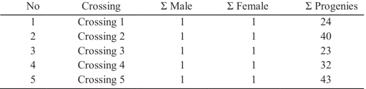
1. Rainbowfish classification according to ANGFA ... 16 2. Sequence of the 61 selected microsatellite primer pairs. ... 49 3. Crossing of M. boesemani for microsatellite marker validation. ... 51 4. Name and main characteristics of the 12 microsatellite markers
validated on 12 Melanotaenia species. ... 54 5. Allele frequencies for each breeding pair (crossing) and its
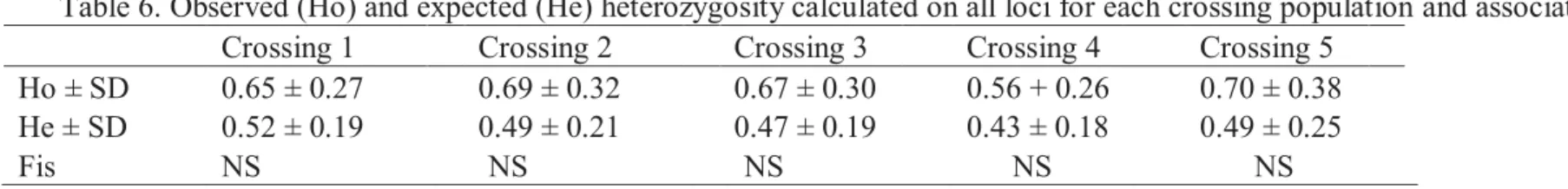
corresponding offspring at the 12 microsatellite loci ... 55 6. Observed (Ho) and expected (He) heterozygosity calculated on all loci
for each crossing population and associated Fis... 57 7. Fst analysis each crossing family M. boesemani ... 58 8. List of all investigated populations (valid and new species in West
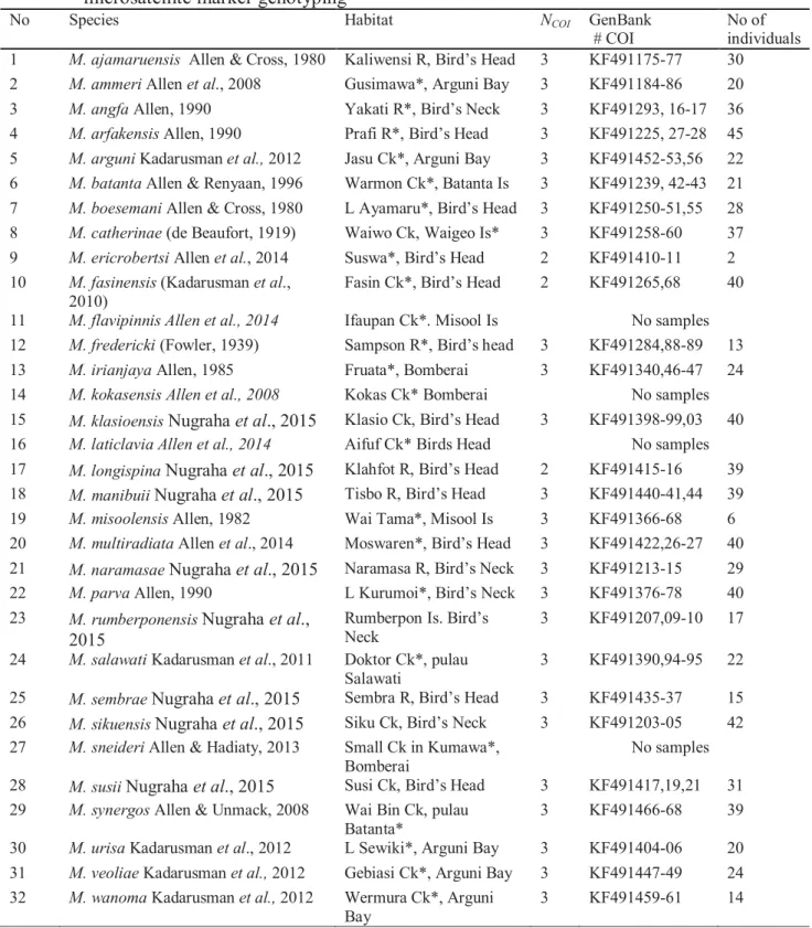
Papua) ... 64 9. Melanotaenia species used in barcode (COI) phylogenetic analysis
and in the microsatellite marker genotyping ... 66 10. Characteristics of the microsatellite markers on Melanotaenia
boesemani and on the 28 cross-amplified species. ... 74
11. Sample size, allele diversity and average observed (Ho) and expected
(He) heterozygosity per population across loci ... 75 12. Locus by locus and multilocus Fis values for each population. ... 76 13. Pairwise Fst values between species from the cluster “Central
Ayamaru Plateau” and M. arfakensis. ... 81 14. Pairwise Fst values between species composing the cluster “Southern
Ayamaru Plateau”, all valid species from “Central Ayamaru plateau
cluster and M. arfakensis ... 81 15. Pairwise Fst values between species composing the cluster “Bird’s
Neck”. ... 82 16. List of M. boesemani farmers in Jakarta and Bekasi. ... 96 17. Number of alleles per locus for each population. ... 98 18. Sample size (N), average observed (Ho) and expected (He)
heterozygosity, and multilocus Fis values for each wild population and
strain. ... 99 19. Population differentiation based on pairwise Fst estimates. ... 99 20. Allelic frequency for each population... 99
11 1. GENERAL INTRODUCTION
The ornamental fish hobby has continuously grown over the last decades. Some endemic fish from Indonesia have been very successfully sold abroad, like arowana (genus Scleropages) from Borneo island, botia (Chromobotia macracanthus) from Borneo and Sumatera islands, Tiger barb (Puntigrus tetrazona) from Sumatera island, and rainbowfish (genus Melanotaenia) from Papua. Talking about rainbowfish (Melanotaeniidae), at the moment there are only 3 species cultivated in Indonesia, namely Glossolepis incisus Weber, 1908, Iriatherina werneri Meinken 1974, and Melanotaenia boesemani Allen & Cross, 1980. Gerald Allen, an ichtiologist from Australia, was the first to perform deep researches on rainbowfish. In addition to Gerald Allen, an Indonesian research team from the Ministry of Maritime Affairs and Fisheries (Sorong Fisheries Academy and Research and Development Center for Ornamental Fish Culture), in collaboration with French researchers (Institut De Recherche pour le Dévelopement - IRD), has conducted researches and species descriptions of this family through several expeditions conducted since 2007.
The first species of rainbowfish was scientifically described in 1843 from a collection of freshwater fishes collected in King River (Northen Australia) and was named Atherina nigrans. In 1862, Thomas Gill proposed to place this species into a new genus called Melanotaenia; Melanotaenia nigrans then became the first member (i.e. nominal species) of this genus. The subfamily Melanotaeniidae was subsequently created by Gill in 1894.More recently, Munro (1964) claimed that
Melanotaeniidae must represent an independant family. Between 1843 and 1964,
there were about 30 species described as belonging to this family. Over the last four decades, many expeditions were conducted in New Guinea, and led to the discovery of an impressive level of rainbowfish diversity. Today, this family includes 102 species in 7 genera following Nugraha et al., (2015) and Allen et al., (2015a, b):
Cairnsichthys (1 species), Chilatherina (11 species), Glossolepis (9 species), Iriatherina (1 species), Melanotaenia (78 species), Pelangia (1 species) and Rhadinocentrus (1 species) (Eschmeyer 2014; Allen et al., 2014a.b). The greatest
12 species, while there are 13 species in Australia, and only 2 species spread between the two biogeographic regions (Allen & Hadiaty 2013).
Melanotaeniidae spread out throughout the island of New Guinea, as well as
the large islands around the western part of the region, including Raja Ampat, Aru and Yapen islands. This fish can also be found throughout Northern Australia, along the east to the south coast of Murray Darling drainage, and in the Western part of Australia (Allen, 1991). Melanotaeniidae belong to the most popular freshwater fish from the island of New Guinea and northern Australia (Eschmeyer, 2014). They represent the most speciose group of pure freshwater fishes within the Australia-New Guinea region. They typically possess a compressed body covered by relatively large scales, two separate dorsal fins (the first with 3-7 spines and the second with a single spine and 6-22 segmented rays), a long-based anal fin, and no lateral line (Allen et
al., 2008). The family is characterized by relatively small (usually less than 10 cm)
and often brightly colored fish. Sexual dimorphism is often apparent and males tend to be deeper-bodied and more vividly colored than females (Allen, 1991). Generally, they are locally abundant and occupy the full spectrum of freshwater habitats including arid-zone waterholes, swamps, rainforest streams, karst rocks and oxbow mountainous lakes and streams in the mountains.
The Boesman’s rainbowfish Melanotaenia boesemani Allen and Cross (1980) is certainly the most popular species of rainbowfish both in Indonesia and abroad (Figure 1). This species is very different from most other rainbowfish because it harbors different colors between the anterior and posterior. The color of anterior (front) is bright blue and the color of posterior (back) is bright yellow. This species was first described by Allen and Cross (1980), from specimens caught by Marinus Boeseman in 1954-1955, which were kept in the museum Rijksmuseum van
13 Figure 1. Melanotaenia boesemani (photo: Dr. Laurent Pouyaud)
This species is native from two distinct lakes in West Papua (i.e. Ayamaru and Uter), distant by around 30 km and separated by limestone mountains (Kadarusman, 2012). Because of its popularity, the species has been greatly exploited in its natural habitat and is currently in the verge of extinction. Since 2004, this species is included in the CITES list as an endangered species (IUCN, 2013), and theoretically only fishery products are now allowed to be exported. However, Allen reported in 2007 that around 60,000 males of M. boesemani were caught monthly and exported for trading (Allen, 2007). Although Melanotaenia boesemani has been domesticated by Indonesian farmers since 1983, the number of farms that exploit this species in Indonesia is still very low. Besides, farmers claim that there is a decline in the production, in terms of quantity and quality, such as a higher proportion of females than males per spawning, a loss of body coloration, lower growth rate and fecundity, or frequent morphological abnormalities. They interpret this as a consequence of a loss of genetic variability and inbreeding.
In this context, my work aimed at gathering new genetic information that would be useful for the aquaculture and conservation of the Melanotaeniidae family. Specifically, the objectives of the research were: 1) to develop new microsatellite DNA markers from the endangered M. boesemani, 2) to evaluate the genetic diversity of wild populations of Melanotaenia and refine their taxonomy, 3) to describe the geographic origins of M. boesemani reared by ornamental fish farmers in Indonesia, and to evaluate the genetic diversity and inbreeding pressure resulting from this domestication. My PhD work was a continuation of the PhD realized by Dr. Kadarusman in the frame of the Lengguru program. Among other activities, this
14 scientific program consists in describing and characterizing the genetic structure and diversity of Rainbowfishes in West Papua, through scientific expeditions jointly conducted by the Institut de Recherche pour le Développement (France), the Academy of Fisheries of Sorong (West Papua, Indonesia), the Aquaculture Research and Development Center for Ornamental Fish (Java, Indonesia), and the Indonesian Institute of Sciences (LIPI).
The present manuscript is divided into five sections: the first one consists in a bibliographic review; the next three ones present the results relating to 1) the development and succesfull validation of 12 polymorphic microsatellite DNA markers from Melanotaenia boesemani; 2) the use of these markers for analyzing the genetic diversity of wild populations of Melanotaenia from West Papua; 3) their use to investigate the genetic diversity and origin of reared strains of M. boesemani in Indonesia. Finally, a general discussion followed by perspectives will constitute the last section of this manuscript.
15 II. LITERATURE REVIEW
2.1. RAINBOWFISH DISTRIBUTION AND DIVERSITY
Rainbowfishes are very famous for ornamental trade since the 1930s (Tappin, 2010), because of their vivid coloration that is reminiscent of a rainbow. They are endemic from New Guinea and Australia. The first rainbowfish (Melanotaenia
nigrans) was scientifically described in 1843 from the collection of freshwater fish
obtained in the northern region of Australia from the King River, near Victoria Settlement - Northern Territory-Australia. In 1964, Ian Munro determined that Rainbowfishes belonged to their own family. Gerald Allen, an ichthyologist from Western Australian Museum, gave a very important contribution to the systematics of Rainbowfishes inhabiting Australia and New Guinea with the description of dozen of new species and and several new genera. There are many endemic species of rainbowfish in various systems of river and lake waters with close phylogenetic and biogeography relationships both in Australia and New Guinea (Zhu et al., 1994; McGuigan et al., 2000; Unmack, 2001). Taxonomy of Melanotaeniidae has therefore a long story. There were as many as 30 species that had been discovered between 1843 and 1964 but several expeditions in New Guinea over the past four decades led to the description of more than 40 new additional species (Kadarusman
et al., 2010). Melanotaeniidae is believed to be part of the Atheriniforme order,
which has evolved from the ancestors of atherinoids at sea (Allen, 1980).
There are currently 2 families of rainbowfish that have been described until now, and which are distributed on the islands of Australia and New Guinea (the region of Indonesia and Papua New Guinea) (Table 1): the family Melanotaeniidae, which includes 7 genera and 102 species, and the family Pseudomugilidae, composed of 18 species spread into 3 genera (Table 1). Melanotaenia is the largest genus of the Melanotaeniidae family (Ivantsoff et al., 1991). There are thirty-two species that are endemic from the Vogelkop Province (Nugraha et al., 2015). The Vogelkop province belongs to the territory of West Papua, Indonesia and biogeographically should include for Melanotaeniids the western side of Lengguru Range (i.e. all watersheds flowing to Arguni Bay), the Bomberai and the Birds Head
16 Peninsulas, the Birds Head Isthmus joining both peninsulas and the four major Raja Ampat islands (i.e. Misool, Batanta, Salawati, Waigeo) (Kadarusman et al., 2012a).
Table 1.
Rainbowfish classification according to ANGFAhttp://rainbowfish.angfaqld.org.au/Melano.htm (download 4 September 2015)
A. Melanotaeniidae
a) Cairnsichthys
1. rhombosomoides (Nichols & Raven, 1928)
b) Chilatherina 1. alleni Price, 1997 2. axelrodi Allen, 1980 3. bleheri Allen, 1985 4. bulolo (Whitley, 1938) 5. campsi (Whitley, 1956) 6. crassispinosa (Weber, 1913) 7. fasciata (Weber, 1913) 8. lorentzi (Weber, 1908)
9. pagwiensis Allen & Unmack, 2012 10. pricei Allen & Renyaan, 1996 11. sentaniensis (Weber, 1908)
c) Glossolepis
1. dorityi Allen, 2001 2. incisus Weber, 1908 3. kabia (Herre, 1935)
4. leggetti Allen & Renyaan, 1998 5. maculosus Allen, 1981
6. multisquamata (Weber & de Beaufort, 1922) 7. pseudoincisus Allen & Cross, 1980
8. ramuensis Allen, 1985
9. wanamensis Allen & Kailola, 1979
d) Iriatherina
1. werneri Meinken, 1974
e) Melanotaenia
1. affinis (Weber, 1908)
2. ajamaruensis Allen & Cross, 1980
3. albimarginata Allen, Hadiaty, Unmack & Erdmann, 2015 4. ammeri Allen, Unmack & Hadiaty, 2008
5. angfa Allen, 1990 6. arfakensis Allen, 1990
7. arguni Kadarusman, Hadiaty & Pouyaud in Kadarusman, Hadiaty, Segura, Setiawibawa, Caruso & Pouyaud, 2012
8. aruensis Allen, Hadiaty, Unmack & Erdmann, 2015 9. australis (Castelnau, 1875)
10. batanta Allen & Renyaan, 1998 11. boesemani Allen & Cross, 1980 12. catherinae (de Beaufort, 1910) 13. caerulea Allen, 1996
17 14. corona Allen, 1982
15. duboulayi (Castelnau, 1878) In Allen G.R. (1980) 16. eachamensis Allen & Cross, 1982
17. ericrobertsi Allen, Unmack & Hadiaty, 2014 18. exquisita Allen, 1978
19. fasinensis Kadarusman, Sudarto, Paradis & Pouyaud, 2010 20. flavipinnis Allen, Hadiaty & Unmack, 2014
21. fluviatilis (Castelnau, 1878) 22. fredericki (Fowler, 1939) 23. goldiei (Macleay, 1883) 24. gracilis Allen, 1978 25. herbertaxelrodi Allen, 1980 26. irianjaya Allen, 1985 27. iris Allen, 1987
28. japenensis Allen & Cross, 1980 29. kamaka Allen & Renyaan, 1996
30. klasioensis Kadarusman, Hadiaty & Pouyaud in Nugraha, Kadarusman, Hubert, Avarre, Hadiaty, Slembrouck, Carman, Sudarto, Ogistira & Pouyaud, 2015
31. kokasensis Allen, Unmack & Hadiaty, 2008
32. kolaensis Allen, Hadiaty, Unmack & Erdmann, 2015 33. lacustris Munro, 1964
34. lakamora Allen & Renyaan, 1996
35. laticlavia Allen, Unmack & Hadiaty, 2014
36. longispina Kadarusman, Avarre & Pouyaud in Nugraha, Kadarusman, Hubert, Avarre, Hadiaty, Slembrouck, Carman, Sudarto, Ogistira & Pouyaud, 2015
37. maccullochi Ogilby, 1915 38. mairasi Allen & Hadiaty, 2011
39. manibuii Kadarusman, Slembrouck & Pouyaud in Nugraha, Kadarusman, Hubert, Avarre, Hadiaty, Slembrouck, Carman, Sudarto, Ogistira & Pouyaud, 2015
40. maylandi Allen, 1982 41. misoolensis Allen, 1982 42. monticola Allen, 1980 43. mubiensis Allen, 1996
44. multiradiata Allen, Unmack & Hadiaty, 2014
45. naramasae Kadarusman, Nugraha & Pouyaud in Nugraha, Kadarusman, Hubert, Avarre, Hadiaty, Slembrouck, Carman, Sudarto, Ogistira & Pouyaud, 2015
46. nigrans (Richardson, 1843) 47. ogilbyi Weber, 1910
48. oktediensis Allen & Cross, 1980 49. papuae Allen, 1981
50. parkinsoni Allen, 1980 51. parva Allen, 1990 52. patoti Weber 1907
18 54. pierucciae Allen & Renyaan, 1996
55. pimaensis Allen, 1980
56. praecox (Weber & de Beaufort, 1922) 57. pygmaea Allen, 1978
58. rubripinnis Allen & Renyaan, 1998
59. rubrivittata Allen, Unmack & Hadiaty, 2015 60. rubrostriata (Ramsay & Ogilby, 1886)
61. rumberponensis Kadarusman, Ogistira & Pouyaud in Nugraha, Kadarusman, Hubert, Avarre, Hadiaty, Slembrouck, Carman, Sudarto, Ogistira & Pouyaud, 2015
62. salawati Kadarusman, Sudarto, Slembrouck & Pouyaud, 2011 63. sembrae Kadarusman, Carman & Pouyaud in Nugraha,
Kadarusman, Hubert, Avarre, Hadiaty, Slembrouck, Carman, Sudarto, Ogistira & Pouyaud, 2015
64. senckenbergianus Weber, 1911 65. sexlineata (Munro, 1964)
66. sikuensis Kadarusman, Sudarto & Pouyaud in Nugraha, Kadarusman, Hubert, Avarre, Hadiaty, Slembrouck, Carman, Sudarto, Ogistira & Pouyaud, 2015
67. sneideri Allen & Hadiaty, 2013 68. splendida inornata (Castelnau, 1875) 69. splendida splendida (Peters, 1866) 70. splendida tatei (Zietz, 1896)
71. susii Kadarusman, Hubert & Pouyaud in Nugraha, Kadarusman, Hubert, Avarre, Hadiaty, Slembrouck, Carman, Sudarto, Ogistira & Pouyaud, 2015
72. sylvatica Allen, 1997
73. synergos Allen & Unmack, 2008 74. trifasciata (Rendahl, 1922)
75. urisa Kadarusman, Setiawibawa & Pouyaud in Kadarusman, Hadiaty, Segura, Setiawibawa, Caruso & Pouyaud, 2012
76. utcheensis McGuigan, 2001
77. vanheurni (Weber & de Beaufort, 1922)
78. veoliae Kadarusman, Caruso & Pouyaud in Kadarusman, Hadiaty, Segura, Setiawibawa, Caruso & Pouyaud, 2012
79. wanoma Kadarusman, Segura & Pouyaud in Kadarusman, Hadiaty, Segura, Setiawibawa, Caruso & Pouyaud, 2012
80. wokamensis Allen, Hadiaty, Unmack & Erdmann, 2015
f) Pelangia 1. mbutaensis Allen, 1998 g) Rhadinocentrus 1. ornatus Regan, 1914 B. Pseudomugilidae a) Kiunga 1. ballochi Allen, 1983 2. bleheri Allen, 2004 b) Pseudomugil 1. connieae (Allen, 1981)
19 2. cyanodorsalis Allen & Sarti, 1983
3. furcatus Nichols, 1955 4. gertrudae Weber, 1911 5. inconspicuus Roberts, 1978 6. ivantsoffi Allen & Renyaan, 1999 7. majusculus Ivantsoff & Allen, 1984 8. mellis Allen & Ivantsoff, 1982 9. novaeguineae Weber, 1908 10. paludicola Allen & Moore, 1981 11. paskai Allen & Ivantsoff, 1986
12. pellucidus Allen, Ivantsoff & Renyaan, 1998 13. reticulatus Allen & Ivantsoff, 1986
14. signifer Kner, 1866 15. tenellus Taylor, 1964
c) Scaturiginichthys
1. vermeilipinnis Ivantsoff, Unmack, Saeed & Crowley, 1991
Rainbowfish family is one of the most important taxas of freshwater fish in Papua.The ancestral forms of these fish taxa has been believed to inhabit Australia since the time of Gondwana which has a close kinship with the sea herring fish, Atheriniformes (Sparks & Smith, 2004). Rainbowfish phylogenetic studies in New Guinea and Australia have been done first by McGuigan et al., (2000) using a small part of the cytochrome b gene (351 bp bases long). Results of this study reported that there were 3 phylogenetic clades according to 3 distinct biogeographic regions: (a) Bird’s Head, (b) northern part of New Guinea, and (c) southern part of New Guinea plus Australia.
Rainbowfish live in the territory of Australia and New Guinea. In the Australian region, rainbowfish habitat spreads between latitudes of 10o41’S (cape york) and 43o39’S (South East cape, Tasmania) and between longitudes of 113o09'E (Steep point) and 153o39'E (Cape Byron). The distance between the latitude of Cape York and the South East Cape (Tasmania) is 3.680 km. The distance in longitude between Steep Point and Cape Byron is about 4.000 km.. The territory of New Guinea, with an area of around 876.800 km2, is located in the south of the equator and in the south-west Pacific, at northeast Australia. The term of New Guinea is used in the concept of freshwater fish habitat that refers to the entire island, which consists of both the Province of Indonesia (West Papua and Papua) and country of Papua New Guinea (PNG). Political boundaries follow the east meridian 141° (141°E).
20 New Guinea has a mountainous cordillera which runs along the central mountain which maximum height is reached at the peak Jaya (5030 meters), the highest point of Papua-New Guinea.
Monophyletic groups of strictly freshwater fishes have special significance in continental biogeography because they require freshwater for dispersal, and thus their distributions are correlated with the evolution of topography and watershed (Lundberg et al. 2000). In general, the fish fauna of New Guinea is closely linked to that of northern Australia. The most diverse taxa are Eleotrididae and Gobiidae of about 115 species, followed by Melanotaeniidae of about 102 species. Around fifty species distributed in southern New Guinea are also found in northern Australia and are limited to these two regions (Lundberg et al. 2000).
2.2. THE MELANOTAENIIDAE FAMILY
Allen (1980) proposed a generic revision of Melanotaeniidae based on osteological characters and recognized Melanotaenia, Glossolepis and Chilatherina as three closely related genera, sharing a rigid fin spine at beginning of first dorsal, anal and pelvic fins (versus soft for the other members of the family). Allen (1998) described Pelangia and stated that this new genus was closely related to Glossolepis, particularly with regards to dentition, and morphology of the premaxillary, pelvic girdle, and pectoral fin.
Melanotaenia (Gill 1862) is the most diverse genus with around 80 species
and sub-species (Allen et al., 2008; Allen et al., 2015a, b; Kadarusman et al., 2012; Nugraha et al., 2015). The genus consists of 15 taxa in Australia and 89 taxa in New Guinea. The species Melanotaenia macculochi (Ogilby 1915) and M. splendida (Peters 1866) are shared in both areas of Australia and New Guinea (Allen & Renyaan 1998).
Between 2007 and 2010, there were several expeditions conducted by IRD and Academy of Fishery Sorong in the frame of Dr. Kadarusman’s PhD: Rainbowfish from West Papua (Melanotaeniidae): Evolution and Systematics. This study contributed to the revision of the taxonomy of rainbowfishes, especially by the
21 identification of new morphological criteria and the use of mitochondrial (CO1, Cyt b and D-loop) and nuclear (S7) molecular markers. Six new species of Melanotaenia (M. arguni, M. fasinensis, M. salawati, M. urisa, M. veoliae, and M. wanoma) were consequently described (Kadarusman et al. 2010; 2011; 2012b).
Based on the PhD thesis of Kadarusman (2012), the biogeography of
Melanotaeniidae distribution consisted of 4 groups (Figure 2):
1. Western New Guinea or Vogelkop Province including the four Raja Ampat islands, the Birds Head and Bomberai Peninsulas, and the western side of the Birds Head Isthmus.
2. The Central part of the Birds Head Isthmus.
3. Northern part of New Guinea (Northern New Guinea). The region covers an area of Great Northern part and the North Eastern Province (Allen 1991). This region includes the islands of Yapen, all streams in the northern part of Weyland Plateau (Wapoga, Siriwo etc) and the north New Guinea (Memberamo, Sepik, Markham etc).
4. Southern New Guinea and Australia. This section covers the Arafuru sea to Triton Lake, Cenderawasih Bay (from Munuari to Nabire) (Figure 2).
These results gave a new view that there is a great diversity of
Melanotaeniidae in the area of New Guinea and Australia, particularly West Papua
22 Figure 2. Map of the Melanotaeniidae distribution based on molecular phylogenies
(Kadarusman 2012)
Specifically, in the Bird’s head region of New Guinea (West Papua Province), 32 species belonging to the genus Melanotaenia have been described, (Nugraha et al. 2015). Based on species composition, Bird’s head Papua is believed to be the center of rainbowfish diversity (Allen 1995).
Kadarusman et al. (2012a) demonstrated that species of Melanotaenia from the area of Bird’s head are monophyletic compared to other clades. The result of the research done by Kadarusman et al. (2012a) showed an unpredictable diversity level, and it also showed that the diversity of Melanotaenia was still underestimated.
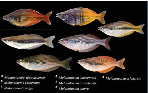
Since 2004, IUCN has identified 7 endangered species from the Bird’s Head in West Papua: Melanotaenia ajamaruensis Allen & Cross 1980; M. angfa Allen 1990; M. arfakensis Allen 1990; M. boesemani Allen & Cross 1980; M. catherinae
(de Beaufort 1910); M. misoolensis Allen 1982; M. parva Allen 1990 (IUCN 2013)
23 Species extinction is generally caused by the introduction of alien species, excessive exploitation of species and illegal logging causing river habitat degradation and lake habitat siltation or dessication, like in the case of Kurumoi and Ayamaru lakes (Allen, 2007; Kadarusman et al. 2010; Tappin, 2010).
Figure 3. Seven endangered Melanotaenia species from Bird’s head peninsula (West papua) – (Photo: Dr. Laurent Pouyaud)
Among the 102 known species of Melanotaeniidae, only three have been domesticated and cultivated in Indonesia, namely Glossolepis inicus weber 1908,
Iriatherina werneri Meinken 1974 and Melanotaenia boesemani Allen & Cross 1980
(Figure 4).
Figure 4. Tree species rainbowfish, have been domesticated and cultivated in Indonesia, namely Glossolepis inicus weber 1908, Iriatherina werneri Meinken 1974 and Melanotaenia boesemani Allen & Cross 1980.
24
Melanotaenia boesemani is incontestably the most famous rainbowfish in the
aquarium hobby. According to Kadarusman (2012), M. boesemani lives in 2 distinct locations, i.e. Ayamaru and Uter lakes. Until now, the exact origin of the strains reared in Indonesia has not been found out.
2. 3. THREATS ON THE MELANOTAENIIDAE
The West Papua Province (115,363 km2; 760,855 people), which encompasses the Vogelkop Peninsula and the adjacent islands of the Raja Ampat, is one of the least economically developed regions in the world. Human activities concentrate mostly in the vicinity of Sorong, Manokwari, Teminabuan and Bintuni. Papua, which includes the province of West Papua and Papua Province, is house of 50% of the Indonesian biodiversity and sinificantly contributes to Indonesia’s status as one of the biologicaly richest countries in the world with Brazil (Supriatna 1999). Currently, Papua is home of 400 species of freshwater fish (Allen 1991). Until now, there have been many unspoiled areas because the access is difficult. The habitat physical topology is very steep as a result of complex geological events. This is probably the reason why new species can still be found every year (Allen 1991; Novotny et al., 2005).
In spite of this, the province of West Papua Indonesia is now facing new challenges. Flora and fauna in this region face a threat of extinction due to human activities such as logging and burning of forests to agricultural areas, transmigration, poaching, mining, etc. All human activities have been increasingly disturbing and destroying the habitat of flora and fauna, particularly freshwater fish. Moreover, since the 1950s, more than thirty species of freshwater fish have been introduced into the waters of New Guinea essentially for human consumption (Allen, 2007). Those species are principally the tilapias Oreochromis mossambicus and O. niloticus, the walking catfish Clarias batrachus, the common carp Cyprinus carpio, the snakehead
25 Rainbowfish species that are threatened with extinction in the region of Australia and New Guinea are: Chilatherina axelrodi Allen, 1980; C. bleheri Allen, 1985; C. bulolo (Whitley, 1938); C. sentaniensis (Weber, 1908); Glossolepis incisus (Weber, 1908); G. maculosus Allen, 1981; G. pseudoincisus (Allen & Cross 1980);
G. ramuensis Allen, 1985; G. wanamensis Allen & Kailola 1979; Kingua ballochi
Allen, 1983; K. bleheri Allen, 2004; Melanotaenia ajamaruensis Allen & Cross 1980; M. angfa Allen, 1990 M. arfakensis Allen, 1990; M. boesemani Allen & Cross 1980; M. catherinae (de Beaufort, 1910); M. corona Allen, 1982, M. eachamensis Allen & Cross 1982; M. exquisita Allen, 1978; M gracilis Allen, 1978; M.
herbertaxelrodi Allen, 1980; M. iris Allen, 1987; M. lacustris Munro, 1964; M. maylandi Allen, 1982; M. misoolensis Allen, 1982; M. monticola Allen, 1980; M ogilbyi Weber, 1910; M. oktediensis Allen & Cross 1980; M. papuae Allen, 1981; M. parva Allen, 1990; M. pimaensis Allen, 1980; M. praecox (Weber & de Beaufort
1922); M. pygmaea Allen, 1978; M. sexlineata (Munro, 1964); M. vanheurni (Weber & de Beaufort 1922); Pseudomugil connieae (Allen, 1981); P. furcatus Nicholas, 1955; P. majusculus Ivantsoff & Allen 1984; P. mellis Allen & Ivantsoff 1982; P.
paskai Allen & Ivantsoff 1986 and Scaturiginichthys vermeilipinnis Ivantsoff,
Unmack, Saeed & Crowley 1991 (Conservation International 2002; IUCN, 2009). The information about the biology and ecology of these fish species in their natural habitat is not much documented. An in-depth study is therefore needed, such as reproductive condition in their natural habitat, water quality, frequency of spawning and habitat preferences. Ecological research on rainbowfish families is very crucial for helping efficiently their conservation.
2. 4. HISTORY OF RAINBOWFISH AS WORLD AQUARIUM PEARLS AND AQUACULTURE
Rainbowfish from New Guinea have been imported to Australia since around the mid-1950s. They have been continuously cultivated, and their farmers had no idea that these fish will be popular for the international aquarium hobby. Some specimens were introduced, including Melanotaenia affinis, M. goldiei, M.
26 Australia in early 1959. In 1982, the Australian and Papua New Guinea rainbowfish were promoted through a book by Gerald Allen and Norbert Cross (Allen and Cross 1982), which resulted in the increase in popularity and curiosity about the rainbowfish species found in New Guinea and Australia. During this period, there was no significant limitation, and there was an important number of different species brought from New Guinea to Australia by private collectors, who then distributed them to hobbyists (Smith, 2007; Tappin, 2010).
The excessive trade of rainbowfish from New Guinea to Australia attracted the attention of the "Advisory Committee on Live Fish (ACOLF)". At the end of 1983, ACOLF prohibited the import of fish into Australia for all freshwater fish species from New Guinea. In the mid-1980s, aquarium fish collectors began collecting species from New Guinea, cultivating and distributing them to the international aquarium hobbyists (Smith et al., 2007). This trend has continued over the last two decades and has grown side by side with the increasing discovery of new species. Ichtyologists and rainbowfish hobbyists have continued to look for these fish to the inland, to collect different shapes and colors, to keep them survive in the aquarium by fulfilling all the physiological needs of the fish outside their natural habitat. However, when these fish are kept in aquarium, they do not produce bright colors as those living in their natural habitat. This resulted in the widespread sales of these fish under 5 cm in size, in which the colors have not come out well (Tappin, 2010).
Rainbowfish from Australia were domesticated for the first time as decoration in the aquarium, as reported by Albert Gale in early 1915, in his book entitled "Aquarian nature Studies and Economic Fish farming made known the
hobby of keeping Australian freshwater fishes". At the beginning of the 1920s and
1930s, ornamental fish aquarium hobbyists were set up in major cities in Europe such as Germany. The delivery used ship, and there was no change of oxygen. When they got in the country of destination, the fish were in poor condition, but there were some survivors and they could be successfully cultivated and saved by the hobbyists. Thus this became the community of hobbyists in European countries.
27 During this period, these fish were known as sunfish. In January 1934, the magazine "National Geographic" published an article written by Walter H Chute, and then the director of the Shed Aquarium in Chicago USA called these as "Tropical Fish Immigrants Reveal New Nature Wonders". This raised the reference of Australian rainbowfish. The earliest record was found in the Aquarium German magazine "Wochenschrift für Aquarien und Terrarienkunde" in September 1931 by Erich Henzelmann, who wrote an article about 'Regenbogenfisch' Melanotaenia nigrans. The earliest reference to the name of a rainbowfish that had been found in Australia was in a report entitled "The Aquarium and Terrarium Society of Queensland". On March 6, 1932, Amandus Rudel reported the results of his expedition and mentioned all the fish with the name "rainbowfish", while the name "Sunfish" were designated only for Rhadinocentrus ornatus which he described as "Moreton Island Sunfish" (Tappin, 2010).
In 1927 Amandous Rudel, a founder of Aquarium and Terrarium Society of Queensland, introduced Australian rainbowfish on the international community, by sending Melanotaenia duboulayi (Castelnau, 1878) to Germany. These fish were then raised in the aquarium of Berlin and then the fish were promoted to America. Amandous Rudel also introduced another rainbowfish for the international aquarium hobbyists. In 1934, he sent 12 specimens of Melanotaenia maccullochi, which were gathered near the river Cairns, northern Australia, and then sent to Fritz Mayer in Hamburg, Germany. Four specimens arrived and lived safely, and they developed into two pairs. These fish have become one of the most popular aquarium fish originating from Australia. This news was written in a German aquarium magazine "Wochenschrift für Aquarien und Terrarienkunde" in May 1935. Fritz Mayer wrote about the fish breeding techniques, translated by FH Stoye in the book "The Aquarium" in December 1936 (Tappin, 2010).
Until now, commercial species of rainbwfish in Indonesia are: Glossolepis
insicus (from lake Sentani, Papua province), Iriatherina werneri (from Merauke,
Papua province), Melanotaenia boesemani (from South Sorong, West Papua province). Until now there have been 15 groups of rainbowfish farmers in Jakarta and Bekasi region.
28
a) b)
c) d)
e) f)
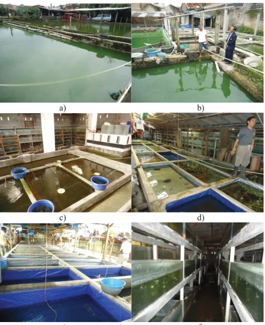
Figure 5. Captive breeding of M. boesemani in Indonesian farms
Rainbowfish aquaculture in Indonesia began in 1983 in Mr Gusi farm. He got founder population of M. boesemani in his farm from exporters of ornamental fish in Jakarta, Indonesia. In that decade, M. boesemani became very famous because of publications, especially that by Allen and Cross (1980) on the description of M.
boesemani. Overfishing of M. boesemani males then started to affect Ayamaru Lake,
the natural habitat of M. boesemani (Allen 2007). Rainbowfish cultivation in Indonesia is carried out indoor and outdoor, involves hatcheries and grow-out ponds (Fig 5 ab), with semi-natural water bodies (Fig 5 c,d,e) and indoor aquaria installations (fig 5f). According to the quarantine authorities in Jakarta, producers
29 export nearly 2,500 live M. boesemani per week, at a price of around 1 US$ per individual. Though there are no official numbers, this is unlikely to account for the totality of M. boesemani that are exported worldwide from Indonesia.
1.5. MAJOR MOLECULAR MARKERS USED IN FISH GENETIC
POPULATION STUDIES
A genetic marker is a measurable character of Mendelian inheritance (Liu and Cordes, 2004) and is considered ideal when polymorphic (variable between individuals), discriminatory (different related individuals), multiallelic (has many alleles on the same locus), codominant (heterozygotes are visible), not epistatic (independent of the expression of other markers), neutral (whatever the allele present at the locus, the fitness of the individual is the same), reproducible from one experiment to the next, to handle large scale and economical. The distribution of markers over the whole genome is also a criterion to remember. The main sources of molecular markers are from either a sequence polymorphism (eg: substitution, insertion, deletion) or a polymorphism number of repetitions units (microsatellites and minisatellites). There are two major types of genetic markers that are widely used to characterize fish populations, namely mitochondrial markers and microsatellite markers.
2.5.1. MITOCHONDRIAL DNA MARKERS
Mitochondrial DNA is a popular marker for study of evolution like phylogenetic inference, identification of species, phylogeography, and analysis of population structure. Characteristics of mitochondrial DNA are small genome with simple structure and organization, ubiquitous presence, high copy number, thereby easy to isolate, effective haploidy in DNA sequences, maternal inheritance, lack of recombination in introns or other non coding sequence, mosaic molecule with aster and slower evolving DNA regions allowing to design conserved primers and to
30 address phylogenetic questions at various taxonomic levels (Zhang & Hewiit 1996; Harrison, 1989).
For identification of species and evolution studies, maternal inheritance has important consequences, because only a fraction of the population (half if the sex ratio is 1:1) pass on their mtDNA to offspring and the effective population size for mtDNA is smaller than that for nuclear genes. In stochastic processes, it will be particularly important to determine frequencies of mtDNA genotypes. Genetic affinities defined by mtDNA genotypes reflect matriarchal phylogeny. MtDNA is suited for studying population patterns including founder populations/founder events. Because females transmit many copies of the mtDNA molecule to each offspring, a new mtDNA variant arises as mutation in a single molecule within a single cell lineage (Harrison, 1989).
MtDNA analysis has been used in three ways in this context; (1) to measure genetic variation within populations, especially ones thought to have declined recently, (2) to identify evolutionarily divergent sets of populations, including the resolution of evolutionarily significant units, and (3) to assess conservation value of populations or areas from an evolutionary or phylogenetic perspective. Mitochondrial DNA variation is more sensitive in population phenomena such as bottlenecks and hybridizations. Sex-specific differences in gene flow could also be revealed by contrasting nuclear with mitochondrial DNA. And also mtDNA is intensively studied and sequences in some parts of the molecule are highly conserved across species (Okumus & Ϛiftei, 2003).
A prerequisite for managing biodiversity is the identification of populations with independent evolutionary histories. Such groupings are variously categorized as species. MtDNA phylogenies can provide unique insights into population history (Avise et al., 1987) and can suggest hypotheses about the boundaries of genetically divergent groups (e.g. cryptic species). Several studies about cryptic species and DNA barcoding have been succesfully implemented in the identification of previously described species. DNA barcoding studies have focused on the identification of pre-defined species (Hebert et al. 2003). Applying mtDNA analyses on rainbowfish, Kadarusman et al. (2012) succesfully described the cryptic diversity in Indo-Australian Rainbowfishes. As much as 14 species of rainbowfish have been
31 discovered in populations exhibiting private barcode clusters diverging from their nearest neighbor by K2P distances similar to those observed among valid species characterized by diagnostic morphological characters. DNA barcoding was not only effective for the identification of species, but it proved to be effective for the discovery of provisional cryptic diversity awaiting further screening through integrative approaches as previously predicted by other species like butterfly (Hebert
et al. 2004), diptera (Smith et al. 2007) and reef fishes (Hubert et al. 2012) .
In spite of many advantages, mtDNA marker shows several drawbacks for genetic studies, such as non-neutrality, non-constant molecular clock, length and sequence heteroplasmy and even mitochondrial bottleneck (Stewart & Larsson, 2014).
2.5.2. MICROSATELLITE MOLECULAR MARKERS (SSR / SIMPLE SEQUENCE REPEAT)
The selection of microsatellite molecular genetic markers was found in the 1980s. They represent a unique type of tandemly repeated genomic sequences, which are abundantly distributed across genomes and demonstrate high levels of allele polymorphism. They are also called simple sequence repeats (SSRs). They are codominant markers with relatively small size, around 20 repetitions of base maximum in length, and can be easily amplified by polymerase chain reaction. They are ubiquitous in prokaryotes and eukaryotes, present even in the smallest bacterial genomes. The majority of microsatellites (30–67%) found are dinucleotides. In the genomes of vertebrates, (AC)n is the most common dinucleotide motif. It is 2.3-fold more frequent than (AT)n, the second most general type of dinucleotides (Tóth et al., 2000). In all vertebrates, (G+C)-rich motives (e.g., CCG, CAG) are the most common among trinucleotides. Microsatellites can be found anywhere in the genome, both in protein coding and noncoding DNA (Tóth et al., 2000). In eukaryotic organisms, SSRs have been shown to be in excess in noncoding regions compared to a random distribution pattern (Metzgar et al., 2000).
32
Applications of microsatellites include: genetic mapping, individual DNA identification and parentage assignment, phylogeny, population and conservation genetics, molecular epidemiology and pathology, quantitative trait loci mapping, marker-assisted selection. Also, the use of microsatellite markers may benefit the genetic dissection of complex and quantitative traits in order to map, identify and eventually clone and characterize the candidate genes controlling economically important traits. In aquaculture, SSRs represent the markers of choice for genetic monitoring of farmed stocks in view of breeding programs through the analysis of genetic variablity and pedigree structure to design beneficial crosses, select genetically improved stocks, minimize inbreeding and increase selection response
(Davis & DeNise, 1998; Knibb, 2000). Microsatellite-based techniques are applied in genome scans and quantitative trait loci (QTL) mapping in natural populations to search for the genetic basis of adaptive selection and biodiversity in an increasing number of species (Rogers & Bernatchez, 2005).
Microsatellites are highly abundant in various eukaryotic genomes including all aquaculture species studied to date. In most of the vertebrate genomes, microsatellites make up a few percent of the genomes in terms of the involved base pairs, depending on the compactness of the genomes (Zhan et al., 2009). In fish, one microsatellite was found every 1.87 kb of DNA. For comparison, in the human genome, one microsatellite was found every 6 kb of DNA (Beckmann & Weber 1992). It is reasonable to predict that in most aquaculture fish species, one microsatellite should exist every 10 kb or less of the genomic sequences, on average (Wright 1993; Duran et al., 2009). Their high polymorphism, together with their PCR-based analysis, have made them one of the most popular genetic markers (Duran et al., 2009; Boris et al., 2011). Some microsatellite loci have very high numbers of alleles per locus (>20), making them very useful for applications such as parent-offspring identification in mixed populations, while others have lower numbers of alleles and may be more suited for population genetics and phylogeny (Al-Atiyat et al., 2012). Primers developed for one species will often cross-amplify microsatellite loci in closely related species (Boris et al., 2011). Genotyping of microsatellite markers is usually straightforward (Castoe et al., 2010).
33 Microsatellite markers are co-dominant markers, which means they can distinguish heterozygous and homozygous alleles. They are usually distributed throughout the genome, and they are independent markers (Chambers & MacAvoy, 2000), neutral (any allele present at this locus has the same selective value on all individuals), and can be used from one experiment to another experiment, at an affordable cost (Goldstein & Schlötterer 1998). In the case of high allelic diversity with repetitions of mono, di, tri, tetra, penta and hexa nucleotides, the theory is based on the concept of neutrality, and the variability can differentiate individuals, populations and different species. This characteristic is very important for population genetics studies, and other genetic studies such as inbreeding pressure analysis of heredity or genetic mapping (Vartia et al., 2014; Bruford & Wayne 1993; Brockmann et al., 1994; Knapik et al., 1998; Goldstein et al., 1999; Primmer et al., 2000).
Microsatellites consist of four models: 1) Perfect microsatellites consist of a single continuously repeated motif without being distracted by other repeated motifs or patterns, like ctctctctctctctctctct. 2) Imperfect microsatellites are microsatellites in which one or more repetitions carries base pairs that are not in accordance with the structure of repetition, like ctctctctctgtctct. 3) Interrupted microsatellites are microsatellites with the insertion of a small number of base pairs that do not fit the structure of repetition, like ctctctctctgggctctctct. 4) Compound microsatellites consist of two or more adjacent microsatellites with different repetition types, for example ctctctctctctgatgatgatgatgatgat (Goldstein and Schlötterer, 1998; Jarne and Lagoda, 1996).
Microsatellites are very abundant in eukaryotic genomes because of their process of mutations. Mutations in microsatellite loci usually involve a change in one repetition, but sometimes mutations invole multiple repetition units. There are two processes of mutation that affect the formation of microsatellite loci, namely: 1) Unequal crossing over (UCO), occurring during the process of meiosis, and 2) Slipped-strand mispairing (SSM), and occurring due to mistakes/errors during DNA replication (Goldstein and Schlötterer, 1998).
34 The microsatellite mutation models as addressed by Estoup and Cournet (1998) in Goldstein and Schlötterer (1998) are: 1) Infinite allele models (IAM), each mutation can create new alleles at random and assume that microsatellite mutations can create an infinite number of repeating units, and the sequence of the original alleles is not present in the population 2) Stepwise mutation models (SMM) is a gradual mutation when microsatellite mutates. They loss or gain of a single tandem repeat, and hence alleles may possibly mutate towards allele state already present in the population . 3) K-allele models (KAM): this is the classic model. In this model, all alleles have equivalent mutation probability because alleles in this model can mutate with equal probability to mutate to one of other alleles (K-1) (where in if there are 8 replications, then the mutation that has occurred is 7 replications, in which the 7 replications is the new mutation allele), 4) Two phase models (TPM) is an extension of the SMM mutation. Microsatellite mutations occur because of the increased or reduced X replications. Opportunities to microsatellite mutations involve more than one unit (Goldstein and Schlötterer, 1998; Anmarkrud et al., 2008).
The key feature of SSRs as molecular markers is their hypermutability and, hence, their hypervariability in species and populations. SSR grow very fast with a high mutation rate of 10-2 - 10-6 per locus per gamete per generation, when compared with point mutations in the gene encoding regions (Anmarkrud et al., 2008). This high level of mutation leads to a high polymorphism. The abundance of microsatellites in the genome occurs because replication slippage are not the same in sister chromatid exchange, nucleotide substitutions and duplication events as well as crossovers in the process of meiosis. Many factors might be important for the mutational processes in microsatellites, such as allele size, motif size, gender, and G/C content. Mutation pattern also depends on the specific location in the genome,
e.g functional potential of the product when transcribed, as well as the effectiveness
of various repair enzymes. In addition, microsatellite mutation rate is also influenced by the pattern of stabilization and potential secondary structure (Anmarkrud et al., 2008).
35 A very high degree of polymorphism is the most striking character of microsatellites, but not all of microsatellites are polymorphic at the same point and in the same time. Microsatellite mutations may occur during replication of chromosomes, either as part of mitosis or meiosis, and can also occur during DNA synthesis. Therefore, the frequency of microsatellite mutations increases rapidly when the cells are under stress and when the cells undergo active repair due to damage. Each mutation that occurs during meiosis or mitosis in early cleavage embryo will develop well, and if it is not selected during the development process, it will form a new microsatellite allele. In contrast, mutations that occur in the cell, as somatic mutations, will not affect other cells. There are many factors affecting the level of microsatellite mutation that makes the locus and microsatellite motif abundant (Jarne and Lagoda 1996). Besides being multiallelic and codominant at a single locus, SSRs offer several advantages over other molecular markers. For instance genetic variability can be easily detected by PCR using primers designed around the SSR repeats and by analyzing length fragments, and they are transferrable across closely related species.
Among the mechanism of this mutation, an error in the replication plays a major role in producing new alleles in the SSR loci; therefore, it increases the diversity of SSR. Replication error is the most important mechanism in the formation of SSR. During DNA replication, longer stretches of repeated units pose more of a problem to DNA polymerase than do shorter stretches making longer alleles more prone to slipped-strand mispairing. Interaction in replication errors and crossovers is an important factor to the diversity of repetition motifs and the number of SSR repetitions, such as (CA) n, (CT) n, and (CA) n (TA).
Replication error in chromosomes in each species is different depending on the rate of recombination of these species and SSR motif models in the patterns of evolution that occurs in these loci (Li et al., 2003). The imperfect SSR pattern makes the pattern more complex than that from the perfect SSR (Orti 'et al., 1997). Consequently, a genome can evolve faster than other genomes, such as in the study of Li et al., (2003) in Triticum dicoccoides. SSR sequences are more abundant and longer in vertebrates than in invertebrates, and among the vertebrates, the SSR motifs