HAL Id: hal-03078485
https://hal.univ-grenoble-alpes.fr/hal-03078485
Submitted on 16 Dec 2020
HAL is a multi-disciplinary open access archive for the deposit and dissemination of sci-entific research documents, whether they are pub-lished or not. The documents may come from teaching and research institutions in France or abroad, or from public or private research centers.
L’archive ouverte pluridisciplinaire HAL, est destinée au dépôt et à la diffusion de documents scientifiques de niveau recherche, publiés ou non, émanant des établissements d’enseignement et de recherche français ou étrangers, des laboratoires publics ou privés.
Interplay of Protein Disorder in Retinoic Acid Receptor
Heterodimer and Its Corepressor Regulates Gene
Expression
Tiago Cordeiro, Nathalie Sibille, Pierre Germain, Philippe Barthe, Abdelhay
Boulahtouf, Frédéric Allemand, Rémy Bailly, Valérie Vivat, Christine Ebel,
Alessandro Barducci, et al.
To cite this version:
Tiago Cordeiro, Nathalie Sibille, Pierre Germain, Philippe Barthe, Abdelhay Boulahtouf, et al.. Inter-play of Protein Disorder in Retinoic Acid Receptor Heterodimer and Its Corepressor Regulates Gene Expression. Structure, Elsevier (Cell Press), 2019, 27 (8), pp.1270-1285.e6. �10.1016/j.str.2019.05.001�. �hal-03078485�
Interplay of protein disorder in retinoic acid receptor heterodimer and its
1
corepressor regulates gene expression
2 3
Tiago N. Cordeiro1,2, Nathalie Sibille1, Pierre Germain1, Philippe Barthe1, Abdelhay 4
Boulahtouf3, Fréderic Allemand1, Rémy Bailly1, Valérie Vivat4, Christine Ebel5, 5
Alessandro Barducci1, William Bourguet1, Albane le Maire1,6,*, Pau Bernadó1,7,* 6
7
1Centre de Biochimie Structurale (CBS). CNRS, INSERM, Université de Montpellier. 8
29, rue de Navacelles. 34090 Montpellier, France 9
2Instituto de Tecnologia Química e Biológica, Universidade Nova de Lisboa, 2790-10
157 Oeiras, Portugal 11
3IRCM, INSERM, ICM, Univ Montpellier, 34298 Montpellier, France 12
4NovAliX, 67400 Illkirch, France 13
5IBS, Univ. Grenoble Alpes, CEA, CNRS, 38000 Grenoble, France
14
6Brazilian Biosciences National Laboratory (LNBio), Brazilian Center for Research in 15
Energy and Materials (CNPEM), 13083-970 Campinas, Sao Paulo, Brazil. 16
7 Lead contact 17
*Correspondence: albane.lemaire@cbs.cnrs.fr; pau.bernado@cbs.cnrs.fr 18 19 20 Manuscript 1 2 3 4 5 6 7 8 9 10 11 12 13 14 15 16 17 18 19 20 21 22 23 24 25 26 27 28 29 30 31 32 33 34 35 36 37 38 39 40 41 42 43 44 45 46 47 48 49 50 51 52 53 54 55 56 57 58 59 60
Summary
21
In its unliganded form, the retinoic acid receptor (RAR) in heterodimer with the 22
retinoid X receptor (RXR) exerts a strong repressive activity facilitated by the 23
recruitment of transcriptional corepressors in the promoter region of target genes. By 24
integrating complementary structural, biophysical, and computational information, we 25
demonstrate that intrinsic disorder is a required feature for the precise regulation of 26
RAR activity. We show that structural dynamics of RAR and RXR H12 regions is an 27
essential mechanism for RAR regulation. Unexpectedly, we found that, while mainly 28
disordered, the corepressor N-CoR presents evolutionary conserved structured regions 29
involved in transient intramolecular contacts. In the presence of RXR/RAR, N-CoR 30
exploits its multivalency to form a cooperative multi-site complex that displays an 31
equilibrium between different conformational states, that can be tuned by cognate 32
ligands and receptor mutations. This equilibrium is key to preserve the repressive 33
basal state while allowing the conversion to a transcriptionally active form. 34
35
Introduction
36
Nuclear Receptors (NRs) are transcription factors that have a direct role in 37
regulating the expression of ligand-responsive genes. This regulatory capacity of NRs 38
occurs through their ability to recognize specific sequences in the promoters of their 39
target genes and their relationships with the RNA polymerase II holocomplex as well 40
as the chromatin environment that surrounds these genes (Roeder, 1998). Like many 41
other members of the NR family, retinoic acid receptors (NR1B1 (RARα), NR1B2 42
(RARβ) and NR1B3 (RARγ)) form heterodimeric complexes with the retinoid X 43
receptors (NR2B1 (RXRα), NR2B2 (RXRβ) and NR2B3 (RXRγ)) and function as 44
ligand (retinoic acid)-regulated transcription factors (Gronemeyer et al., 2004; Perissi 45 1 2 3 4 5 6 7 8 9 10 11 12 13 14 15 16 17 18 19 20 21 22 23 24 25 26 27 28 29 30 31 32 33 34 35 36 37 38 39 40 41 42 43 44 45 46 47 48 49 50 51 52 53 54 55 56 57 58 59 60
and Rosenfeld, 2005). In fact, RXR/RAR heterodimers may act either as repressors or 46
activators of gene transcription depending on their ligation status that in turn 47
determines the ability of these DNA-bound receptors to recruit coregulators (either 48
corepressors or coactivators) to target gene promoters (Perissi and Rosenfeld, 2005). 49
Coactivator recruitment is usually ligand-dependent, whereas corepressors interact in 50
most cases with unliganded (apo) receptors. 51
RARs and RXRs have a conserved modular structure with an N-terminal 52
activation function (AF-1), a central DNA-binding domain (DBD) and a C-terminal 53
ligand-binding domain (LBD) (Germain et al., 2006). The multifunctional LBD is 54
responsible for ligand binding and dimerization and contains a ligand-dependent 55
activation function (AF-2), which corresponds to coregulator interaction surfaces that 56
can be modulated by natural (e.g. retinoic acid) or pharmacological ligands 57
(Gronemeyer et al., 2004). Over the years, different classes of synthetic ligands have 58
been generated to induce or repress gene transcription. While agonists enhance the 59
recruitment of coactivators and destabilize interaction with corepressors, thus 60
inducing the transcription of target genes, inverse agonists do the opposite, decreasing 61
the basal transcriptional activity of apo-receptors (Germain et al., 2002; le Maire et 62
al., 2012). Neutral antagonists inhibit both interactions and block the receptor in an 63
inactive conformation. 64
In the absence of ligand or in the presence of inverse agonists, RARα (named 65
RAR hereafter) exhibits strong repressive activity that is brought about by the 66
recruitment of corepressors (Glass and Rosenfeld, 2000; McKenna et al., 1999; 67
Germain et al., 2002; Germain et al., 2009; le Maire et al., 2010). The two main 68
corepressors, Nuclear receptor CoRepressor (N-CoR/NCoR1/RIP13) (Hörlein et al., 69
1995) and the Silencing Mediator of Retinoic acid receptor and Thyroid hormone 70 1 2 3 4 5 6 7 8 9 10 11 12 13 14 15 16 17 18 19 20 21 22 23 24 25 26 27 28 29 30 31 32 33 34 35 36 37 38 39 40 41 42 43 44 45 46 47 48 49 50 51 52 53 54 55 56 57 58 59 60
receptors (SMRT/NCoR2/TRAC) (Chen and Evans, 1995), have been shown to reside 71
in, or recruit, high molecular weight complexes that display histone deacetylase 72
activity (Heinzel et al., 1997; Nagy et al., 1997). Deacetylated histones are associated 73
with silent regions of the genome, and it is generally accepted that histone acetylation 74
and deacetylation shuffle nucleosomal targets between a relaxed and condensed 75
chromatin configuration, the former being requisite for transcriptional activation. 76
From a mechanistic point of view, ligand binding to receptors induces a 77
rearrangement of the C-terminal region of the LBD, the so-called helix H12, leading 78
to corepressor dissociation and coactivator recruitment. Coactivators, such as those of 79
the TIF-2/SRC-1/RAC3 (p160) family, mediate the interaction of coactivator 80
complexes with NRs. CBP, p300, P/CAF, and some p160 coactivators themselves are 81
reported to act as histone acetyltransferases (HATs) (Glass and Rosenfeld, 2000; 82
Lonard and O’Malley, 2007). They are capable of acetylating specific residues in the 83
N-terminal tails of different histones, a process that is believed to play an important 84
role in the opening of chromatin during transcription activation (Chen and Evans, 85
1995; Imhof et al., 1997). 86
Like many proteins in signaling pathways, coregulators are mainly disordered 87
proteins that act as a platform where multiple proteins attach to perform activities 88
linked to gene transcription (Csizmok et al., 2016; Hegyi et al., 2007). These 89
interactions are mediated by Short Linear Motifs (SLiMs) that are embedded in the 90
sequence and that enable the simultaneous recognition of multiple partners (Van Roey 91
et al., 2014). Regarding the interaction with NRs, two major conserved 92
corepressor/NR recognition motifs (CoRNR box 1-2) LxxI/HIxxxI/L have been 93
identified in SMRT and N-CoR (Hu and Lazar, 1999; Nagy et al., 1999; Perissi et al., 94
1999). The boxes, which are close in corepressor sequences, define the NR interaction 95 1 2 3 4 5 6 7 8 9 10 11 12 13 14 15 16 17 18 19 20 21 22 23 24 25 26 27 28 29 30 31 32 33 34 35 36 37 38 39 40 41 42 43 44 45 46 47 48 49 50 51 52 53 54 55 56 57 58 59 60
domain (NID). The co-existence of two NR interaction motifs in the NID of 96
corepressors (multivalency) raises the question of the actual mode of binding to 97
RXR/RAR heterodimer, namely, whether binding occurs to a single LBD (assymetric 98
model) or simultaneously to both LBDs using the two helical motifs (deck model). 99
Important information regarding the molecular mechanisms that regulate the 100
alternative interactions of RAR LBD, with either class of cofactors has been decoded 101
by crystallographic studies using short coregulator-derived peptides (Bourguet et al., 102
2000; le Maire et al., 2010; Pogenberg et al., 2005). Whereas H12 is primarily 103
involved in the interaction with coactivators, the discovery of a specific interface 104
between RAR and a fragment (CoRNR1) of N-CoR revealed that a secondary-105
structure transition affecting H11 plays a master role in corepressor association and 106
release. The constitutive interaction of RAR with corepressors involves: (i) the 107
formation of an antiparallel β-sheet between β-strand S3 of the receptor and β-strand 108
β1 of the corepressors, and (ii) the binding of the four-turn α-helix α1 of the 109
corepressors to the coregulator groove of RAR. Agonist binding induces the S3 to 110
H11 secondary-structure switch due to the stabilization of the H11 conformation, as 111
observed in the crystal structure of RAR in complex with the synthetic agonist 112
AM580 (le Maire et al 2010). On the contrary, in the structure of RAR in complex 113
with the inverse agonist BMS493 (le Maire et al., 2010), the RAR–corepressor 114
interaction is strengthened by the stabilization of the β-sheet S3–β1 interface. 115
However, multi-protein complexes containing intrinsically disordered 116
segments, such as the NID of N-CoR, have an extraordinary structural heterogeneity, 117
which poses significant technical challenges for their structural characterization. 118
Indeed, X-ray crystallography is best suited for studies of rigid folded domains and 119
tightly bound complexes. The application of solution Nuclear Magnetic Resonance 120 1 2 3 4 5 6 7 8 9 10 11 12 13 14 15 16 17 18 19 20 21 22 23 24 25 26 27 28 29 30 31 32 33 34 35 36 37 38 39 40 41 42 43 44 45 46 47 48 49 50 51 52 53 54 55 56 57 58 59 60
(NMR), which has been extensively used to study disordered proteins (Dyson and 121
Wright, 2004; Jensen et al., 2013), encounters severe limitations when characterizing 122
large biomolecular complexes. Lower resolution methods can synergistically 123
complement these high-resolution techniques. In particular, small angle X-ray 124
scattering (SAXS) offers a source of structural and dynamic information for highly 125
flexible biomolecules (Bernadó and Svergun, 2012; Cordeiro et al., 2017a). Hybrid 126
approaches, which integrate information from these different techniques into 127
computational tools are the most promising strategy for the structural characterization 128
of highly dynamic proteins and complexes in solution. Here, we report on the 129
structural and dynamic details of the RXR/RAR heterodimer, the disordered NCoR 130
NID, and the highly flexible complex that they form, by integrating solution 131
techniques and computational methods. 132
Our study reveals that, in addition of the two CoRNR motifs, a conserved 133
region of the NID is partially structured and forms transient intramolecular contacts. 134
Furthermore, we show that NID binds to RXR/RAR through both CoRNR motifs in a 135
highly cooperative manner inducing an equilibrium between several conformational 136
states. Perturbations on the individual CoRNR/NR affinities using RAR ligands and 137
point mutations have a global effect on the structure, dynamics and thermodynamics 138
of the complex. Moreover, we demonstrate that although both receptors contribute to 139
the interaction with N-CoR NID, RAR plays a dominant role over RXR. Thus, we 140
report novel insights into the structural basis of the recruitment of corepressors by 141
RXR/RAR heterodimer, emphasizing the interplay of N-CoR and H12 helix disorder-142
to-order transitions in the regulation of NR-mediated gene transcription. 143 144 145 1 2 3 4 5 6 7 8 9 10 11 12 13 14 15 16 17 18 19 20 21 22 23 24 25 26 27 28 29 30 31 32 33 34 35 36 37 38 39 40 41 42 43 44 45 46 47 48 49 50 51 52 53 54 55 56 57 58 59 60
Results
146
Disorder in RXR/RAR H12 helices is modulated by ligands and mutations. The
147
most remarkable observation of the multiple crystallographic structures of RXR and 148
RAR reported so far is the conformational variability of their C-terminal helices H12 149
depending on the ligands and cofactors bound (Bourguet et al., 1995; Bourguet et al., 150
2000; Chandra et al., 2017; Germain et al., 2002; Germain et al., 2009; le Maire et al., 151
2010; Pogenberg et al., 2005; Renaud et al., 1995; Sato et al., 2010). Nuclear 152
Magnetic Resonance (NMR) and fluorescence anisotropy have also shown the 153
plasticity of these helices in solution depending on the ligation state (Lu et al., 2006; 154
Nahoum et al., 2007). Here, we have used SAXS to validate these previous 155
observations and to analyze other non-crystallized conditions. 156
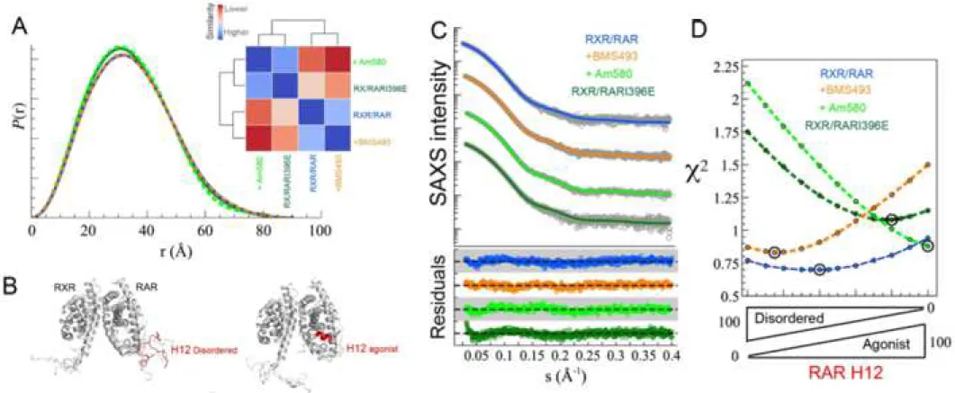
SAXS data indicate that unliganded RXR/RAR heterodimer is a globular 157
particle in solution with a radius of gyration, Rg of 26.6 ± 0.4 Å and a maximum
158
intramolecular distance, Dmax of 89.0 ± 3.0 Å (Table S1; Fig. 1A). Molecular weight
159
estimation suggests that the particle is a heterodimer, in line with Sedimentation 160
Velocity Analytical Ultracentrifugation (SV-AUC) experiments, s20w = 3.78 ± 0.13 S 161
and f/fmin= 1.30 ± 0.05 (Fig. S2). The smooth asymmetrical pair-wise distance 162
distribution, P(r), suggests the presence of moderate flexibility in the RXR/RAR 163
heterodimer (Fig. 1A). Crystallographic structures of RXR/RAR (PDB entries 1DKF 164
(Bourguet et al., 2000), 1XDK (Pogenberg et al., 2005), and 3A9E (Sato et al., 2010) 165
(Table S2)), all obtained in the presence of agonist or antagonist of RXR and RAR, 166
were not consistent with measured SAXS data (χi2 = 2.55, 2.34 and 1.83, 167
respectively). We hypothesized that the observed discrepancy can be attributed to two 168
reasons: firstly, the missing flexible regions, including the N- and C-termini and the 169
loops connecting helices 2 and 3 in both RXR and RAR (Fig. S1B), and secondly, the 170 1 2 3 4 5 6 7 8 9 10 11 12 13 14 15 16 17 18 19 20 21 22 23 24 25 26 27 28 29 30 31 32 33 34 35 36 37 38 39 40 41 42 43 44 45 46 47 48 49 50 51 52 53 54 55 56 57 58 59 60
positions of RXR and RAR H12 helices that are in antagonist or agonist-bound 171
conformations in the X-ray structures. To validate this hypothesis and based on 172
several crystallographic structures, we built three ensemble models for the RXR/RAR 173
heterodimer in which both H12 helices were maintained disordered, in agonist or in 174
antagonist conformation (see Fig. S1 and methods section for details). SAXS curves 175
derived from these ensemble models were compared with the experimental one. As 176
observed in Figure 1 and Figure S1, the average scattering profile computed from the 177
ensemble model with disordered H12 fragments yielded an excellent agreement to the 178
experimental curve (χ2 = 0.77), in accordance with the high propensity of RXR and 179
RAR H12 sequences to be disordered, in particular in their C-terminal part (Fig. 180
S1A). The other two alternative ensembles with ordered H12 regions displayed a 181
small but significant decrease in the agreement to the experimental curve, with χ2 of 182
1.37 and 2.44 for the antagonist and agonist positions, respectively (Fig. S1). These 183
results indicate that in the unliganded form, H12 helices of both RXR and RAR are 184
disordered, and highlight the sensitivity of SAXS measurements and analysis to 185
minute structural changes in the heterodimer. 186
We have exploited this sensitivity to monitor the structural changes in 187
RXR/RAR induced by the binding of two selective RAR ligands, BMS493 (RAR 188
inverse agonist) and Am580 (RAR agonist). The presence of these two ligands 189
induces subtle but noticeable differences in the resulting curves (Fig. 1, Fig. S1 and 190
Table S1). In the presence of BMS493, the observed Rg, 26.5 ± 0.3 Å, is similar to the
191
one measured for the unliganded RXR/RAR heterodimer. Conversely, the presence of 192
Am580 induces a compaction of the particle, with a Rg of 25.6 ± 0.2 Å. We have used
193
the ensemble models based on the available X-ray structures to understand the 194
structural bases of these differences. Concretely, two ensembles of the heterodimer 195 1 2 3 4 5 6 7 8 9 10 11 12 13 14 15 16 17 18 19 20 21 22 23 24 25 26 27 28 29 30 31 32 33 34 35 36 37 38 39 40 41 42 43 44 45 46 47 48 49 50 51 52 53 54 55 56 57 58 59 60
were built in which RXR H12 was maintained disordered whereas RAR H12 was 196
assumed disordered or placed in agonist position (see methods section). The average 197
curves from these ensembles were linearly combined with that of the disordered H12 198
RAR to optimally describe the measured SAXS curves. SAXS curve of RXR/RAR in 199
the presence of BMS493 is nicely described (χ2 = 0.87) with models consisting in a 200
major contribution (85%) of a fully disordered RAR H12 helix (Fig. 1). Conversely, 201
in the presence of Am580, SAXS data are in agreement with the 100% of RAR H12 202
folded in the agonist position (χ2 = 0.88) (Fig. 1). 203
Similarly, we have analyzed the conformational changes of RAR H12 helix in 204
the RARI396E point mutant in the context of the heterodimer (RXR/RARI396E). 205
This point mutation, which was designed based on the crystal structure of the 206
complex between RARα and a corepressor peptide, is expected to destabilize the 207
RAR β-strand S3 conformation and favor a helical conformation in H11, thus 208
mimicking agonist-induced conformational change (le Maire et al., 2010). 209
Interestingly, the SAXS analysis indicates that the main heterodimeric species of this 210
mutant has the RAR H12 in a compact agonistic disposition although a certain 211
population (~20%) of disordered H12 was also observed (χ2 = 1.11, Fig. 1D). Not 212
surprisingly, the P(r) functions of the mutant RXR/RARI396E and Am580-bound 213
heterodimer are similar, whereas the unliganded RXR/RAR resembles more the 214
BMS493-bound RXR/RAR (Fig. 1A). 215
In summary, our structural analysis shows that H12 helices of unliganded 216
RXR/RAR heterodimer are mainly disordered in solution, and that the flexibility of 217
RAR H12 can be modulated by the presence of specific ligands or mutation. These 218
observations substantiate the pivotal role of H12 region as a modulator of RAR 219 activity. 220 1 2 3 4 5 6 7 8 9 10 11 12 13 14 15 16 17 18 19 20 21 22 23 24 25 26 27 28 29 30 31 32 33 34 35 36 37 38 39 40 41 42 43 44 45 46 47 48 49 50 51 52 53 54 55 56 57 58 59 60
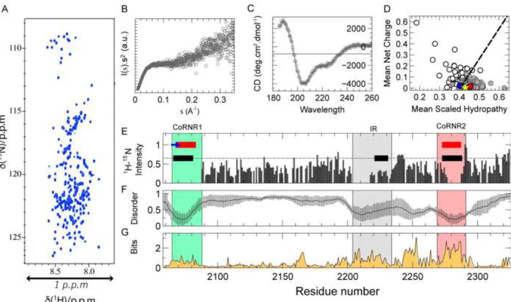
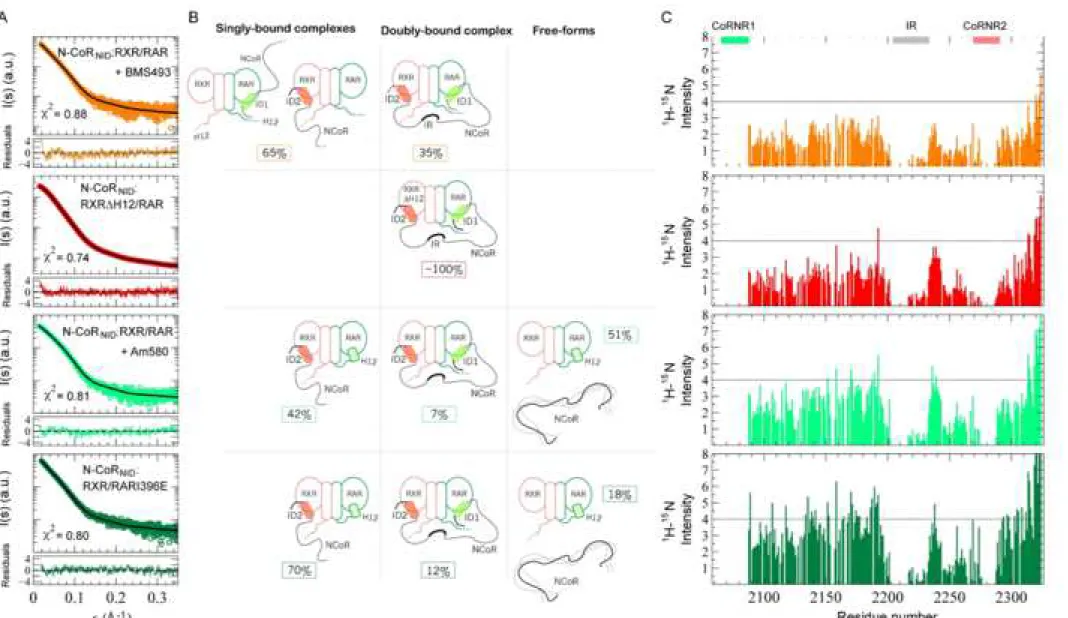
N-CoRNID is a disordered protein with evolutionarily conserved and partially
221
structured elements. We produced and characterized a large fragment of the nuclear
222
receptor corepressor N-CoR (Hörlein et al., 1995) spanning from residue Gln2059 to 223
Glu2325. This fragment, N-CoRNID from now on, corresponds to the nuclear 224
interaction domain (NID) of N-CoR and encompasses the two nuclear receptor 225
binding motifs involved in the interaction of the protein with RXR/RAR heterodimer, 226
CoRNR1 (from 2065 to 2088) and CoRNR2 (from 2269 to 2291) (Fig. 2). The 227
biophysical characterization of N-CoRNID unambiguously indicates that the protein 228
behaves as an Intrinsically Disordered Protein (IDP) (Table S3, Fig. S2 and Fig. 2). 229
Concretely, N-CoRNID displays a reduced 1H dispersion in NMR spectra (Fig. 2A), 230
and the Kratky plot does not present a clear maximum (Fig. 2B). Moreover, the s20w 231
and frictional ratio measured by SV-AUC, 2.37 ± 0.19 S and 1.53 ± 0.13, are not 232
compatible with a globular protein of this size (≈29 kDa) (Fig. S2). Interestingly, far-233
UV Circular Dichroism (CD) measured on N-CoRNID presents features that suggest 234
the presence of helical regions (Fig. 2C). Secondary structure is manifested by a shift 235
in the negative maximum at 205 nm, rather than at 198 nm for a pure random coil 236
profile, the negative shoulder near 220 nm, which is more pronounced than that 237
observed for fully disordered proteins, and the positive signal at 190 nm (Bienkiewicz 238
et al., 2002). 239
We performed the NMR study of N-CoRNID to identify structural features at 240
the residue level (Fig. 2A). The resonances of backbone nuclei of N-CoRNID were 241
assigned using standard triple resonance spectra at high spectrometer field. Out of the 242
241 expected 1H-15N HSQC backbone correlation peaks, only 183 could be 243
unambiguously assigned (Fig. S3). The identified correlations were either non-244
visible peaks or had extremely low intensities precluding assignment. When mapping 245 1 2 3 4 5 6 7 8 9 10 11 12 13 14 15 16 17 18 19 20 21 22 23 24 25 26 27 28 29 30 31 32 33 34 35 36 37 38 39 40 41 42 43 44 45 46 47 48 49 50 51 52 53 54 55 56 57 58 59 60
the missing peaks on the amino acid sequence of N-CoRNID, they clustered in three 246
non-consecutive regions of the protein (Fig. 2E and Fig. S3B). Interestingly, two of 247
these clusters were centered in the consensus NR binding domains CoRNR1 and 248
CoRNR2 and extended towards both flanking regions (Fig. 2E). Furthermore, a third 249
region, spanning from 2204 to 2234, also displayed absence or systematic decrease of 250
NMR intensities. Within this third region, which will be named Intermediate Region 251
(IR) from now on, no correlation peaks could be assigned for the residues 2204-2217 252
and 2222-2223. We performed 1H-15N HSQC experiments at different temperatures 253
(283, 288 and 293 K) but the number of peaks in the spectra did not change. Based on 254
these observations, we attributed the absence or decrease of NMR intensities in these 255
three regions to the formation of transient secondary structural elements that 256
experience chemical exchange processes in the μs-ms time-scale inducing severe 257
broadening of the signals. The presence of partially structured regions was 258
substantiated by the analysis of N-CoRNID sequence using several disorder prediction 259
servers (Fig. 2F). All predictors applied coincided in identifying CoRNR1 and 260
CoRNR2 as helices and their respective flanking regions as partially structured, both 261
motifs named as ID1 and ID2, respectively, from now on. Interestingly, disorder 262
predictors also identify the IR as partially structured, although to a lesser extent 263
(Buchan et al., 2013). 264
A sequence conservation bioinformatics analysis of N-CoRNID fragments from 265
multiple eukaryotic organisms indicates that ID1 and the large C-terminal region 266
(LCR, 2190 to 2295), which encompasses IR and ID2, are evolutionarily conserved 267
(Fig. 2G). On the contrary, the N-terminal region of the N-CoRNID, with the exception 268
of ID1, is poorly conserved as typically observed in IDPs (Ota and Fukuchi, 2017). 269
Interestingly, the LCR presents a sequence composition that is closer to globular 270 1 2 3 4 5 6 7 8 9 10 11 12 13 14 15 16 17 18 19 20 21 22 23 24 25 26 27 28 29 30 31 32 33 34 35 36 37 38 39 40 41 42 43 44 45 46 47 48 49 50 51 52 53 54 55 56 57 58 59 60
proteins according to the charge hydropathy plot (Uversky and Gillespie, 2000), a 271
behaviour that is different from the N-terminus (Fig. 2D). 272
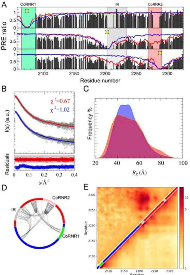
N-CoRNID presents intramolecular transient contacts between conserved,
co-273
evolved and partially structured IR and ID2 regions. The presence of local
274
compaction and long-range contacts in the partially structured and conserved C-275
terminal region of N-CoRNID was explored using SAXS, Paramagnetic Relaxation 276
Enhancement (PRE) NMR experiments, which report on distance-depending induced 277
relaxation on NMR active nuclei, and Molecular Dynamics simulations (MD). In 278
addition to the native Cys2074, which sits in the middle of the ID1 motif, Ser2213 279
(preceding the IR) and Ser2288 (succeeding ID2) were mutated to cysteines. Note that 280
for these two mutants, the native Cys2074 was mutated into serine (C2074S) to have 281
only one cysteine at a time in the entire sequence of N-CoRNID.After incorporating a 282
PROXYL stable radical on each of the cysteine residues independently, PRE-ratios 283
were measured for the three samples. When the paramagnetic moiety was introduced 284
in native Cys2074, no distal effects were observed, indicating that the N-terminal 285
region of N-CoRNID does not present long-range interactions with the rest of the 286
protein (Fig. 3A). Conversely, PRE data measured in the two point mutants, S2213C 287
and S2288C, provide a different picture of N-CoRNID. For both single-cysteine 288
variants, a substantial decrease in intensity is observed for 1H-15N HSQC peaks from 289
the region between the IR and ID2 indicating the presence of extensive long-range 290
contacts (Fig. 3A). Interestingly, PREs measured in this region display a bell-shape 291
with stronger PRE effects in the proximity of both partially structured regions. 292
Moreover, PRE profiles for both mutants in the LCR are very similar suggesting a 293
direct interaction between the IR and the ID2 motifs that also affects the connecting 294
region (Fig. 3A). The compactness of N-CoRNID observed by PRE was substantiated 295 1 2 3 4 5 6 7 8 9 10 11 12 13 14 15 16 17 18 19 20 21 22 23 24 25 26 27 28 29 30 31 32 33 34 35 36 37 38 39 40 41 42 43 44 45 46 47 48 49 50 51 52 53 54 55 56 57 58 59 60
by SAXS. A simple random coil model built with Flexible-Meccano (Bernado et al., 296
2005) could not reproduce the experimental SAXS curve (Fig. 3B). Indeed, the 297
theoretical ensemble turned out to be more extended than N-CoRNID in solution, with 298
Rg of 50.4 Å and 47.2 ± 1.2 Å for the theoretical and experimental SAXS curves,
299
respectively. 300
PRE and SAXS data were used to further characterize the structural 301
compaction observed in N-CoRNID.Large ensembles of conformations were built with 302
Flexible-Meccano to which multiple explicit dispositions of the PROXYL moiety 303
were attached to the native or engineered cysteine residues. Subsequently, the 304
theoretical averaged PRE ratios were computed as previously described (Salmon et 305
al., 2010), and compared with the three experimental PRE profiles (see methods for 306
details). Not surprisingly, the random coil model only presented contacts in the 307
vicinity of the paramagnetic sites, and therefore did not reproduce the experimental 308
profiles especially in the C-terminal region (Fig. 3A, blue lines). In order to interpret 309
the long-range contacts observed in the LCR, a structurally biased model was built 310
based on previous experimental and bioinformatics observations. Concretely, 311
conformations with at least two contacts of 15 Å between residues from distal
312
regions of the LCR presenting partial structuration (average disorder below 0.5, Fig.
313
2F), evolutionary conservation (Bits 1.6, Fig. 2G), or co-evolution (Fig. 3D), were
314
selected from a large ensemble of random coil conformations. This filtered ensemble
315
resulted too compact, and it was further refined using the SAXS curve to yield a
sub-316
ensemble compatible with the scattering profile (Fig. 3B, C), which was subsequently
317
used to compute the PRE values for the three PROXYL-tagged N-CoRNID constructs
318
and compared with the experimental ones. The resulting theoretical PRE profiles
319
displayed an excellent agreement with the experimental ones (Fig. 3A, red lines). Not
320 1 2 3 4 5 6 7 8 9 10 11 12 13 14 15 16 17 18 19 20 21 22 23 24 25 26 27 28 29 30 31 32 33 34 35 36 37 38 39 40 41 42 43 44 45 46 47 48 49 50 51 52 53 54 55 56 57 58 59 60
surprisingly, a systematic decrease of the PRE values in the LCR compared to the
N-321
terminal region was observed. More interestingly, PRE fluctuations in the connecting
322
region between the IR and the ID2 region were nicely reproduced for the S2213C and
323
S2288C mutants.
324
In order to further investigate the formation of these long-range contacts, we 325
performed MD simulations of N-CoRNID based on a coarse-grained model that was 326
specifically designed for IDPs (Smith et al., 2014) and takes into account sequence-327
specific electrostatic and hydrophobic interactions. The resulting conformational 328
ensemble, which was not biased by experimental data, revealed that N-CoRNID 329
sequence is poised to form transient yet noticeable interactions in the C-terminal 330
region. Importantly, the contact matrix derived from coarse-grained MD is in good 331
agreement with the PRE-derived ensemble (Fig. 3E). These results substantiate the 332
presence of transient intramolecular contacts between distal regions within the C-333
terminal region of N-CoRNID. Moreover, these regions present relatively stable 334
secondary structural elements and a high level of evolutionary conservation. 335
RXR/RAR heterodimer interacts with the corepressor mainly through RAR and
336
in a cooperative manner. After characterizing the individual partners, we
337
investigated the complex between RXR/RAR and N-CoRNID. The affinities of 338
RXR/RAR heterodimer for NCoR peptides encompassing either CoRNR1 or 339
CoRNR2 motifs and for the N-CoRNID fragment were measured by fluorescence 340
anisotropy and thermophoresis, respectively. As shown in Figure 4, formation of the 341
RXR/RAR heterodimer does not modify the binding capacity of individual RXR and 342
RAR monomers for isolated CoRNR peptides. In fact, the RXR/RAR heterodimer 343
binds the two isolated CoRNR peptides with nearly the same affinity, 1.68 and 1.47 344
μM for CoRNR1 and CoRNR2, respectively (Fig. 4B). These affinity values are very 345 1 2 3 4 5 6 7 8 9 10 11 12 13 14 15 16 17 18 19 20 21 22 23 24 25 26 27 28 29 30 31 32 33 34 35 36 37 38 39 40 41 42 43 44 45 46 47 48 49 50 51 52 53 54 55 56 57 58 59 60
similar to those measured previously (le Maire et al., 2010) for the unliganded RAR 346
monomer (1.40 and 1.55 μM for peptides encompassing CoRNR1 and CoRNR2, 347
respectively) (Fig. 4A). The slight preference observed for CoRNR1 is induced by the 348
ability of this motif to form a β-sheet interface with S3 of RARα, which is further 349
stabilized in the presence of the inverse agonist BMS493 with an affinity of 0.17 μM. 350
Conversely, in its monomeric form, RXR presents a moderate affinity (22.5 μM) for 351
CoRNR2, and no measurable interaction with CoRNR1 (Fig. 4A). As expected, the 352
presence of the RAR-selective agonist Am580 or the RARI396E mutation cause a 353
noticeable decrease in the affinity of RXR/RAR for both N-CoR peptides with a 354
stronger effect on CoRNR1 (Fig. 4B). Conversely, the RAR inverse agonist BMS493 355
efficiently increases the binding affinity of RXR/RAR for CoRNR1, but has not much 356
effect on the interaction with CoRNR2 (Fig. 4A-B). These observations suggest a 357
sequential and directional mechanism in which, in the context of N-CoRNID, 358
comprising the two CoRNR motifs, the main anchoring point would involve RAR on 359
the heterodimer side and CoRNR1 on the N-CoR side. This primary contact would 360
then enable a second lower-affinity interaction between RXR and CoRNR2. 361
Microscale thermophoresis measurements further substantiated the 362
cooperativity and directionality of the interaction. The affinity of RXR/RAR 363
heterodimer for N-CoRNID was found to be much higher than for individual peptides 364
with a value of 0.21 ± 0.09 μM, to be compared to 1.68 and 1.47 μM for CoRNR1 365
and CoRNR2, respectively (Fig. 4B-C). The inverse agonist BMS493 or deletion of 366
RXR helix H12 (RXRΔH12), which are known to enhance the interaction of RAR 367
with CoRNR1 (Germain et al., 2009; le Maire et al., 2010) (Fig. 3A) and that of RXR 368
for CoRNR2 (Hu and Lazar, 1999), respectively, were shown to increase significantly 369
the overall affinity of the heterodimer for N-CoRNID (Fig. 4C). In contrast, the RAR 370 1 2 3 4 5 6 7 8 9 10 11 12 13 14 15 16 17 18 19 20 21 22 23 24 25 26 27 28 29 30 31 32 33 34 35 36 37 38 39 40 41 42 43 44 45 46 47 48 49 50 51 52 53 54 55 56 57 58 59 60
agonist Am580, or the RAR mutation I396E induced a strong decrease of the 371
affinities with Kd values of 8.6 ± 3.70 μM or 4.42 ± 1.86 μM, respectively, due to the 372
weakening of the interaction of RAR with CoRNR1 in both cases. The observation 373
that the perturbation of individual anchoring points has severe consequences on the 374
affinity of RXR/RAR for N-CoRNID demonstrates the cooperativity and directionality 375
of the complex. 376
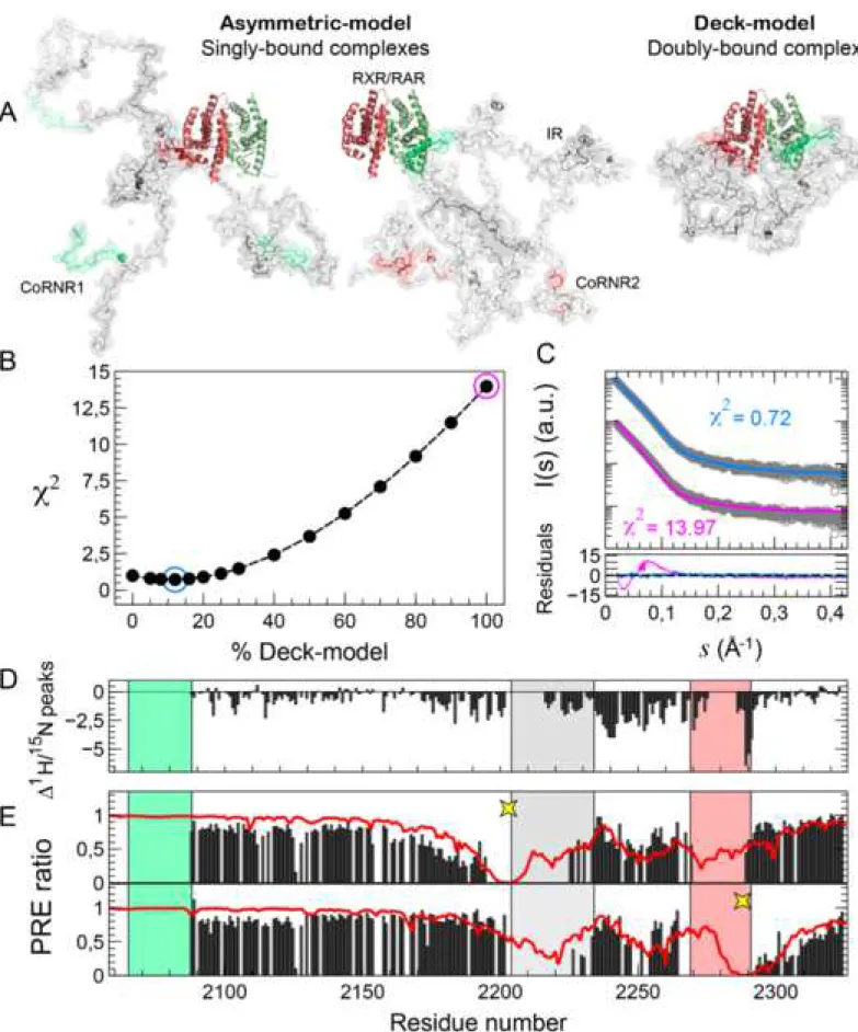
N-CoRNID forms a transient multi-site complex with RXR/RAR. Affinity
377
measurements indicate that N-CoRNID binds cooperatively to RXR/RAR heterodimer. 378
To unveil the structural bases of this cooperativity, we combined SAXS and NMR to 379
study the N-CoRNID:RXR/RAR heterotrimeric complex. 380
SAXS data indicates that the complex is monodisperse with a Rg and Dmax
381
values of 48.4 ± 1.1 Å and 194 ± 10 Å, and with a stoichiometry of 1:1:1 according to 382
SV-AUC and mass spectrometry data (Table S3, Fig. S2 and Fig. S4). Moreover, the 383
Kratky representation with elevated baseline at high s, and asymmetric P(r) function 384
suggest that N-CoRNID:RXR/RAR has a significant degree of flexibility (Fig. S5). 385
The observation of cooperative effects prompted towards the possibility that 386
the two binding regions of N-CoRNID, CoRNR1 and CoRNR2, could simultaneously
387
interact with RXR/RAR forming the so-called deck model (le Maire and Bourguet, 388
2014). The latter scenario is in contrast with the asymmetric model, where a single 389
corepressor binding side would interact with the RXR/RAR heterodimer as observed 390
for the complex between RXR/RAR and a fragment of the coactivator Med1 (Rochel 391
et al., 2011). Ensembles of conformations were built for both the deck and the 392
asymmetric binding modes (see SI Appendix for details), integrating previous 393
knowledge of the system. This includes the directionality of the complex derived from 394
affinity measurements, and two crystallographic structures of RAR and RXR with 395 1 2 3 4 5 6 7 8 9 10 11 12 13 14 15 16 17 18 19 20 21 22 23 24 25 26 27 28 29 30 31 32 33 34 35 36 37 38 39 40 41 42 43 44 45 46 47 48 49 50 51 52 53 54 55 56 57 58 59 60
corepressor peptides that were used to define the atomic details of the interacting sites 396
(Table S2) (le Maire et al., 2010; Zhang et al., 2011). H12 helices from both RXR and 397
RAR were considered disordered based on our SAXS analysis (see above). Finally, 398
the non-interacting regions of N-CoRNID were assumed to adopt random coil 399
conformations based on the NMR data (see below). Theoretical SAXS curves were 400
computed from large ensembles of structures for both interacting modes yielding 401
notably different profiles (Fig. S6). These curves were optimally combined to derive 402
the relative population of both interacting modes by minimizing the agreement to the 403
experimental curve. An excellent agreement (χi2 = 0.88) to the experimental SAXS 404
curve of N-CoRNID:RXR/RAR was obtained when a 85/15 ratio for the
405
asymmetric/deck models was used (Fig. 5A, B). This result indicates that both 406
binding modes coexist in solution although the asymmetric model is the major species 407
in the absence of ligand. The similarity of the scattering curves between the two 408
alternative asymmetric models (N-CoRNID bound to RAR through ID1 or N-CoRNID 409
bound to RXR through ID2) precludes the identification of the major asymmetric 410
species. 411
In order to understand the interaction at the residue level, we performed NMR 412
experiments by adding an equimolar amount of RXR/RAR to a 15N-isotopically 413
labeled N-CoRNID sample. The NMR 1H-15N HSQC spectrum of N-CoRNID presents 414
correlation peaks that are superimposable to these obtained in the absence of 415
heterodimer, and no correlation peaks shifted upon the addition of the heterodimer 416
(Fig. S7). This observation indicates that the conformational properties of N-CoRNID 417
are equivalent in the free and in the bound states, and that N-CoRNID remains mostly 418
disordered when bound to the RXR/RAR heterodimer. However, in the presence of 419
RXR/RAR, a systematic decrease in peak intensities is observed for N-CoRNID when 420 1 2 3 4 5 6 7 8 9 10 11 12 13 14 15 16 17 18 19 20 21 22 23 24 25 26 27 28 29 30 31 32 33 34 35 36 37 38 39 40 41 42 43 44 45 46 47 48 49 50 51 52 53 54 55 56 57 58 59 60
compared to the free form (Fig. S7 and Fig. 2E). We attribute this observation to the 421
perturbation of the hydrodynamic properties of N-CoRNID upon binding to the 422
heterodimer that senses the presence of a large globular particle and increases the 423
apparent correlation time. Interestingly, the intensity decrease is not homogeneously 424
distributed along the protein, and two regions of the protein can be clearly 425
distinguished. The N-terminal region connecting ID1 and the IR presents moderate 426
intensity decreases that are slightly larger in the proximity of both partially structured 427
regions. Conversely, the region connecting the IR with ID2 displays a more important 428
intensity reduction. The transient binding of ID2 to RXR as demonstrated by SAXS 429
and the change in the dynamic regime for the intra-molecular interactions could 430
explain the enhanced intensity reduction in the C-terminus. To explore this 431
phenomenon, we performed PRE experiments for the complexes formed by the 432
previously described N-CoRNID cysteine mutants and the heterodimer RXR/RAR. The 433
incorporation of radical moieties on the N-CoRNID S2213C and S2288C point mutants 434
induced a sequence-dependent decrease on the PRE ratios of residues placed at their 435
flanking regions, a phenomenon which is typically observed in IDPs (Fig. 5D, E). 436
Interestingly, for both mutants a small but systematic increase in the PRE ratios was 437
found for the N-terminal residues when compared with the PRE data of the free forms 438
(Fig. 3A). This is most probably caused by the reduced conformational exploration in 439
N-CoRNID when bound to the heterodimer, which limits sporadic contacts of the C-440
terminal region with the N-terminus. Compared with the free form of N-CoRNID, the 441
presence of the heterodimer causes a PRE-ratio increase in the region connecting the 442
IR and ID2, and partially suppresses the above-described bell-shape of PRE ratios. 443
This increase is not homogeneous and the region around 2250 presents strong PRE 444
effects. These observations indicate that the population of conformations experiencing 445 1 2 3 4 5 6 7 8 9 10 11 12 13 14 15 16 17 18 19 20 21 22 23 24 25 26 27 28 29 30 31 32 33 34 35 36 37 38 39 40 41 42 43 44 45 46 47 48 49 50 51 52 53 54 55 56 57 58 59 60
long-range contacts in the C-terminus is diminished in the bound form. This last 446
observation is coherent with the existence of a minor population of the deck model 447
that conformationally restricts N-CoRNID, and partially hampers intra-molecular 448
contacts involving ID2. 449
Cognate ligands and mutations modulate the conformational equilibrium in the
450
complex. Thermophoresis experiments demonstrated that the affinity of N-CoRNID for 451
RXR/RAR can be finely tuned by the addition of cognate ligands or by mutations at 452
the recognition sites of both RXR and RAR. We applied SAXS and NMR to explore 453
the structural bases of this affinity modulation. 454
The addition of the RAR inverse agonist BMS493 reinforced the interaction of 455
RXR/RAR with N-CoRNID through CoRNR1 (Fig. 4B and 4C). The analysis of the 456
SAXS curve of the ternary complex in the presence of BMS493 indicates that the 457
overall size of the particle is slightly reduced with respect to the unliganded form of 458
the complex, Rg = 47.5 ± 1.0Å (Table S3). The characterization of that curve in terms
459
of atomistic ensemble models indicated that the asymmetric model is still the major 460
species but the population of the doubly-bound deck model increases up to 35% (Fig. 461
6A and 6B). A more extreme situation was observed when the complex was formed 462
with RXRΔH12/RAR that strongly reinforces the overall affinity with N-CoRNID by 463
increasing the interaction of RXR with ID2 (Fig. 4B and 4C). This RXR H12 deletion 464
renders the RXR hydrophobic groove more accessible and has been reported to 465
significantly increase the interaction of RXR with corepressor(Hu and Lazar, 1999; 466
Schulman et al., 1996; Zhang et al., 2011). In that situation, the SAXS curve, which 467
presented a smaller Rg of 42.0 ± 2 Å, could be explained with the only presence of the
468
deck model (Table S3, Fig. S5 and Fig. 6A and 6B). The 15N N-CoR
NID NMR 469
intensities measured for both complexes, upon addition of RXR/RAR in the presence 470 1 2 3 4 5 6 7 8 9 10 11 12 13 14 15 16 17 18 19 20 21 22 23 24 25 26 27 28 29 30 31 32 33 34 35 36 37 38 39 40 41 42 43 44 45 46 47 48 49 50 51 52 53 54 55 56 57 58 59 60
of BMS493 and of RXRΔH12/RAR, displayed a systematic decrease with respect to 471
the unliganded N-CoRNID:RXR/RAR (Fig. 6C). We attributed this observation to the 472
increase of the overall correlation time sensed by N-CoRNID when the population of 473
the deck model increases. This is especially significant in the region close to ID2 that 474
fits well with the higher interaction of ID2 with RXRΔH12. Compared with the 475
unliganded (Fig. S7), the region connecting the IR and ID2 in the N-476
CoRNID:RXRΔH12/RAR displays a less prominent decrease of intensities, indicating 477
that some of the structural and/or dynamical properties that cause the decrease in the 478
native complex are abolished by the formation of a doubly-bound state. 479
We also performed SAXS experiments of the complex N-CoRNID:RXR/RAR
480
in the presence of the RAR agonist Am580, and of the complex N-481
CoRNID:RXR/RARI396E. Affinity measurements indicated that both conditions 482
diminished the interaction of RXR/RAR with N-CoRNID through CoRNR1 (Fig. 4B
483
and 4C). The analysis of the SAXS data using explicit models indicated that the 484
decrease of the interaction has severe structural consequences for both complexes. On 485
one hand, the SAXS curve measured on N-CoRNID:RXR/RAR in the presence of 486
Am580 can only be described (χ2 = 0.81) if large populations, 51%, of unbound N-487
CoRNID and RXR/RAR are invoked (Fig. 6A and 6B), showing that Am580 partially
488
breaks the complex by diminishing the interaction of CoRNR1 with RAR. Similarly, 489
the SAXS curve measured for the complex N-CoRNID:RXR/RARI396E is optimally
490
described using a combination of unbound species, asymmetric complex and a low 491
percentage of deck complex. 15N N-CoR
NID NMR intensities measured in these two 492
conditions were also coherent with SAXS observations (Fig. 6C). The general 493
increase in these intensities is in line with the presence of a weaker complex with 494
lower dragging forces and smaller apparent correlation time. 495 1 2 3 4 5 6 7 8 9 10 11 12 13 14 15 16 17 18 19 20 21 22 23 24 25 26 27 28 29 30 31 32 33 34 35 36 37 38 39 40 41 42 43 44 45 46 47 48 49 50 51 52 53 54 55 56 57 58 59 60
Mammalian two-hybrid experiments confirm the interaction in cellular context.
496
Mammalian two-hybrid experiments were performed to validate in a cellular context 497
the cooperativity and the directionality of the interaction between N-CoRNID and 498
RXR/RAR heterodimer, deduced from the measurements of in vitro binding constants 499
and from the SAXS/NMR modelling. Two-hybrid analyses were performed in COS 500
cells with chimeras containing GAL4 DNA-binding domain fused to a N-CoR 501
fragment encompassing ID1 and ID2 (from 1629 to 2453 termed Gal-NCoR) or either 502
CoRNR1 or CoRNR2 of N-CoR (Gal-CoRNR1 and Gal-CoRNR2, respectively), and 503
the LBD of RXRα or of RARα fused to the activation domain of VP16 (termed VP16-504
RXR or VP16-RAR, respectively). 505
First, we confirmed that wt RXR/RAR heterodimer interact with both 506
CoRNR1 and CoRNR2 (Fig. 7A), with a slightly better effectiveness for CoRNR1. 507
However, the interaction of RXR/RAR heterodimer with the longer N-CoR fragment 508
(equivalent to N-CoRNID) is significantly stronger than the individual CoRNR regions. 509
Therefore, in a cellular environment the two CoRNR motifs in N-CoR bind to the 510
RXR/RAR heterodimer in a cooperative manner and may induce a stronger affinity. 511
In addition, the selective RAR agonist TTNPB is able to release CoRNR1 and the N-512
CoR fragment from RXR/RAR heterodimer (Fig. 7A). Importantly, a significant 513
interaction of CoRNR2 is measured even in the presence of TTNPB which only 514
targets RAR likely reflecting the interaction of RXR with the CoRNR2 motif (Fig. 515
7A). The directionality of the interaction between N-CoRNID and RXR/RAR 516
heterodimer was further confirmed with two-hybrid experiment with the N-CoR 517
fragment (Fig. 7B). Whereas RXR alone interacts very weakly with N-CoR, in 518
agreement with previous observations of our group (Germain et al., 2002) and with 519
our fluorescence anisotropy data on RXR monomer and CoRNR peptides (Fig. 4), 520 1 2 3 4 5 6 7 8 9 10 11 12 13 14 15 16 17 18 19 20 21 22 23 24 25 26 27 28 29 30 31 32 33 34 35 36 37 38 39 40 41 42 43 44 45 46 47 48 49 50 51 52 53 54 55 56 57 58 59 60
addition of RARα allows to measure a significant interaction between the RXR/RAR 521
heterodimer and Gal-NCoR. Interestingly, the addition of the RAR-selective agonist 522
TTNPB was sufficient to decrease this interaction (Fig. 7B). Moreover, RAR alone 523
can efficiently recruit N-CoR (Fig. 7C). All these observations confirm that RAR is 524
indispensable for the corepressor recruitment by RXR/RAR heterodimer. On the other 525
side, the role of RXR for the recruitment of N-CoR was subsequently addressed in a 526
cellular environment (Fig. 7C). Relative to RAR alone, addition of RXR yielded a 527
slight but significant increase of the N-CoR interaction confirming an active role of 528
RXR in the heterodimeric form (Fig. 7C), and thus the cooperativity in the 529
interaction. This effect was more pronounced for the RXRΔH12 deletion mutant (Fig. 530
7C), in agreement with MST experiments and SAXS modelling. We reasoned that the 531
RXR H12 deletion may generate a new interaction surface for N-CoR. Contrary to the 532
wt heterodimer, TTNPB does not reduce N-CoR interaction with RXRΔH12/RAR 533
heterodimer (Fig. 7C). This observation indicates that RXRΔH12/RAR retains the 534
ability to interact with N-CoR, through the RXR hydrophobic groove. Two-hybrid 535
experiments with RXRΔH12/RAR heterodimer and Gal-CoRNR1 and Gal-CoRNR2 536
clearly demonstrated that CoRNR2 interacts with the more accessible groove of RXR 537
as TTNPB was unable to reduce CoRNR2 association, and that CoRNR1 interacts 538
with RAR as its binding is impaired by addition of TTNPB (Fig. 7D), confirming the 539
directionality in the interaction of N-CoR with RXRΔH12/RAR. 540
541
Discussion
542
In this study we have integrated multiple structural, biophysical and cell-543
biology techniques to decipher the molecular bases of transcriptional repression of the 544
retinoic acid nuclear receptor by the corepressor N-CoR. Our results demonstrate that 545 1 2 3 4 5 6 7 8 9 10 11 12 13 14 15 16 17 18 19 20 21 22 23 24 25 26 27 28 29 30 31 32 33 34 35 36 37 38 39 40 41 42 43 44 45 46 47 48 49 50 51 52 53 54 55 56 57 58 59 60
the molecular mechanism relies on the interplay of the flexible elements found in 546
RXR/RAR heterodimer and the intrinsically disordered N-CoR. 547
We have exploited the high sensitivity of SAXS to probe minute structural 548
changes that perturbe the shape of the RXR/RAR LBDs heterodimer in the absence or 549
in the presence of various RAR ligands and point mutations. The main structural 550
changes of the heterodimer reside in the conformation and position of helices H12 of 551
each monomer. Our analyses demonstrate that helices H12 of RAR and RXR are 552
better described as disordered in the absence of ligand, in line with previous NMR 553
and amide hydrogen/deuterium (H/D) exchange experiments for a number of NRs 554
including RXR (Johnson et al., 2000; Kallenberger et al., 2003; Lu et al., 2006). 555
These results invalidate the initial hypothesis of allosteric inhibition of RXR by a 556
subset of nuclear receptors, such as RAR and THR. It was suggested that, in 557
unliganded RXR/RAR and RXR/THR heterodimers, H12 of RXR docks to the 558
coregulator-interaction site of the partner (Westin et al., 1998; Zhang et al., 1999). 559
The lack of structural order of RAR helix H12 was also deduced from SAXS 560
data measured on RXR/RAR heterodimer in the presence of RAR inverse agonist 561
(BMS493), in agreement with the absence of density for RAR H12 in the crystal 562
structure of the complex formed by RARα-LBD, the corepressor N-CoRNR1 and the 563
inverse agonist (le Maire et al., 2010). These two forms of RXR/RAR heterodimers, 564
unliganded and in complex with the inverse agonist, bind corepressors, confirming 565
that the disordered helices H12 are not directly involved in corepressor binding. 566
Nevertheless, the disordered and highly flexible RXR and RAR helices H12 may 567
sterically screen the interaction with corepressors. Indeed, RXR H12 and RAR H12 568
deletion mutants recruit corepressors more efficiently than wt RXR and RAR (le 569
Maire et al., 2010; Zhang et al., 1999), probably due to a higher accessibility of the 570 1 2 3 4 5 6 7 8 9 10 11 12 13 14 15 16 17 18 19 20 21 22 23 24 25 26 27 28 29 30 31 32 33 34 35 36 37 38 39 40 41 42 43 44 45 46 47 48 49 50 51 52 53 54 55 56 57 58 59 60
LBD interaction region for the corepressors. Conversely, addition of RAR agonist 571
(Am580) induces a compaction of the heterodimer that fits with RAR helix H12 572
adopting the agonist position, as observed in crystal and solution structures of RAR in 573
the presence of agonists (Bourguet et al., 2000; Egea et al., 2001; Klaholz et al., 2000; 574
le Maire et al., 2010). Our SAXS analysis of the mutant RARI396E, for which no 575
structural data had been reported before, and whose mutation was suggested to induce 576
the S3 strand to H11 helix transition (le Maire et al., 2010), also provokes a main 577
rearrangement of RAR helix H12 that is compatible with an agonist conformation. 578
The transition from a disordered state to an ordered and active conformation of RAR 579
helix H12 is induced by the binding of an agonist that efficiently triggers corepressor 580
release. Importantly, this release is dependent of the proportion of agonist 581
conformation of this helix. With this analysis we confirm that the disorder to order 582
transition of helix H12 conformation is a key mechanism of corepressor release and 583
coactivator recruitment by RXR/RAR, contrary to the region corresponding to 584
H11/S3 in RAR which is the master regulator of corepressor association to RAR (le 585
Maire et al., 2010). 586
Our extensive experimental and computational analysis of N-CoRNID 587
demonstrates that it is a disordered protein presenting local and long-range structural 588
phenomena that are directly linked to its function. With the exception of the CoRNR1 589
motif, the N-terminal region presents the prototypical spectroscopic features of a 590
random coil. Moreover, bioinformatics analyses on this region indicate a poor 591
evolutionary conservation and the absence of co-evolutionary interactions with other 592
parts of the protein has often been observed in disordered regions without direct 593
functional roles (Ota and Fukuchi, 2017). Conversely, the C-terminal region including 594
the CoRNR2 motif presents multiple structural features and is evolutionary 595 1 2 3 4 5 6 7 8 9 10 11 12 13 14 15 16 17 18 19 20 21 22 23 24 25 26 27 28 29 30 31 32 33 34 35 36 37 38 39 40 41 42 43 44 45 46 47 48 49 50 51 52 53 54 55 56 57 58 59 60
conserved. In solution, CoRNR1 and CoRNR2 are preformed molecular recognition 596
elements (MOREs) (Mohan et al., 2006) that mediate the interaction with the 597
heterodimer. The lack of NMR information for these regions is probably due to 598
intermediate exchange regime processes linked to the transient formation of 599
secondary structures. Very interestingly, NMR has unveiled a new region, IR, placed 600
between the two NR interaction domains that is partially ordered and highly 601
conserved in eukaryotes. These two observations suggest a relevant functional role for 602
IR. Indeed, N-CoRNID experiences transient long-range tertiary interactions between 603
the IR and ID2 that we have identified and structurally characterized using PREs. The 604
accurate description of experimental PREs was achieved when the long-range 605
interaction between co-evolved residues in partially structured regions of the C-606
terminal region of N-CoRNID was used. This long range contact is partially impaired 607
when N-CoR interacts with NRs as demonstrated by our NMR analysis of N-CoRNID
608
in complex with the RXR/RAR heterodimer. The formation of the complex would 609
liberate the IR that would become available to interact with other proteins of the 610
repressive macromolecular complex. 611
A number of structural and biophysical studies have already revealed the 612
complex interactions between NR heterodimers and coactivators (Chandra et al., 613
2017; Osz et al., 2012; Pavlin et al., 2014; Pogenberg et al., 2005; Rochel et al., 2011; 614
de Vera et al., 2017; Zheng et al., 2017). However, the interaction of NRs with 615
corepressors that hampers gene transcription in the basal state are poorly understood. 616
Our study, which included solution-state structural methods along with biochemical, 617
biophysical and computational approches, reveals for the first time the atomistic 618
details in terms of ensembles of the interaction between RXR/RAR heterodimer and 619
N-CoR. Our study shows a cooperative interaction of both CoRNR1 and CoRNR2 620 1 2 3 4 5 6 7 8 9 10 11 12 13 14 15 16 17 18 19 20 21 22 23 24 25 26 27 28 29 30 31 32 33 34 35 36 37 38 39 40 41 42 43 44 45 46 47 48 49 50 51 52 53 54 55 56 57 58 59 60
motifs with RXR/RAR as the binding affinity of N-CoRNID is stronger than the 621
individual interactions. In addition, the interaction has a defined directionality: N-622
CoRNID is recruited primarily to RAR through CoRNR1 enabling CoRNR2 to 623
subsequently bind to RXR for which it has a moderate affinity. Importantly, the other 624
configuration would not produce a cooperative binding as CoRNR1 does not interact 625
with RXR. 626
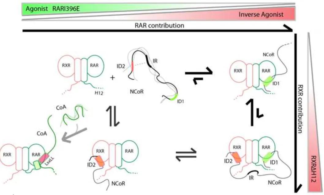
Taken toghether, our data suggest the mechanistic model that is depicted in 627
Figure 8. In the absence of ligand, it exists an equilibrium between a major population 628
of assymetric binding of N-CoRNID to RXR/RAR and a minor population of doubly
629
bound N-CoRNID (deck binding mode) in which both CoRNR boxes simultaneously 630
interact with the heterodimer, accounting for the cooperativity of the interaction. The 631
major species of the assymetric binding mode can be reasonably assigned to N-632
CoRNID binding to RAR through the CoRNR1 motif, which presents the strongest 633
local affinity. Our observations through mammalian two hybrid experiments 634
substantiate with this model, also suggesting that the structural phenomena probed in 635
vitro also occurs in cells. The addition of RAR ligands or point mutations in either
636
RXR and RAR modify the strenght of the individual interactions and, consequently 637
change the cooperativity and the equilibrium between the assymetric and the deck 638
binding modes. Concretely, the addition of a RAR inverse agonist, which strenghtens 639
the interaction of CoRNR1 with RAR, or the use of a heterodimer with the truncated 640
RXR H12 (RXR H12), which increases the interaction of CoRNR2 with RXR, 641
produce a notable increase of the overall affinity of the complex. This increase is a 642
consequence of an enhancement of the cooperativity by the equilibrium displacement 643
towards the deck model as observed in the SAXS and NMR analyses. Conversely, 644
when weakening RAR binding site using a RAR agonist or the RARI396E mutant, 645 1 2 3 4 5 6 7 8 9 10 11 12 13 14 15 16 17 18 19 20 21 22 23 24 25 26 27 28 29 30 31 32 33 34 35 36 37 38 39 40 41 42 43 44 45 46 47 48 49 50 51 52 53 54 55 56 57 58 59 60
there is a descrease in the overall affinity of RXR/RAR for N-CoRNID, as a 646
consequence of the reduction of the population of the deck complex in favour of the 647
assymetric binding mode that eventually dissociates. Therefore, the equilibrium 648
between the two interaction modes has relevant consequences for gene transcription, 649
the assymetric mode facilitating coregulator swapping from corepressor to 650
coactivator. Thus, despite the high overall affinity of the corepressor complex in the 651
deck binding mode, the low affinity of the individual CoRNR motifs for the LBDs 652
permits effective coregulator binding versatility upon environmental perturbations. 653
The multivalency of coregulator proteins has important consequences in the 654
thermodynamics of the interaction and the kinetics of the transition between repressed 655
and active states. Multivalency is specially relevant for NR regulation as coactivators 656
and corepressors have been described to contain several LBD interaction boxes with 657
slightly different individual affinities. As a consequence, the number of coexisting 658
assembly states increases dramatically, complexifying the regulation mechanism. In 659
this context, the integrative approach applied in the present study will offer the 660
opportunity to disentangle this complexity, structurally characterize the individual 661
states and address the thermodynamics of NR gene transcription regulation. 662
663
Acknowledgments
664
The CBS and the Grenoble Instruct center (ISBG; UMS 3518 CNRS-CEA-UGA-665
EMBL) are members of the France-BioImaging (FBI) and the French Infrastructure 666
for Integrated Structural Biology (FRISBI), supported by the French National 667
Research Agency (ANR-10-INBS-04-01 and ANR-10-INBS-05, respectively). The 668
Grenoble Instruct center is also supported by the Grenoble Alliance for Integrated 669
Structural Cell Biology GRAL (ANR-10-LABX-49-01) within the Grenoble 670 1 2 3 4 5 6 7 8 9 10 11 12 13 14 15 16 17 18 19 20 21 22 23 24 25 26 27 28 29 30 31 32 33 34 35 36 37 38 39 40 41 42 43 44 45 46 47 48 49 50 51 52 53 54 55 56 57 58 59 60
Partnership for Structural Biology (PSB). We acknowledge the use of BioSAXS 671
BM29 beamline at ESRF-Grenoble and the use of NanoTemper equipment of 672
Laboratory of Spectroscopy and Calorimetry (LEC) at LNBio, CNPEM (Campinas, 673
Brasil). We acknowledge the financial support from the TGIR-RMN-THC Fr3050 674
CNRS, the ANR GPCteR (ANR-17-CE11-0022 to NS), the Labex EpiGenMed, an 675
«Investissements d’avenir» program (ANR-10-LABX-12-01), the CNPq Programa 676
Ciencia Sem Fronteiras (BJT 300143/2015-0 to ALM), the CNPq Programa Universal 677
(420416/2016-1 to ALM), from FEDER-COMPETE2020 and FCT (Project LISBOA-678
01-0145-FEDER-007660 to TNC) and from the ANR (ANR-14-ACHN-0016 to AB 679 and RB).. 680 681 Author contributions 682
T.C., W.B., P.G., N.S., A.lM. and Pa.B. designed research. T.C., Ph.B., F.A., V.V., 683
C.E., P.G., N.S., Al.B. R.B., and A.lM. performed experiments. Ab.B. provided 684
reagents. T.C., W.B., N.S., A.lM. and Pa.B. analyzed data and wrote the manuscript. 685
686
The authors declare no conflict of interest. 687
688
Main figure titles and legends
689
Figure 1: Modulation of RAR H12 disorder. (A) Pairwise distance distribution
690
functions, P(r), computed from experimental SAXS curves of wt RXR/RAR
691
heterodimer (blue), in the presence of the ligands BMS493 (orange) and Am580 (light
692
green), and for the RXR/RARI396E mutant (dark green). The inset is a heat-map
693
showing their pairwise Kolmogorov-Smirnov-based similarities (KS). The scale is
694
relative from low (red) to high (blue) KS values. (B) Ensemble models (5 695 1 2 3 4 5 6 7 8 9 10 11 12 13 14 15 16 17 18 19 20 21 22 23 24 25 26 27 28 29 30 31 32 33 34 35 36 37 38 39 40 41 42 43 44 45 46 47 48 49 50 51 52 53 54 55 56 57 58 59 60