HAL Id: hal-03095034
https://hal-univ-lyon1.archives-ouvertes.fr/hal-03095034
Submitted on 4 Jan 2021
HAL is a multi-disciplinary open access
archive for the deposit and dissemination of
sci-entific research documents, whether they are
pub-lished or not. The documents may come from
L’archive ouverte pluridisciplinaire HAL, est
destinée au dépôt et à la diffusion de documents
scientifiques de niveau recherche, publiés ou non,
émanant des établissements d’enseignement et de
Dose-dependent genomic DNA hypermethylation and
mitochondrial DNA damage in Japanese tree frogs
sampled in the Fukushima Daiichi area
K. Gombeau, J.M. Bonzom, I. Cavalié, V. Camilleri, D. Orjollet, N. Dubourg,
K. Beaugelin-Seillers, J.P. Bourdineaud, T. Lengagne, O. Armant, et al.
To cite this version:
K. Gombeau, J.M. Bonzom, I. Cavalié, V. Camilleri, D. Orjollet, et al.. Dose-dependent genomic
DNA hypermethylation and mitochondrial DNA damage in Japanese tree frogs sampled in the
Fukushima Daiichi area. Journal of Environmental Radioactivity, Elsevier, 2020, 225, pp.106429.
�10.1016/j.jenvrad.2020.106429�. �hal-03095034�
Dose-dependent genomic DNA hypermethylation and
1
mitochondrial DNA damage in Japanese tree frogs
2
sampled in the Fukushima Daiichi area
3 4
Abstract 5
The long-term consequences of the nuclear disaster at the Fukushima Daiichi Nuclear 6
Power Plant (FDNPP) that occurred on March 2011, have been scarcely studied on wildlife. 7
We sampled Japanese tree frogs (Dryophytes japonicus), in a 50 –km area around the FDNPP 8
to test for an increase of DNA damages and variation of DNA methylation level. The ambient 9
dose rate ranged between 0.4 and 2.8 µGy h-1 and the total estimated dose rate absorbed by
10
frogs ranged between 0.4 and 4.9 µGy h-1. Frogs from contaminated sites exhibited a dose
11
dependent increase of global genomic DNA methylation level (5-mdC and 5-hmdC) and of 12
mitochondrial DNA damages. Such DNA damages may indicate a genomic instability, which 13
may induce physiological adaptations governed by DNA methylation changes. 14
This study stresses the need for biological data combining targeted molecular methods and 15
classic ecotoxicology, in order to better understand the impacts on wildlife of long term 16
exposure to low ionizing radiation levels. 17
18
Keywords 19
Japanese tree frog- Dryophytes japonicus ; Fukushima ; DNA methylation ; mitochondrial 20
DNA damage ; ionizing radiation 21
22 23
BACKGROUND 24
The nuclear disaster at the Fukushima Daiichi Nuclear Power Plant (FDNPP) which occurred 25
on March 11 2011 was rated at the highest level (7) on the international nuclear disaster scale. 26
It is considered as the second major accident in the history after the Chernobyl NPP accident, 27
which occurred on April 26, 1986, with vast amounts of artificial radionuclides released into 28
the environment. Ten to fifteen % (400-630 PBq) and 35 % (58 PBq) of the total iodine and 29
cesium isotopes were indeed emitted by the Chernobyl NPP accident, respectively1,2.
30
Subsequent dose rates absorbed by non-human biota have been estimated to be high shortly 31
after the accident (e.g. 20 mGy h-1 for macroalgae for the northern drainage channel near the
32
FDNPS site) but fell rapidly. However for the late phase after the accident a potential risk of 33
effects on individuals of certain species, especially mammals, may exist in areas of relatively 34
high deposition2 with some estimated values being above the generic benchmark value of 10
35
µGy h-1 recommended for the protection of ecosystems3.
36
Long term consequences of this ionizing radiation exposure on wild animals have been scarcely 37
studied to date. In birds, a decrease of abundance was linked with the increase of ambient4,5 or 38
absorbed radiological dose rate6. In wild monkeys from the forests of Fukushima City, the
39
number of white blood cells was negatively correlated with muscle radiocesium concentration, 40
suggesting an impairment of the immune system7. The same authors found also smaller head
41
size for monkey foetuses after the FDNPP accident8. Several studies on the endemic pale grass
blue butterfly (Zizeeria maha)9-14 reported morphological abnormalities as soon as in May
43
2011, such as deformation of the eyes, wings, palps, and colouration anomalies on the wings, 44
which were not observed at control sites. Interestingly, more severe morphological 45
abnormalities were observed in the two successive generations raised in laboratory controlled 46
conditions9. The molecular processes involved in this increase of biological effects through
47
generations are unknown, but the authors hypothesized the implication of genetic or epigenetic 48
mechanisms. In parallel with these results, other studies have found no or weak effects of low 49
doses in radiation contaminated fields, leading to a debate or even a controversy, about effects 50
from low dose exposure in wildlife15-19.
51
Among the biological impairments inducible by chronic exposure to low levels of ionizing 52
radiation (IR), those targeting DNA are the most susceptible to trigger deleterious effects over 53
generations, if the germline is affected. Such a link between increase of reprotoxicity and DNA 54
damages was demonstrated in a laboratory study where 3 generations of parthenogenetic 55
microcrustaceans have been continuously irradiated with gamma radiation20. Increase in
56
mutation rate and DNA damages have also been observed in several organisms (bacteria, plants 57
and animals) from the Chernobyl Exclusion Zone (CEZ)21-26but the long-term phenotypic
58
consequences of these mutations are difficult to predict. For example, strongly affected 59
populations of organisms such as pine trees or small mammals seemed to recover rapidly after 60
the Chernobyl NNP accident despite high DNA damages26, which may indicate the involvement
of adaptation processes. The molecular mechanisms involved in these radio-adaptive processes 62
may involve specific genetic selection at loci leading to biochemical changes that underpin 63
adaptation, as already observed for fish27, birds22 and small mammals28 exposed to
64
radionuclides. Although this selection phenomenon is largely observed, some data have shown 65
that rapid adaptation towards radionuclides in organisms cannot be explained only by increased 66
mutation rates, but could also be due to non-genetic changes in the activity of functional genes 67
that reveal the action of epigenetic mechanisms on gene structure and regulation26, 29.
68
Epigenetic changes are defined as ‘the study of mitotically and/or meiotically heritable 69
changes in gene function that cannot be explained by changes in gene sequence’30. The three
70
main identified epigenetic mechanisms are histone modification, non-coding RNA and DNA 71
methylation, this latter being the most studied epigenetic mechanism31. DNA methylation is
72
involved in several biological functions such as development, regulation of gene expression32,33,
73
chromosomal stability34 and organisms capacity to cope with environmental stress35. Studies
74
conducted on pine trees exposed in Chernobyl have shown that chronic exposure to low levels 75
of IR increase genomic DNA methylation and mutagen resistance, suggesting that DNA 76
methylation plays a role in DNA molecule stabilization36. DNA methylation is also involved in
77
DNA repair mechanisms following radiation-induced damage by exposure to high levels of 78
IR37 (absorbed doses from 5 to 60 Gy).
In this general context, we hypothesize here that organisms exposed to IR in Fukushima may 80
present an increase of DNA damages and a variation of DNA methylation levels. We therefore 81
sampled an amphibian, the Japanese tree frog (Dryophytes japonicus), chosen because they may 82
be exposed to high levels of radioactivity from various environmental sources (e.g. food, tree, 83
water, sediment, soil). In addition, frogs are among the 12 Reference Animals and Plants of the 84
International Commission on Radiological Protection. Tree frogs were sampled in 85
contaminated area of the Fukushima Prefecture at three contaminated sites (with ambient dose 86
rates up to 2.8 µGy h-1) and at one control site (0.38 µGy.h-1). We assessed the biological effect
87
induced by the chronic exposure to FDNPP releases through the quantification of both 88
mitochondrial DNA (Mt DNA) damage and global genomic DNA methylation level. In parallel, 89
based on a quantification of activity concentrations in different living environment (water, 90
sediment, soil) and in the organisms, we estimated the internal, external and total dose rate 91
(TDR) for each frog, in order to evaluate the contribution of internal dose rate to the total dose 92
rate. Using linear mixed effect model analyses, we tested for a relationship between TDR, age, 93
body condition index (BCI) of each frog and the observed biological impairments. 94
95
MATERIALS AND METHODS 96
In situ sampling. The in situ sampling campaign was carried out in 2013 during the Japanese
98
tree frog breeding season (June and July). Four sites were sampled (Fig. 1), one with a low 99
ambient level (mean±standard deviation) of radioactivity (Nihonmatsu town, control site: 100
0.38±0.16 µGy h-1) and three contaminated sites near Iitate town R1 (2.76±0.86 µGy h-1), R2
101
(2.67±0.89 µGy h-1) and R3 (2.63±0.76 µGy h-1). Ten calling male frogs were sampled in each
102
site and dissected to collect the tibia muscle. This tissue was chosen because it is composed by 103
similar cell types (supposed to have rather homogenous DNA methylation pattern) and provides 104
a sufficient amount of biological material. Collected tissue was quick frozen and stored at -80 105
°C until further analysis. Whole frogs and samples of the surrounding media (water, sediment, 106
soil) were taken to determine activity concentrations and calculate internal and external 107
radiological doses. 108
The complete measures of radionuclide activity concentrations and dose rate measurement 109
and calculation are provided in Table S1 and the procedures used to capture frogs in the 110
Supporting information file available online. 111
112
Estimation of the total dose rate (TDR). The total dose rate is the sum of internal dose rate 113
(absorbed from food, soil ingestion etc) and external dose rate. These components of the total 114
dose rates are calculated using Dose Coefficients (DCs), which enable to convert activity 115
concentrations (Bq kg-1) into dose rate (Gy per unit of time). DCs were calculated both for
116
Commenté [A1]: Jean-Marc, peux-tu me confirmer STP que c’est
internal and external exposure of frogs using the EDEN v3 software, considering shapes, 117
element composition of organisms (i.e. individual frog) and their environmental exposure 118
sources (i.e. water and soil) for the two radionuclides detected in frog samples (134Cs, 137Cs)
119
(Figure 2, Table S1). These DCs were then combined to radionuclide activity concentrations 120
measured in the collected samples (i.e. whole body, water, soil and sediment samples, expressed 121
in Bq per unit of mass or volume) to estimate the TDR absorbed by each frog. The individual 122
TDR estimated for each frog is provided in Figure 2 and Table S2. The whole process is detailed 123
in the Supporting information file available online. 124
125
Estimation of the body condition index (BCI). Body condition is a scale of the energy 126
stores, and has important implications for fitness. It is frequently estimated as a body condition 127
index (BCI) using length and mass measurements, because it is itself difficult to measure 128
directly. The body size of each frog was measured from the snout to the vent using a digital 129
calliper, rounded to the nearest 0.1 mm. The body mass was measured with a digital scale and 130
rounded to the nearest 0.1 g. The BCI was estimated as the residuals of the log-log least-squares 131
linear regression of body mass against snout-vent length. Using this method, a positive value 132
of BCI indicates a fat animal and a negative value indicates a skinny animal. 133
Skeletochronology analysis. The skeletochronological analysis is used for age calculation 135
using the tibiofibular, considered as one of the best long bones to measure age in hylid frogs. It 136
was performed following previously published procedures38. Briefly, muscle and skin were
137
removed and the bone decalcified in 4% (v/v) nitric acid for 1-4 h (depending on the size of the 138
bone), and washed in running tap water for 12 h. Cross sections of the diaphyseal region of the 139
bone were obtained using a freezing microtome (Microtom heidelberg HM330), stained with 140
Ehrlich’s haematoxylin, and analysed with a light microscope (Olympus CX40). Since annual 141
periodicity in lines of arrested growth (LAGs) has been previously demonstrated39, we used
142
these marks to estimate the age of our individuals (in years). The results corresponding to BCI 143
and age for each sampling site are presented in Table S2. 144
Genomic DNA extraction. Genomic DNA was extracted using the DNAeasy Blood and 145
Tissue Kit (Qiagen, Les Ulis, France), according to the manufacturer’s protocol, from the tibia 146
muscle of frogs (n=10). Following the extraction, 1 µL of sample was used to determine DNA 147
concentration and purity (ratio 260/280 ~ 1.8 and 260/230 between 1.8 - 2.2) using the 148
NanoDrop 2000 (Thermo Scientific, Villebon sur Yvette, France). 149
150 151
Genome-wide DNA methylation measured with HPLC–MS/MS. The 2-deoxycytosine 152
(dC), 5-methyl-2-deoxycytosine (5-mdC), and 5-hydroxymethyl-2-deoxycytosine (5-hmdC) 153
nucleosides were detected and quantified with mass spectrometry in the positive ionization 154
mode using the so-called multiple reaction monitoring mode (mrm) with transitions m/z = 228 155
→ m/z = 112, m/z =242 → m/z = 126, and m/z = 258 → m/z = 142, respectively40. These
156
measurements were made with a TSQ Quantum Ultra electrospray ionization tandem mass 157
spectrometer (Thermo Fisher Scientific Inc., Illkirch, France). The conditions for DNA 158
digestion were similar to those described previously41 and provided in the Supporting
159
information file available online. The levels of methylated cytosines (expressed as the 160
percentage of 5-mdC relative to dC) and hydroxymethylated cytosines (expressed as the 161
percentage of 5-hmdC relative to 5-mdC) measured in each sampling site are presented in Table 162
S2 and Figure 2. 163
164
Assessment of mitochondrial DNA damage. The method chosen to assess DNA damages 165
is focused on mitochondrial DNA damages. It enables to measure DNA damages in frozen 166
samples, in opposition to other methods such as the classical COMET assay which is better 167
suited to analyse genotoxicity in fresh samples. It is a PCR-based technic which provides the 168
average number of lesions in the Mt DNA42,43. Mt DNA is used instead of nuclear DNA as the
169
tree frog genome is not fully sequenced and Mt DNA is the best known DNA sequence in this 170
species. However, it must be kept in mind that mitochondrial DNA does not have the same level 171
of DNA damage repair as genomic DNA, hence DNA damage is more prevalent in this 172
organelle. To measure Mt DNA damage, the efficiency of synthesis of two PCR products was 173
analysed using : (i) a long fragment sizing 10.7 kb, called mitochondrial long fragment (MtLF); 174
(ii) a short fragment sizing 180 bp, called mitochondrial short fragment (MtSF, comprised in 175
the MtLF’s sequence, see Fig. S2). Briefly, two sets of PCRs were run on the same DNA 176
sample, the first PCR targeting the MtLF and the second the MtSF. Following PCRs, samples 177
were diluted and incubated with Picogreen® (Life Technologies, Saint Aubin, France) to allow 178
the quantification of the PCRs efficiency by fluorescence. Then, for each biological sample, the 179
fluorescence data obtained during the three analytical replicates were used to calculate the 180
normalized average number of lesions over 10.7 kbp. The results obtained for each sampling 181
site are presented in Figure 2 and Table S2. The complete protocol used to perform the analysis 182
of the MtDNA damage is provided in the Supporting information file available online. 183
184
Statistical analyses. All statistical comparisons between sampling sites were performed 185
using 10 biological replicates per group and using the R software version 3.2.4. The statistical 186
differences presented in the Table 1, corresponding to the comparison of the results obtained 187
between each sampling sites, were obtained using the following tests. To evaluate the dose-188
response relationship, we proceeded to statistical modelling. A linear mixed effects model was 189
used considering the TDR, the age or the BCI as fixed effects to evaluate their influence on the 190
biological impacts observed (i.e. mitochondrial DNA (Mt DNA) damage, and methylation 191
level). Additionally, to take into account the random variability across the sampling sites, these 192
sites were modelled as random effect on the intercept. The results of these analyses are 193
presented in Table 1. The complete protocol used to perform the statistical modelling is 194
provided in the Supporting information file available online. 195
196
RESULTS 197
198
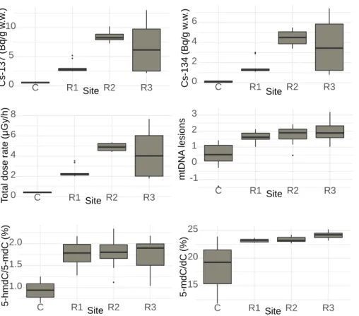
Radiocesium isotopes (134Cs and 137Cs) were detected in frogs sampled in the FDNPP
199
accident impacted area (Figure 2). The highest concentrations were found in frogs of the R3 200
site, with values of activity concentrations going up to 13 and 7.4 Bq g-1 w.w. respectively for
201
137Cs and 134Cs (wet weight, whole body) (Table S2). The average frog’s age was of 3 years at
202
each site (range of 2-5 years) (Table S2). 203
204
Internal DR is the main contributor to the TDR received by frogs. The mean (±SD) TDR 205
at the contaminated sites was higher than the TDR at control site (control site: 0.41±0.04 µGy 206
h-1 ; site R1: 2.4±0.54 µGy h-1 ; site R2: 4.9±0.38 µGy h-1 ; site R3: 4.3±2.40 µGy h-1 (Figure
207
2, Table S2)). Additionally, within each sampling site the internal DR represented the highest 208
contribution to the TDR (58, 67, 90 and 83 % in frogs from the control site, contaminated sites 209
R1, R2 and R3, respectively) (Table S2). 210
211
Hypermethylation of genomic DNA. We quantified the average global levels of both the 212
methylated (5-mdC) and the hydroxymethylated (5-hmdC) forms of cytosine in frogs’ genomic 213
DNA. An increase of the average level of 5-mdC (26 to 31 %) was observed in frogs from the 214
three contaminated sites as compared to the control site (Figure 2). Similarly, a doubling of the 215
level of 5-hmdC was observed in frogs from all three contaminated sites as compared to levels 216
from the control site (1.77, 1.78 and 1.75 % in frogs from sites R1, R2 and R3, respectively, 217
versus 0.94 % in frogs from the control site). 218
219
Increase of Mt DNA damage. In frogs from the control site, the average number of lesions 220
to the Mt DNA was of 0.5 lesions/10.7 kbp (Figure 2). The number of lesions to DNA increased 221
in frogs from the contaminated sites up to 4 times as compared to controls (3.5, 3.7 and 4.2 fold 222
higher than in controls for sites R1, R2 and R3, respectively). 223
224
Correlation between TDR and biological factors on one hand, and global DNA 225
methylation and Mt DNA damages. The relationships between the frog’s TDR and the 226
observed biological responses (DNA methylation and damage) were the most significant among 227
fixed effects (Table 1). The results indicated a significant positive correlation between the frogs’ 228
TDR and both the average level of 5-hmdC (padj < 0.001) and the level of Mt DNA damages
(padj < 0.001). To the contrary, for 5-mdC, the relationship with the frogs’ TDR was not
230
significant (padj = 0.0748). We also observed a significant positive relationship between the
231
average level of 5-hmdC and the frogs’ age (padj < 0.001). Finally, a significant positive
232
relationship was detected between the BCI and the level of 5-mdC (padj = 0.0149).
233 234
DISCUSSION 235
The radiocesium activity concentrations measured in frogs in this study are of a few Bq g-1
236
w.w. These values are in the range of activity concentrations measured in 5 frog species sampled 237
in the Fukushima Prefecture in August and September 2012 at similar air dose rate44. In
238
addition, the Concentration Ratio estimated between water and frogs (CRfrog-water) (Table S1) in
239
our study, ranges from 400 to 1400 L kg-1 w.w., which is in adequacy with the mean CR whole
240
organism-water value estimated at 580 L kg-1 w.w. for adult frogs from the Fukushima Prefecture
241
from April 2012 to April 201645.
242
The mean TDRs absorbed by frogs in the different sampling sites were all lower than the 10 243
µGy h-1 generic screening value that below which 95% of all species should be protected from 244
ionising radiation. The calculation of the TDR highlighted a major contribution of the internal 245
exposure, representing from 58 % to 90% of TDR in contaminated sites. In addition, the 246
calculated TDR is higher than the ambient dose rate. This is an important result since most of 247
the studies carried out in Chernobyl and Fukushima area are based on the ambient DR to assess 248
biological effects4,5,9-14,24,46, which may under-estimate the total dose rate absorbed by wildlife.
249
As already underlined in other works, such comprehensive calculation of total absorbed dose 250
rates is important to assess thresholds of toxicity in wildlife and compare them to toxicity 251
thresholds or benchmarks obtained under laboratory control conditions3,6.
252
253
This study focusing on Japanese tree frogs exposed in situ following the FDNPP accident 254
shows significant positive correlations between TDR, mitochondrial DNA damages and DNA 255
methylation levels in frogs. These molecular responses may be more sensitive than responses 256
observed at higher biological organisation levels. Indeed, in a previous study on five frog 257
species (Rana japonica, R. ornativentris, R. tagoi tagoi, Pelophylax porosus porosus, and 258
Dryophytes japonicus) sampled in the radiocontaminated area around Fukushima, the 259
histological examination of ovaries and testes using conventional microscopy, failed to detect 260
any overt aberrations in the morphology of germ cells in the testes and ovaries of frogs44. To
261
the contrary, chromosomal aberrations and malformed cells in bone marrow were observed in 262
R. temporaria L. in radiocontaminated areas in Belarus in a period of 3-7 years after the 263
Chernobyl accident26. In laboratory experiments, chromosomal aberrations, a decreased
264
reproductive capacity and an increase in male number were also observed in 4 generations of 265
frogs produced from gametes exposed to 1.5-3.5 Gy of X-rays47. These different results could
be explained by several factors, including the different levels (dose and dose rates) and types 267
of radiocontaminants studied. 268
269
Our study highlights a dose-dependent correlation between TDR and 5-hmdC levels, and a 270
strong but not statistically significant trend to cytosine hypermethylation in radiocontaminated 271
area (Table 1). 5hmC is a product of 5mC oxidation during the process of DNA demethylation, 272
but it also plays an important role in gene expression, pluripotency of stem cells, stress response, 273
disease progression and aging48. As such, it is interesting to note that there is also a trend for a
274
positive correlation between 5-hmC and age (Table 1, p=0.0503), underlining the reliability of 275
5hmC as biomarker of aging48. However, as aged frogs may also have been exposed to higher
276
total doses just after the FDNPP accident, it cannot be ruled out that this increase of 5hmC 277
could also reflect the past exposure of frogs. Nonetheless, changes in global DNA methylation 278
have already been observed in several organisms exposed to IR, such as plants29, 48-52 or fish53.
279
Hence, we hypothesize that the changes in methylation levels observed in our data could be 280
implicated in a physiological response or a phenotypic plasticity leading to adaptation of frogs 281
exposed chronically to IR. 282
At the whole-genome scale, hypermethylation of cell cycle and detoxification gene promoters 283
have been observed in offspring of irradiated humans long term after radiation exposure54 while
284
the hypomethylation of gene coding for stress protein hsp70 was observed in offspring of 285
irradiated microcrustaceans55. DNA methylation is used to predict adverse effects in terms of
286
tumour recurrence as some gene-specific DNA methylations are predictors of response to 287
radiotherapy56. Such a whole-genome study of DNA methylation would be useful to better
288
understand the mechanisms involved in the response of Japanese tree frogs to IR. 289
The mechanisms of radiation-induced changes in DNA methylation remain largely unknown 290
but some hypotheses are proposed by Miousse et al. (2018)56 : the most plausible scenario is an
291
effect of IR on DNA methyltransferases, as decreases of DNA methyltransferases’ levels of 292
mRNA and proteins have been observed. Additionally, IR has been shown to affect several 293
microRNAs that specifically target DNA methyltransferases57. Further studies at higher
294
biological organisation levels are needed in order to demonstrate that these processes do occur 295
on wild animals and contribute to phenotypical changes or adaptive process in contaminated 296
area. 297
Our results show a significant positive correlation between the frogs’ TDR, 5-hmdC but also 298
the average number of lesions to the Mt DNA (Table 1). Epigenetic mechanisms and genome 299
stability are closely linked58. Epigenetic marks such as DNA methylation are involved in several
300
aspects that can enhance genetic stability (e.g. transcriptional repression of repetitive elements 301
by DNA methylation preventing homologous recombination or chromatin condensation 302
induced by DNMTs (DNA methyltransferases)). This favourable role of DNA methylation in 303
increasing genomic DNA stability59 has been proposed as a potential mechanism for radiation
adaptation in plants exposed at Chernobyl36,60 or to environmental stress in general61. There is
305
likely an “epigenetic advantage” to phenotypic switching by epigenetic inheritance, rather than 306
by gene mutation, as an epigenetically-inherited trait can arise simultaneously in many 307
individuals62. However, genomic destabilization can also occur through DNA hypermethylation
308
of DNA repair genes, leading to reduced expression of genes required for genetic stability, 309
which has been evidenced in cancer. 310
While the issue of inheritance of epigenetic characters in humans is still a matter of 311
controversy, the transmission of acquired states can occur in plants and animals62,63. As such, it
312
may contribute to evolution61, underlining the importance of integrating the study of
313
(epi)genetic molecular markers in ecological risk assessment, to assess for effects of long term 314
chronic exposure to IR. 315
316 317
318
Figure 1. Map of ambient dose rate (µGy h-1) in the Fukushima area during the sampling period
319
(June and July 2013). The location of the four sites (control site C, contaminated sites R1, R2 320
and R3) are indicated on the map by the black dots. 321
322
324
Figure 2. Boxplots of Cs-137 and Cs-134 activity concentrations, total dose rates, mitochondrial 325
DNA lesions and DNA methylation in frogs from the four sites (control site C, contaminated 326 sites R1, R2 and R3, n=10). 327 0 5 10 C R1 Site R2 R3 Cs -13 7 (Bq /g w .w .) 0 2 4 6 C R1 Site R2 R3 Cs -13 4 (Bq /g w .w .) 0 2 4 6 8 C R1 Site R2 R3 T otal dose rate (µ Gy /h) 15 20 25 C R1 Site R2 R3 5 -m dC /dC (%) 1.0 1.5 2.0 C R1 Site R2 R3 5 -hm dC /5 -m d C (%) -1 0 1 2 3 C R1 Site R2 R3 m tD N A les ions
Table 1: linear mixed-effects models for methylated cytosine, hydroxymethylated cytosine and mitochondrial DNA (MtDNA) lesions. 328
Model Factor Estimate Standard Error df t-value P value
Methylated cytosine (5-mdC) Fixed effects
(Intercept) 23.023434 0.4621987 26 49.81285 <0.0001
Age -0.038076 0.1067110 26 -0.35682 0.9932
Body Condition Index 0.369256 0.1275974 26 2.89391 0.0149
Total Dose Rate 0.715193 0.3085177 26 2.31816 0.0773
Hydroxymethylated cytosine (5-hmdC) Fixed effects
(Intercept) 1.5273975 0.06559786 26 23.284257 <0.001
Age 0.1079019 0.04391403 26 2.457117 0.0513
Body Condition Index 0.0819409 0.06029821 26 1.358928 0.4924
Total Dose Rate 0.2967815 0.05422058 26 5.473595 <0.001
MtDNA lesions Fixed effects
(Intercept) 1.4754382 0.1214342 26 12.150109 <0.0001
Age 0.0323862 0.1246704 26 0.259774 0.998040
Body Condition Index 0.2143823 0.1450854 26 1.477629 0.440104
Total Dose Rate 0.5287266 0.1352902 26 3.908093 <0.001
ASSOCIATED CONTENT
Supporting information. The protocols used to capture frogs and determine radionuclides
concentrations, to calculate the DCs and TDRs, the HPLC-MS/MS and Mt DNA damage methods are provided in supporting information. Raw data corresponding to individual internal,
external and total DRs, HPLC-MS/MS and Mt DNA damage are also provided in
supplementary information.
REFERENCES
1 IRSN. Fukushima, one year later—initial analyses of the accident and its consequences. (Report IRSN/DG/2012-0032012. Institut de Radioprotection et de Surete Nucleaire, 2012).
2 UNSCEAR. Annex A: Levels and effects of radiation exposure due to the nuclear accident after the 2011 great east-Japan earthquake and tsunami. UNSCEAR 2013 report: sources, effects and risks of ionizing radiation. New York: United Nations; 2014. 3 Garnier-Laplace, J., Beaugelin-Seiller, K. & Hinton, T. G. Fukushima Wildlife Dose Reconstruction Signals Ecological Consequences. Environ Sc Technol 45, 5077-5078 (2011).
4 Møller, A. P., Mousseau, T. A., Nishiumi, I. & Ueda, K. Ecological differences in response of bird species to radioactivity from Chernobyl and Fukushima. Journal of Ornithology, 1-10 (2015a).
5 Møller, A. P., Nishiumi, I. & Mousseau, T. A. Cumulative effects of radioactivity from Fukushima on the abundance and biodiversity of birds. Journal of Ornithology, 1-9 (2015b).
6 Garnier-Laplace, J., Beaugelin-Seiller, K., Della-Vedova, C., Métivier, J-M., Ritz, C., Mousseau, T. A., Møller, A. P. Radiological dose reconstruction for birds reconciles outcomes of Fukushima with knowledge of dose-effect relationships. Scientific reports 5, 16594 (2015).
7 Ochiai, K., Hayama, S-I., Nakiri, S., Nakanishi, S., Ishii, N., Uno, T., Kato, T., Konno, F., Kawamoto, Y., Tsuchida, S.,. Low blood cell counts in wild Japanese monkeys after the Fukushima Daiichi nuclear disaster. Scientific reports 4 (2014).
8 Hayama, S.-I. et al. Small head size and delayed body weight growth in wild Japanese monkey fetuses after the Fukushima Daiichi nuclear disaster. Scientific reports 7, 3528 (2017).
9 Hiyama, A. et al. The biological impacts of the Fukushima nuclear accident on the pale grass blue butterfly. Scientific Reports 2, 570, doi:10.1038/srep00570 (2012).
10 Hiyama, A. et al. The Fukushima nuclear accident and the pale grass blue butterfly: evaluating biological effects of long-term low-dose exposures. BMC Evol Biol 13, 1471-2148 (2013).
11 Hiyama, A. et al. Spatiotemporal abnormality dynamics of the pale grass blue butterfly: three years of monitoring (2011–2013) after the Fukushima nuclear accident. BMC Evolutionary Biology 15, 15 (2015).
12 Iwata, M., Hiyama, A. & Otaki, J. M. System-dependent regulations of colour-pattern development: a mutagenesis study of the pale grass blue butterfly. Scientific Reports 3, 2379, doi:10.1038/srep02379 (2013).
13 Nohara, C. et al. Ingestion of radioactively contaminated diets for two generations in the pale grass blue butterfly. BMC Evolutionary Biology 14, 193, doi:10.1186/s12862-014-0193-0 (2014).
14 Taira, W., Nohara, C., Hiyama, A. & Otaki, J. M. Fukushima's biological impacts: the case of the pale grass blue butterfly. J Hered 105, 710-722 (2014).
15 Beresford NA, Horemans N, Copplestone D, et al. Towards solving a scientific controversy - The effects of ionising radiation on the environment. J Environ Radioact. 2020;211:106033. doi:10.1016/j.jenvrad.2019.106033
16 Fuller N, Ford AT, Lerebours A,Gudkov DI, Nagorskaya LL, Smith JT. Chronic radiationexposure at Chernobyl shows no effect on genetic diversityin the freshwater crustacean, Asellus aquaticus thirty yearson. Ecol Evol. 2019;9:10135–10144. https://doi.org/10.1002/ece3.5478
17 Lerebours A, Gudkov D, Nagorskaya L, et al. Impact of Environmental Radiation on the Health and Reproductive Status of Fish from Chernobyl. Environ Sci Technol. 2018;52(16):9442-9450. doi:10.1021/acs.est.8b02378
18 Bonzom JM, Hättenschwiler S, Lecomte-Pradines C, Chauvet E, Gaschak S, Beaugelin-Seiller K, Della-Vedova C, Dubourg N, Maksimenko A, Garnier-Laplace J, Adam-Guillermin C (2016). Effects of radionuclide contamination on leaf litter decomposition in the Chernobyl exclusion zone. Science of The Total Environment 562: 596-603 19 Lecomte-Pradines C, Bonzom JM, Della-Vedova C, Beaugelin-Seiller K, Villenave C5,
Gaschak S, Coppin F, Dubourg N, Maksimenko A, Adam-Guillermin C, Garnier-Laplace J (2014). Soil nematode assemblages as bioindicators of radiation impact in the Chernobyl Exclusion Zone Sci Total Environ. 2014 Aug 15;490:161-70. doi: 10.1016
20 Parisot, F., Bourdineaud, J.-P., Plaire, D., Adam-Guillermin, C. & Alonzo, F. DNA alterations and effects on growth and reproduction in Daphnia magna during chronic exposure to gamma radiation over three successive generations. Aquatic Toxicology 163, 27-36, doi:http://dx.doi.org/10.1016/j.aquatox.2015.03.002 (2015).
21 Bonisoli-Alquati, A. et al. DNA damage in barn swallows (Hirundo rustica) from the Chernobyl region detected by use of the comet assay. Comparative Biochemistry and Physiology Part C: Toxicology & Pharmacology 151, 271-277, doi:http://dx.doi.org/10.1016/j.cbpc.2009.11.006 (2010).
22 Ellegren, H., Lindgren, G., Primmer, C. R. & Møller, A. P. Fitness loss and germline mutations in barn swallows breeding in Chernobyl. Nature 389, 593-596 (1997). 23 Møller, A. P. & Mousseau, T. A. Strong effects of ionizing radiation from Chernobyl
24 Pomerantseva, M. D., Ramaiya, L. K. & Chekhovich, A. V. Genetic disorders in house mouse germ cells after the Chernobyl catastrophe. Mutation Research/Fundamental and Molecular Mechanisms of Mutagenesis 381, 97-103 (1997).
25 Ryabokon, N. I. & Goncharova, R. I. Transgenerational accumulation of radiation damage in small mammals chronically exposed to Chernobyl fallout. Radiation and environmental biophysics 45, 167-177 (2006).
26 Geras'kin, S. A., Fesenko, S. V. & Alexakhin, R. M. Effects of non-human species irradiation after the Chernobyl NPP accident. Environment International 34, 880-897 (2008).
27 Theodorakis, C.W., Shugart, L.R., 1997. Genetic ecotoxicology .2. population genetic structure in mosquitofish exposed in situ to radionuclides. Ecotoxicology 6 (6), 335e354. https://doi.org/10.1023/a:1018695231565.
28 Theodorakis, C.W., Bickham, J.W., Lamb, T., Medica, P.A., Lyne, T.B., 2001. Integration of genotoxicity and population genetic analyses in kangaroo rats (Dipodomys merriami) exposed to radionuclide contamination at the Nevada Test Site, USA. Environ. Toxicol. Chem. 20 (2), 317e326. https://doi.org/10.1897/1551-5028(2001)020<0317:iogapg>2.0.co;2.
29 Kovalchuk, O. et al. Genome hypermethylation in Pinus silvestris of Chernobyl--a mechanism for radiation adaptation? Mutat Res 529, 13-20 (2003).
30 Russo, V. E. A., Martienssen, R. A. & Riggs, A. D. Epigenetic Mechanisms of Gene Regulation. (1996).
31 Bird, A. P. The essentials of DNA methylation. Cell 70, 5-8 (1992).
32 Attwood, J. T., Yung, R. L. & Richardson, B. C. DNA methylation and the regulation of gene transcription. Cell Mol Life Sci 59, 241-257 (2002).
33 Guo, J. U., Su, Y., Zhong, C., Ming, G.L., Song, H. Hydroxylation of 5-methylcytosine by TET1 promotes active DNA demethylation in the adult brain. Cell 145, 423-434, doi:10.1016/j.cell.2011.03.022 (2011).
34 Robertson, K. D. & Jones, P. A. DNA methylation: past, present and future directions. Carcinogenesis 21, 461-467 (2000).
35 Rando, O. J. & Verstrepen, K. J. Timescales of genetic and epigenetic inheritance. Cell 128, 655-668 (2007).
36 Kovalchuk, I., Abramov, V., Pogribny, I. & Kovalchuk, O. Molecular aspects of plant adaptation to life in the Chernobyl zone. Plant Physiol 135, 357-363, doi:10.1104/pp.104.040477 (2004).
37 Pogribny, I., Raiche, J., Slovack, M. & Kovalchuk, O. Dose-dependence, sex- and tissue-specificity, and persistence of radiation-induced genomic DNA methylation changes. Biochem Biophys Res Commun 320, 1253-1261, doi:10.1016/j.bbrc.2004.06.081 (2004).
38 Castanet, J. and E. Smirina, Introduction to the skeletochronology method in amphibians and reptiles. Annales des Sciences Naturelles (Zoologie), 1990. 11 p. 191– 196.
39 Smirina, E.M., Age determination and longevity in amphibians. Gerontology 1994. 40: p. 133-146.
40 Pereira, S. et al. Low doses of gamma-irradiation induce an early bystander effect in zebrafish cells which is sufficient to radioprotect cells. PLoS One 9, e92974, doi:10.1371/journal.pone.0092974 (2014).
41 Ravanat, J. L., Douki, T., Duez, P., Gremaud, E., Herbert, K., Hofer, T., Lasserre, L., Saint-Pierre, C., Favier, A., Cadet, J. Cellular background level of 8-oxo-7,8-dihydro-2'-deoxyguanosine: an isotope based method to evaluate artefactual oxidation of DNA during its extraction and subsequent work-up. Carcinogenesis 23, 1911-1918 (2002). 42 Hunter, S. E., Jung, D., Di Giulio, R. T. & Meyer, J. N. The QPCR assay for analysis
of mitochondrial DNA damage, repair, and relative copy number. Methods 51, 444-451 (2010).
43 Santos, J. H., Meyer, J., Mandavilli, B. S. & Van Houten, B. in DNA Repair Protocols Vol. 314 Methods in Molecular Biology™ (ed DarylS Henderson) Ch. 14, 183-199 (Humana Press, 2006).
44 Matsushima, N., Ihara, S., Takase, M. et al. Assessment of radiocesium contamination in frogs 18 months after the Fukushima Daiichi nuclear disaster. Sci Rep 5, 9712 (2015). 45 Tagami, K., Uchida, S., Wood, M.D. et al. Radiocaesium transfer and radiation exposure
46 Wickliffe, J. K. et al. Mitochondrial DNA heteroplasmy in laboratory mice experimentally enclosed in the radioactive Chernobyl environment. Radiation research 159, 458-464 (2003).
47 Nishioka, M. Abnormalities in the descendants of Rana japonica produced from irradiated eggs or sperm. Sci. Rep. Lab. Amphib. Biol. 3, 189-384 (1978).
48 Shi, D.-Q., Ali, I., Tang, J. & Yang, W.-C. New Insights into 5hmC DNA Modification: Generation, Distribution and Function. Frontiers in Genetics 8, doi:10.3389/fgene.2017.00100 (2017).
49 Georgieva, M., Rashydov, N. M. & Hajduch, M. DNA damage, repair monitoring and epigenetic DNA methylation changes in seedlings of Chernobyl soybeans. DNA Repair 50, 14-21, doi:https://doi.org/10.1016/j.dnarep.2016.12.002 (2017).
50 Horemans, N. et al. Genome-wide DNA methylation changes in two Brassicaceae species sampled alongside a radiation gradient in Chernobyl and Fukushima. Journal of
Environmental Radioactivity 192, 405-416,
doi:https://doi.org/10.1016/j.jenvrad.2018.07.012 (2018).
51 Ou, X. et al. Spaceflight induces both transient and heritable alterations in DNA methylation and gene expression in rice (Oryza sativa L.). Mutation Research/Fundamental and Molecular Mechanisms of Mutagenesis 662, 44-53, doi:https://doi.org/10.1016/j.mrfmmm.2008.12.004 (2009).
52 Volkova, P. Y. et al. Chronic radiation exposure as an ecological factor: Hypermethylation and genetic differentiation in irradiated Scots pine populations.
Environmental Pollution 232, 105-112,
doi:https://doi.org/10.1016/j.envpol.2017.08.123 (2018).
53 Kamstra, J. H. et al. Ionizing radiation induces transgenerational effects of DNA methylation in zebrafish. Scientific Reports 8, 15373, doi:10.1038/s41598-018-33817-w (2018).
54 Kuzmina, N. S., Lapteva, N. S. & Rubanovich, A. V. Hypermethylation of gene promoters in peripheral blood leukocytes in humans long term after radiation exposure. Environmental Research 146, 10-17, doi:https://doi.org/10.1016/j.envres.2015.12.008 (2016).
55 Trijau, M. et al. Transgenerational DNA Methylation Changes in Daphnia magna Exposed to Chronic γ Irradiation. Environmental Science & Technology 52, 4331-4339, doi:10.1021/acs.est.7b05695 (2018).
56 Miousse, I. R., Kutanzi, K. R. & Koturbash, I. Effects of ionizing radiation on DNA methylation: from experimental biology to clinical applications. International journal of radiation biology 93, 457-469, doi:10.1080/09553002.2017.1287454 (2017). 57 Zinovkina, L. A. Mechanisms of Mitochondrial DNA Repair in Mammals.
58 Putiri, E. L. & Robertson, K. D. Epigenetic mechanisms and genome stability. Clinical epigenetics 2, 299-314, doi:10.1007/s13148-010-0017-z (2010).
59 O'Hagan, H. M. Chromatin modifications during repair of environmental exposure-induced DNA damage: A potential mechanism for stable epigenetic alterations. Environ. Mol. Mutagen. 55, 278-291, doi:10.1002/em.21830 (2014).
60 Bräutigam, K. et al. Epigenetic regulation of adaptive responses of forest tree species to the environment. Ecology and Evolution 3, 399-415, doi:doi:10.1002/ece3.461 (2013). 61 Burggren, W. Epigenetic Inheritance and Its Role in Evolutionary Biology:
Re-Evaluation and New Perspectives. Biology 5, 24 (2016).
62 Heard, E. & Martienssen, Robert A. Transgenerational Epigenetic Inheritance: Myths and Mechanisms. Cell 157, 95-109, doi:https://doi.org/10.1016/j.cell.2014.02.045 (2014).
63 Horsthemke, B. A critical view on transgenerational epigenetic inheritance in humans. Nature Communications 9, 2973, doi:10.1038/s41467-018-05445-5 (2018).
ACKNOWLEDGMENTS
This project was supported by the European project COMET (7th PCRD EURATOM, contract number: Fission-2012-3.4.1-604794), the French Institute of Radioprotection and