HAL Id: hal-01956104
https://hal.archives-ouvertes.fr/hal-01956104
Submitted on 14 Dec 2018
HAL is a multi-disciplinary open access
archive for the deposit and dissemination of
sci-entific research documents, whether they are
pub-lished or not. The documents may come from
teaching and research institutions in France or
abroad, or from public or private research centers.
L’archive ouverte pluridisciplinaire HAL, est
destinée au dépôt et à la diffusion de documents
scientifiques de niveau recherche, publiés ou non,
émanant des établissements d’enseignement et de
recherche français ou étrangers, des laboratoires
publics ou privés.
Distributed under a Creative Commons Attribution - NonCommercial - NoDerivatives| 4.0
International License
To cite this version:
Mateusz Pallach, Flaviana Di Lorenzo, Fabio Alessandro Facchini, Djamel Gully, Eric Giraud,
et al..
Structure and inflammatory activity of the LPS isolated from Acetobacter pasteurianus
CIP103108. International Journal of Biological Macromolecules, Elsevier, 2018, 119, pp.1027-1035.
�10.1016/j.ijbiomac.2018.08.035�. �hal-01956104�
Structure and in
flammatory activity of the LPS isolated from Acetobacter
pasteurianus CIP103108
Mateusz Pallach
a, Flaviana Di Lorenzo
a, Fabio Alessandro Facchini
b, Djamel Gully
d, Eric Giraud
d,
Francesco Peri
b, Katarzyna A. Duda
c,e, Antonio Molinaro
a, Alba Silipo
a,⁎
a
Department of Chemical Sciences, University of Napoli Federico II, Complesso Universitario Monte Sant'Angelo, Via Cintia 4, I-80126 Napoli, Italy
b
Department of Biotechnology and Biosciences, University of Milano-Bicocca, Piazza della Scienza 2, 20126 Milano, Italy
c
Junior Group of Allergobiochemistry, Research Center Borstel, Leibniz Lung Center, 23845 Borstel, Germany
d
IRD, Laboratoire des Symbioses Tropicales et Méditerranéennes (LSTM), UMR IRD/SupAgro/INRA/UM2/CIRAD, TA-A82/J– Campus de Baillarguet, 34398 Montpellier Cedex 5, France
eAirway Research Center North (ARCN), German Center for Lung Research, Germany
a b s t r a c t
a r t i c l e i n f o
Article history: Received 16 May 2018
Received in revised form 6 August 2018 Accepted 7 August 2018
Available online 8 August 2018
Acetobacter pasteurianus is an acetic acid-producing Gram-negative bacterium commonly found associated with plants and plant products and widely used in the production of fermented foods, such as kefir and vinegar. Due to the acid conditions of the bacterium living habitat, uncommon structural features composing its cell envelope are expected. In the present work we have investigated the A. pasteurianus CIP103108 lipopolysaccharide (LPS) structure and immunoactivity. The structure of the lipid A and of two different O-polysaccharides was assessed. Furthermore, immunological studies with human cells showed a low immunostimulant activity of the isolated LPS, in addition to a slight capability to lower the NF-kB activation upon stimulation by toxic LPS.
© 2018 The Authors. Published by Elsevier B.V. This is an open access article under the CC BY-NC-ND license (http:// creativecommons.org/licenses/by-nc-nd/4.0/). Keywords: Lipopolysaccharides (LPS) Gram-negative bacteria Innate immunity MD2-TLR4 Acetobacter pasteurianus 1. Introduction
Gram-negative bacteria can be found in different habitats, many used within human activities as production and processing of tradi-tional foods and beverages, in biotechnology and genetic engineering, in the production of pharmaceuticals [1]. A fascinating example is given by Acetobacter pasteurianus, an acetic acid-producing bacterium [2,3] commonly found associated with plants and plant products and present on sugar-rich substrates such as fruits,flowers and vegetables. A. pasteurianus is used in the production of fermented foods, such as kefir and vinegar [4] and, together with other microorganisms such as Aspergillus oryzae, Saccharomyces cerevisiae and lactic acid bacteria, is used in production process of Japanese black rice vinegar, called kurozu [5], whose consumption is believed to carry significant benefits to human health in cancer prevention and relief of hypertension [6].
A. pasteurianus, as most Gram-negative bacteria, possesses an outer membrane which predominantly contains lipopolysaccharides (LPS) on the outer leaflet [7]. The LPS is an amphiphilic molecule typically
constituted by three different domains in the so called“smooth” form (S-LPS): the glycolipid anchoring the LPS to the outer membrane is called lipid A and is commonly made up of a variously acylated bis-phosphorylated glucosamine disaccharide backbone [8,9]. A core oligo-saccharide (core OS) is attached to the lipid A through the 3-deoxy-D -manno-oct-2-ulosonic acid (Kdo), a sugar marker of Gram-negative bacteria. To the core OS is, in turn, connected a polysaccharide termed O-specific polysaccharide or O-chain [10]. Being the most exposed moi-ety of the LPS, the O-chain is in direct contact with the surrounding en-vironment and is involved in interaction with host cells in case of pathogen and symbiont bacteria [11,12].
The LPS is classified as a microbe associated molecular pattern (MAMP) and it can activate the innate immune response following its recognition by immune receptors referred to as pattern recognition re-ceptors (PRR) [8]. In mammals, LPS is recognized by the Toll-like Recep-tor 4/Myeloid differentiation facRecep-tor-2 (TLR4/MD-2) recepRecep-tor complex [13,14]. The center of LPS immunoactivity lies in the lipid A portion, spe-cifically bound by the TLR4/MD-2 complex and differently activating the production of pro-inflammatory cytokines depending on its fine struc-ture [8]. This event is beneficial to the host, enhancing resistance to in-fecting microbes, nonetheless excessive activation of TLR4/MD-2 signalling by agonistic LPS leads to massive and uncontrolled cytokines releasefinally leading to septic shock and multi-organ failure. Intrigu-ingly, some lipid A showed a weak or no immunopotency and, in
⁎ Corresponding author.
E-mail addresses:mateusz.pallach@unina.it(M. Pallach),flaviana.dilorenzo@unina.it
(F. Di Lorenzo),f.facchini@campus.unimib.it(F.A. Facchini),djamel.gully@ird.fr(D. Gully),
eric.giraud@ird.fr(E. Giraud),francesco.peri@unimib.it(F. Peri),kduda@fz-borstel.de
(K.A. Duda),molinaro@unina.it(A. Molinaro),silipo@unina.it(A. Silipo).
https://doi.org/10.1016/j.ijbiomac.2018.08.035
0141-8130/© 2018 The Authors. Published by Elsevier B.V. This is an open access article under the CC BY-NC-ND license (http://creativecommons.org/licenses/by-nc-nd/4.0/).
Contents lists available atScienceDirect
International Journal of Biological Macromolecules
j o u r n a l h o m e p a g e :h t t p : / / w w w . e l s e v i e r . c o m / l o c a t e / i j b i o m a cing interesting inhibitory effects against potent toxic LPS. 2. Experimental
2.1. Extraction and purification of A. pasteurianus CIP103108 LPS Dried A. pasteurianus CIP103108 cells were washed with ethanol, water and acetone. Afterwards, the hot phenol/water procedure was employed to extract the LPS material [22]. After dialysis against distilled water, each phase, namely the water phase (WP) and phenol phase (PP) underwent enzymatic digestion using DNAse, RNAse and proteinase K. WP and PP were analyzed by 14% sodium deoxycholate-polyacrylamide gel electrophoresis (DOC-PAGE) analysis followed by silver nitrate gel staining. A size exclusion chromatography on a Sephacryl S200 column in denaturing conditions (0.25% DOC at pH 8) was performed to further purify the LPS material. Fractions were lyoph-ilized and DOC was removed by ethanol washing, followed by extensive dialysis against distilled water. Each fraction was analyzed by sodium dodecyl sulfate-polyacrylamide gel electrophoresis (SDS-PAGE). 2.2. Monosaccharide compositional analysis of A. pasteurianus CIP103108 LPS
The neutral sugar constitution was established using acetylated alditols method [23]. An aliquot of sample was hydrolyzed using 2 M TFA solution at 120 °C for 120 min. The so obtained monosaccharides were reduced to alditols using NaBH4and then acetylated twice with
equal amounts of acetic anhydride in pyridine at 85 °C for 10 min. The acetylated alditols were analyzed by gas-liquid chromatography/mass spectrometry (GLC-MS). For quantification 3 μg of xylose as internal standard was added to the sample. Amino sugars were characterized using 16 h hydrolysis in 4 M HCl at 100 °C, followed by reduction and acetylation as described above. The uronic acids composition was char-acterized using the same method as neutral sugars but preceded by ad-ditional reduction of the carboxyl group with NaBD4. Ulosonic acids
were identified using acetylated methyl glycosides method [23]. The sample underwent 16 h methanolysis with 1.25 M HCl/MeOH at 85 °C, therefore was acetylated as described above. Linkage analysis was performed using Ciucanu-Kerek's method [24]. An aliquot of LPS was suspended in DMSO and methylated with iodomethane in presence of NaOH. Therefore, the product was hydrolyzed with 2 M TFA for 2 h at 120 °C, reduced with NaBD4and acetylated with acetic anhydride in
pyridine. Partially methylated acetylated alditols were analyzed by GC–MS.
2.3. Fatty acid compositional analysis of A. pasteurianus CIP103108 LPS Total fatty acid content was characterized as fatty acid methyl esters. An aliquot of LPS was hydrolyzed using 4 M HCl at 100 °C for 4 h. After the sample was neutralized with 5MNaOH for 30 min at 100 °C. Liber-ated fatty acids were extracted with chloroform and methylLiber-ated using diazomethane at room temperature. In order to elucidate the position of hydroxyl group of hydroxy fatty acids 2 h derivatization with N,O-bis-(trimethylsilyl)trifluoroacetamide (BSTFA) at 65 °C was performed. Ester linked fatty acid content was characterized using 30 min hydroly-sis with 0.5MNaOH in water:methanol solution (1:1) at 100 °C for an
the A. pasteurianus CIP103108 LPS, a mild acid hydrolysis with acetate buffer (pH 4.4) for 5 h at 100 °C was performed. The glycolipid part was separated from the hydrophilic carbohydrate part using Blight-Dyer's method [25]. Briefly, a mixture of methanol and chloroform was added to the sample to reach a ratio of chloroform/methanol/hy-drolysate 2:2:1.8 (v/v/v). The mixture was shaken and centrifuged. The water-soluble phase was then applied on a GE Healthcare Life Sci-ences Superdex 75 column eluted with 50 mM ammonium bicarbonate. The chromatography fractions were then monitored by NMR spectros-copy. The lipid A part was also collected, lyophilized and analyzed by MALDI MS.
2.5. MALDI-TOF MS and MS/MS
Reflectron MALDI-TOF MS and MS/MS analysis were performed on an ABSCIEX TOF/TOF™ 5800 Applied Biosystems mass spectrometer equipped with an Nd:YLF laser with aλ of 345 nm, a b500-ps pulse length and a repetition rate of up to 1000 Hz. The lipid A fraction was dissolved in CHCl3/MeOH (1:1, v/v) as previously described [26,27].
The matrix was the trihydroxyacetophenone (THAP) dissolved in CH3OH/0.1% TFA/CH3CN (7:2:1, v/v/v) at a concentration of
75 mg ml−1. Lipid A solution and the matrix solution, both 0.5μl, were deposited on the MALDI plate and dried at room temperature. All of spectra were a result of the accumulation of 1500 laser shots, whereas 6000–7000 shots were summed for the MS/MS data acquisi-tions [28].
2.6. NMR spectroscopy
For structural assignments of isolated polysaccharides1H NMR and
2D NMR spectra were recorded in D2O at 278 K at pD 7 with a
cryoprobe-equipped Bruker 600 DRX spectrometer. Total correlation spectroscopy (TOCSY) experiments were performed with spinlock times of 100 ms using data sets (t1 × t2) of 4096 × 512 points. Rotating-frame Overhauser enhancement spectroscopy (ROESY) and nuclear Overhauser enhancement spectroscopy (NOESY) experiments were performed using data sets (t1 × t2) of 4096 × 512 points with mixing times between 100 and 400 ms. Double-quantum-filtered phase-sensitive correlation spectroscopy (DQF-COSY) experiments were performed using data sets of 4096 × 912 points. The data matrix in all homonuclear experiments was zero-filled in both dimensions to give a matrix of 4 K × 2 K points and was resolution-enhanced in both dimensions using a cosinebell function before Fourier transformation. Coupling constants were determined by 2D phase-sensitive DQF-COSY measurements [29,30]. Heteronuclear single-quantum coherence (HSQC) and heteronuclear multiple-bond correlation (HMBC) experi-ments were performed in the1H-detection mode by single-quantum
coherence with proton decoupling in the13C domain using data sets of 2048 × 400 points. HSQC was performed using sensitivity improve-ment and in the phasensitive mode using echo/antiecho gradient se-lection, with multiplicity editing during the selection step [31]. HMBC was optimized on long-range coupling constants, with a low-pass Jfilter to suppress one-bond correlations, using gradient pulses for selection. Moreover, a 60 ms delay was used for the evolution of long-range corre-lations. HMBC spectra were optimized for 6–15 Hz coupling constants. The data matrix in all the heteronuclear experiments was extended to
2048 × 1024 points by using a forward linear prediction extrapolation [32].
2.7. TLR4 activation assay with HEK-Blue hTLR4 cells
HEK-Blue hTLR4 cells were cultured in DMEM high glucose medium supplemented with 10% fetal bovine serum (FBS), 2 mM glutamine, an-tibiotics and 1× HEK-Blue™ Selection (Invivogen). Cells were detached using a cell scraper, counted and seeded in a 96-well multiwell plate at a density of 4 × 10⁴cells per well. After overnight incubation (37 °C, 5%
CO2, 95% humidity), supernatants were replaced with only DMEM w/o
Phenol Red. The activation assay was conducted stimulating cells with different concentrations (0.1, 1, 10, 100 ng/ml) of A. pasteurianus CIP103108 LPS or E. coli O55:B5 LPS (Sigma-Aldrich) for 16 h. While, the competition assay was performed pre-incubating cells with A. pasteurianus CIP103108 LPS (0.1, 1, 10, 100 ng/ml) for 30 min and then adding cells 1 ng/ml of E. coli O55:B5 LPS. After 16 h of incubation SEAP-containing supernatants were collected and incubated with paranitrophenylphosphate (pNPP) for 2–4 h in the dark at room tem-perature. The wells optical density (OD) was determined using a micro-plate reader set to 405 nm. Regarding activation assay, SEAP levels (OD at 405 nm) were used as indicator of TLR4 pathway activation. For the competition assay, data were normalized with respect to E. coli LPS stimulation and represented as fold decrease.
3. Results
3.1. LPS isolation, purification and compositional analysis
The LPS of A. pasteurianus CIP103108 was isolated using the hot phenol-water procedure [22]; DOC-PAGE analysis revealed the pres-ence of a mixture of R-type LPS (the O-polysaccharide lacking form) and S-type LPS in the extract (Fig. S1). Purification steps, including enzy-matic digestion followed by size exclusion chromatography in denatur-ing conditions, allowed to isolate S-type LPS and R-type LPS (Fig. S1). The sugar compositional analysis of the fraction containing S-LPS re-vealed the occurrence ofL-rhamnose (Rha),D-mannose (Man),D -glucose (Glc),D-galactose (Gal), and, in minor amount, ofD-glucuronic acid (GlcA), D-glucosamine (GlcN), 2,3-diamino-2-3-dideoxy-D
-glucose (DAG), 3-deoxy-D-manno-oct-2-ulosonic acid (Kdo) andD -glycero-D-talo-oct-2-ulosonic acid (Ko). The sugar compositional analy-sis of isolated lipid A showed presence of GlcA, Man, GlcN, DAG and Ko. The compositional analysis in R-LPS revealed the presence of Rha, GlcA, Man, Glc, GlcN, DAG, Kdo and Ko. Methylation analysis of LPS [24] re-vealed the presence of 2,4-disubstituted Rhap, substituted Rhap, 2-substituted Manp, terminal Glcp, 6-2-substituted-Glcp and 2,6-disubsti-tuted and 3-substi2,6-disubsti-tuted Galf. The following fatty acids were also found: 14:0 (3-OH), 18:0 (3-OH) and 16:0. A selective O-deacylation [23] revealed presence of 14:0(3-OH) and 16:0, thus suggesting that 18:0 (3-OH) was the only N-linked acyl chain.
3.2. Isolation, purification of lipid A and O-polysaccharide fractions An aliquot of pure S-LPS underwent a mild acid hydrolysis in order to separate the lipid A moiety from the sugar part, therefore the O-chain containing fraction was recovered and further purified using gel filtration chromatography. NMR spectra and compositional analysis re-vealed the presence of two polysaccharide containing fractions, PS1 and PS2 (Fig. 1). PS2 was elutedfirst and thus possessed higher molecular mass compared to PS1. The two fractions were characterized as follows. 3.3. Structural characterization ofPS1 and PS2
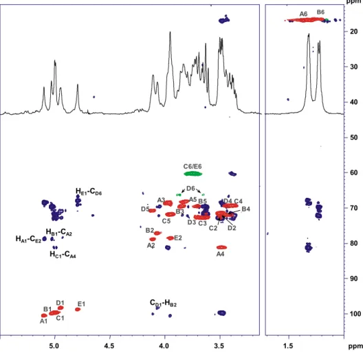
The spin systems and monosaccharide sequence were characterized using a combination of 1D and 2D NMR spectra. The anomeric con figu-ration of the monosaccharides were assigned on the basis of the3J
H1,H2
and1J
C1,H1coupling constants and confirmed by the intra-residual NOE
contacts; the vicinal3J
H,Hcoupling constants and intra residual NOE
con-tacts revealed the relative configuration of the sugar residues. The anomeric region of PS1 revealed the presence of four different anomeric signals (Fig. 1a,Table 1). Spin systems A, B and D at 5.21, 5.07 and 4.97 ppm were identified as β-D-Galf units. The downfield
shift of all carbon signals, the strong downfield shift of carbon C-4 and the intra-residual long-range correlation between positions 1 and 4 found in the HMBC spectrum (Fig. 2) were diagnostic of a furanose ring [33]. Theβ-anomeric configuration was assessed on the basis of
13C chemical shift [34]. Spin system C was identified as an α-D-Glcp;
its anomeric configuration was assigned on the basis of 1
JC1,H1
(Table 1) and the3J
H1,H2(b4 Hz) whereas high3JH3,H4(9.1 Hz) and
intra-residual NOE connectivity of H-3 and H-5 confirmed the gluco configuration.
The downfield shift of carbon resonances suggested glycosylated po-sitions at O-2 and O-6 of A, O-6 of B and O-3 of D. Spin system C was characterized as a terminal Glc residue. The inter-residual connectivity (Fig. S2) along with long-range correlations found on the HMBC spec-trum (Fig. 2) of A1/D3, B1/D3, D1/A6 and C1/A2 allowed to define the saccharide sequence. Residue C was found to be not stoichiometrically linked at position O-2 of the galactofuranose residue A, substituted for about 75%.
The NMR spectrum of PS2 unveiled the presence offive anomeric signals (Figs. 1b and3,Table 2). Spin systems A and B, H-1 at 5.10 and 5.03 ppm respectively (Table 2), were characterized as α-rhamnopyranose units. In both cases, correlations of the ring protons with methyl signals were visible from the TOCSY spectrum. The manno configuration of both spin systems was assigned on the basis of
3J
H1,H2and3JH2,H3coupling constants, theα-configuration from
intra-residual NOE contact of H-1 with H-2 and the chemical shift value of C-5. Analogously, spin systems C and D were assigned as α-glucopyranose units while spin system E was identified as α-mannopyranose. The downfield shift of carbon resonances identified the glycosylated positions: 2 and 4 of A, 2 of B, 6 of D and O-2 of E. Unit C was identified as a terminal residue. The inter-residual NOE contacts observed from the ROESY spectrum (Fig. S3) together with the long-range correlations, derived from the HMBC spectrum (Fig. 3), between A1 with E2; E1 with D6, D1 with B2; B1 with A2 so as C1 with A4 confirmed the following repeating unit for the polysac-charide PS2:
3.4. MALDI MS on A. pasteurianus CIP103108 lipid A
A. pasteurianus CIP103108 lipid A structure was investigated by MALDI-MS and MS/MS. The negative-ion MALDI MS spectrum (Fig. 4) revealed the presence of several cluster of peaks in the mass range m/ z 1543.9–2434.7, indicative of lipid A species with a different acylation degree. Each cluster was characterized by the presence of mass differ-ences of 14 amu (–CH2– unit), attributable to lipid A species differing
by the length of their acyl chains (Fig. 4).
In detail and based on the compositional analysis, cluster of peaks at around m/z 2406.7 matched with hexa-acylated lipid A species with a pentasaccharide sugar backbone containing one hexose (identified as Man), one DAG, one GlcN, one acid (identified as GlcA) and one Ko
unit and acylated by two 14:0 (3-OH), two 18:0 (3-OH) and two 16:0, in accordance with previously reported structure on A. pasteurianus NBRC 3283 lipid A [35]. The cluster at m/z 2168.4 was assigned to penta-acylated lipid A species made up of two 14:0 (3-OH), two 18:0 (3-OH) and one 16:0, thereafter, tetra-acylated lipid A species at m/z 1942.2, de-void of one 14:0 (3-OH) with respect to the species at m/z 2168.4, was also identified. Furthermore, tetra-acylated species lacking one hexose and/or Ko units and minor tri-acylated species were also present in the MALDI spectrum (Fig. 4).
MS/MS experiments were performed in order to characterize the lo-cation of the lipid A acyl chains with respect to the saccharide backbone [26,36]. The negative-ion MS/MS spectrum of precursor ion m/z 1942.2
(Fig. S4 and S5), consistent with a tetra-acylated pentasaccharidic spe-cies, showed an intense peak at m/z 1706.1 attributed to an ion origi-nated from the loss of Ko residue. Peaks at m/z 1766.1 and 1780.1 were assigned to fragments lacking the GlcA and the hexose unit re-spectively. A lipid A fragment devoid of the 16:0 moiety was attributed to the peak at m/z 1686.1. More importantly, peaks originating from the sugar ring fragmentations1,3A
3(m/z 1582.5),1,4A3(m/z 1552.5),0,3A3
(m/z 1393.3) and0,4A3(m/z 1362.4) were very informative as they
clearly demonstrated that the proximal GlcN was decorated by a GlcA and an amide-bound 18:0 (3-OH) moiety whereas the distal DAG was substituted by Ko, hexose and acylated by one 18:0 (3-OH), one 14:0 (3-OH) and one secondary 16:0 fatty acid. Finally, the peak at m/z 1450.0 was ascribed to a fragment ion where both Ko and 16:0 were absent.
MS/MS experiments conducted on precursor ion at m/z 2168.4 (Fig. S6) showed an ion peak at m/z 1932.3 originating from the loss of Ko unit, as well as from the loss of the hexose at m/z 2006.3. Lipid A fragments lacking the Ko and one 14:0 (3-OH) (m/z 1688.2) and one 16:0 (m/z 1432.3) were also identified. Moreover, ions originated from the loss of the hexose unit and 16:0 (m/z 1750.3) and from the loss of the hexose and one 14:0 (3-OH) (m/z 1762.3) were also detected. The absence of an ion originating from the loss of a whole hydroxylated 18:0 or 14:0 bearing a 16:0 suggested that the secondary fatty acid was linked to the acyloxyacyl amides, thus excluding its presence on the proximal GlcN as a substituent of the primary ester-bound 14:0 (3-OH). Furthermore, in support of this hypothesis, a peak was found at
Fig. 1.1
H NMR spectra of (a) PS1 and (b) PS2.
Table 1 Proton (1
H) and carbon (13
C) (italic) NMR shifts of A. pasteurianus CIP103108 PS1. Residue 1 2 3 4 5 6 A 2,6-β-D-Galf 5.21 4.08 4.15 3.86 3.91 3.53/3.78 105.2 86.6 75.0 82.3 69.0 69.3 1 JC1,H1 172.3 Hz 3 JH1,H2 3.4 Hz 3 JH2,H3 9.5 Hz 3 JH3,H4 9.4 Hz 3 JH4,H5 9.4 Hz B 6-β-D-Galf 5.07 4.02 3.97 3.84 3.86 3.77/3.50 106.8 81.2 76.6 82.9 69.2 69.3 C t-α-D-Glcp 4.98 3.47 3.61 3.34 3.67 3.77/3.67 98.1 71.1 72.7 69.4 72.3 60.4 D 3-β-D-Galf 4.97 4.21 4.09 4.00 3.80 3.62/3.57 108.1 79.2 82.6 82.0 70.7 62.7
m/z 1984.4 resulting from the loss of 184 amu arising from the primary O-linked 14:0 (3-OH) and promoted by its free 3-OH group. Unfortu-nately, no ions originating from sugar ring fragmentations were de-tected. In addition, MS/MS spectrum of precursor ion at m/z 2406.7 (Fig. S7), relative to a hexa-acylated lipid A species, presented the ion peak derived from the sugar ring fragmentation1,4A
3at m/z 1791.5
that was fundamental to further corroborate the location of the 16:0 residues as secondary acyl substituents of the DAG unit.
Therefore, the lipid A and O-chain structure from A. pasterianus CIP103108 were as reported inFig. 5.
3.5. Immunological studies on A. pasterianus CIP103108 LPS
Studies on immunological properties of A. pasterianus CIP103108 LPS were performed using HEK-Blue hTLR4 cells. These cells are designed for studying the activation of human TLR4 by monitoring the activation of NF-κB and AP-1 transcription factors. HEK-Blue hTLR4 cells are HEK cells transfected in order to stably express hTLR4, hMD-2 and hCD14 ceptor genes and a secreted embryonic alkaline phosphatase (SEAP) re-porter gene placed under the control of NF-κB and AP-1. Stimulation with a TLR4 ligand activates NF-κB and AP-1 inducing the production and secretion of SEAP in cells culture medium. Levels of SEAP can be eas-ily determined incubating the enzyme with para-nitrophenylphosphate (pNPP). In order to evaluate the capacity of A. pasteurianus CIP 103108 LPS to trigger TLR4-mediated signalling (activation assay) cells were ex-posed to different concentrations (0.1, 1, 10, 100 ng/ml) of this LPS var-iant for 16 h. As a positive control E. coli O55:B5 LPS was used. As expected, E. coli O55:B5 LPS strongly activated TLR4-mediated pathway in a dose-dependent manner. On the contrary, the results obtained re-vealed that A. pasteurianus CIP103108 LPS was incapable to trigger the activation of the same pathway (Fig. S8A). Furthermore, the capability of A. pasteurianus CIP103108 LPS to interfere with E. coli LPS-triggered
TLR4-mediated signalling was also evaluated (competition assay). To investigate this aspect, cells were pre-incubated with different concen-tration of A. pasteurianus CIP103108 LPS (0.1, 1, 10, 100 ng/ml) for 30 min and then exposed to E. coli O55:B5 LPS (1 ng/ml) for 16 h. Results showed that the pre-incubation with A. pasteurianus CIP103108 LPS slightly reduced the capacity of cells to respond to E. coli O55:B5 LPS, as demonstrated by the minor activation of the TLR4-mediated pathway (Fig. S8 B).
4. Discussion
In this work we have focused our attention on the structural charac-terization of the LPS isolated from A. pasteurianus CIP103108. Two novel LPS O-chains were found, PS1 and PS2, both possessing structural anal-ogies with those isolated from other Acetobacter strains. The presence of two different O-chains is not considered as a common feature of LPSs, nevertheless this phenomenon was reported among Burkholderia spe-cies [37]. PS1 possessed a Galf disaccharide repeating unit carrying a non-stoichiometric Glc substitution; it showed subtle compositional similarities with the O-chains isolated from two Acetobacter methanolicus strains, MB58/4 [38] and MB70 [39]. Moreover, we have also found that a similar exopolysaccharide was produced by another acidophilic bacterium, Zymomonas mobilis, which was proven to have relevance with the outer milieu of bacterium (manuscript in prepara-tion). The second polysaccharide PS2 contained a pentasaccharide re-peating unit constituted by a tetrasaccharide skeleton containing glucose, mannose and two rhamnose residues, one of which carrying a terminal glucose as an appendage. A similar O-antigen was reported for Acetobacter tropicalis SKU1100 [40], whose backbone also contained two rhamnose residues, although in anα-(1–3) linkage, instead of theα-(1–2) here reported, with one rhamnose further substituted by a glucose residue. Furthermore, other
O-Fig. 2. Overlapped sections of1
H13
C HMBC (blue) and1
H13
C HSQC (red and green) NMR of PS1. The anomeric signals and key inter-residual long range correlations involving sugar res-idues of PS1 are indicated. Letters are as found inTable 1.
polysaccharides possessing structural similarities were also found in Acetobacter diazotrophicus PAL5 [41]. None of the above structures possessed mannose residues, though for A. diazotrophicus PAL5 contained a manno-configured sugar.
Recently, Hashimoto et al. [35] reported about the capability of A. pasteurianus NBRC 3283 LPS to remain stable in acidic conditions for long periods and correlated this capability to its unique lipid A structure.
Indeed, the authors demonstrated that the LPS was characterized by the occurrence of aD-glycero-D-talo-oct-2-ulosonic acid (Ko), in place of
Kdo, as thefirst sugar of the core OS, directly linked to the lipid A do-main. This was explained with the acid-lability of Kdo that would be de-graded in the acid environmental conditions of bacterium life. Thus, the substitution of Kdo with the acid-stable Ko is considered as an adapta-tion phenomenon necessary for bacterial survival. The presence of Ko has been so far described in the core oligosaccharides from Burkholderia, Acinetobacter, Yersinia, and Serratia species [42], although a direct link-age of Ko to the lipid A has so far reported only for Acinetobacter haemolyticus [43], in which the Kdo is almost completely replaced by Ko residues. Furthermore, A. pasteurianus LPS is devoid of phosphate groups and the lipid A is characterized by tetrasaccharide backbone composed of Man-DAG-GlcN-GlcA. Interestingly, lipid A devoid of phos-phate groups and with a similar sugar skeleton were already reported for other plant associated bacteria belonging to the Alphaproteobacteria family, as Rhizobiaceae and Bradyrhizobiaceae. The lipid A isolated from Rhodopseudomonas palustris [19] possessed nearly the same tetrasaccharide skeleton but with a complete DAG skeleton. Bradyrhizobium strains possess instead anα-(1 → 6)-mannose disac-charide linked to the non-reducing DAG [44,45]. Moreover, a DAG-GlcN disaccharide skeleton was previously reported for Campylobacter jejuni [46] while a DAG backbone has been found in the lipid A of other strains like Azorhizobium caulinodans [47], Phyllobacterium trifolii [48], Leptospira interrogans [49], Acidithiobacillus ferrooxidans [50], Thiobacillus [51], Bartonella [21] and Brucella [52]. The occurrence of this phenomenon is explained by the presence of two enzymes, GnnA and GnnB in the lipid A biosynthesis process which are pivotal for syn-thesis of UDP-D-GlcpN3N from UDP-D-GlcpNAc [53].
Fig. 3. Overlapped sections of1H13C HMBC (blue) and1H\\13C HSQC (red and green) NMR of PS2. The anomeric signals and key inter-residual long range correlations involving sugar
residues of PS2 are indicated. Letters found inTable 2.
Table 2
Values of1H and13C (italic) chemical shifts of A. pasteurianus CIP103108 PS2.
Residue 1 2 3 4 5 6 A 2,4-α-L-Rhap 5.10 4.11 3.97 3.49 3.82 1.32 100.5 78.9 68.6 81.2 68.3 16.9 1J C1,H1 173.4 Hz 3J H1,H2b 1 Hz 3J H2,H32.4 Hz 3J H3,H49.02 Hz B 2-α-L-Rhap 5.03 4.07 3.84 3.45 3.71 1.22 99.9 77.3 69.6 72.0 69.6 16.6 1 JC1,H1 172.0 Hz 3 JH1,H2b 1 Hz 3 JH2,H33.0 Hz 3 JH3,H49.5 Hz C t-α-D-Glcp 5.00 3.49 3.63 3.39 3.94 3.75 99.6 71.5 72.8 69.5 71.9 60.2 1 JC1,H1 174.4 Hz 3 JH1,H23.5 Hz 3 JH2,H39.4 Hz 3 JH3,H49.5 Hz D 6-α-D-Glcp 4.95 3.50 3.69 3.43 4.10 3.65/3.88 98.3 71.5 72.8 69.5 70.8 66.4 1 JC1,H1 174.2 Hz 3 JH1,H22.9 Hz 3 JH2,H39.5 Hz 3 JH3,H49.6 Hz E 2-α-D-Manp 4.80 3.95 3.84 3.38 3.63 3.76 98.7 78.6 67.9 69.4 69.2 60.2 1 JC1,H1 173.2 Hz 3 JH1,H2b 1 Hz 3 JH2,H33.5 Hz 3 JH3,H49.5 Hz
The lipid A is considered as the center of toxicity of the LPS. It is well known that this part of the molecule is recognized by the TLR4/MD-2 re-ceptor complex resulting in immunostimulation activity. A widely known fact is that hexa-acylated bis-phosphorylated lipid A produced by E. coli possesses high affinity to TLR4/MD-2 receptor complex, trig-gering powerful innate immune response. Overstimulation by agonistic LPS can deregulate innate immune system signalling,finally effecting in uncontrolled, massive proinflammatory cytokine release. Nevertheless, it was shown that modifications of the lipid A structure can tune and modulate the innate immune response. Within this frame, research
and investigation of natural LPS able to inhibit the TLR4/MD-2 depen-dent signalling is an important and interesting topic.
The results obtained on TLR4/MD-2 mediated NF-kB activation on A. pasteurianus CIP103108 LPS have shown significantly lower response than E. coli O55:B5 LPS. A weak effect of inhibition of the TLR4/MD-2 mediated activation of NF-kB induced by E. coli O55:B5 LPS was also ob-served. The very weak agonist activity may be explained by lipid A structural features as i) the absence of phosphate groups [54], ii) the presence of penta-, tetra- and tri-acylated species, besides the hexa-acylated form, iii) the occurrence of a DAG unit.
Fig. 4. Reflectron MALDI mass spectrum, recorded in negative polarity, of the A. pasteurianus CIP103108 lipid A. Lipid A species are outlined. In the inset, the proposed structural composition of the peak at m/z 2406.7 is listed.
References
[1] M.P. Doyle, L.R. Steenson, Bacteria in Food and Beverage Production, in: J. Meng, E. Rosenberg, E.F. DeLong, S. Lory, E. Stackebrandt, F. Thompson (Eds.), The Prokary-otes, Springer, Berlin, Heidelberg 2013, pp. 241–256.
[2] I.Y. Sengun, S. Karabiyikli, Importance of acetic acid bacteria in food industry, Food Control 22 (5) (2011) 647–656.
[3] Y. Yamada, P. Yukphan, Genera and species in acetic acid bacteria, Int. J. Food Microbiol. 125 (1) (2008) 15–24.
[4] K. Nanda, M. Taniguchi, S. Ujike, N. Ishihara, H. Mori, H. Ono, Y. Murooka, Character-ization of acetic acid bacteria in traditional acetic acid fermentation of rice vinegar (komesu) and unpolished rice vinegar (kurosu) produced in Japan, Appl. Environ. Microbiol. 67 (2001) 986–990.
[5] M. Hashimoto, T. Matsumoto, M. Tamura-Nakano, M. Ozono, S. Hashiguchi, Y. Suda, Characterization of outer membrane vesicles of Acetobacter pasteurianus NBRC3283, J. Biosci. Bioeng. 125 (4) (2017) 425–431.
[6] M. Hashimoto, K. Obara, M. Ozono, M. Furuyashiki, T. Ikeda, Y. Suda, K. Fukase, Y. Fujimoto, H. Shigehisa, Separation and characterization of the immunostimulatory components in unpolished rice black vinegar (kurozu), J. Biosci. Bioeng. 116 (6) (2013) 688–696.
[7] X. Wang, P.J. Quinn, Endotoxins: lipopolysaccharides of gram-negative bacteria, in: X. Wang, P.J. Quinn (Eds.), Endotoxins: Structure, Function and Recognition, Sub-cellular Biochemistry, 53, Springer, Dordrecht Heidelberg London New York 2010, pp. 3–25.
[8] A. Molinaro, O. Holst, F. Di Lorenzo, M. Callaghan, A. Nurisso, G. D'Errico, A. Zamyatina, F. Peri, R. Berisio, R. Jerala, J. Jiménez-Barbero, A. Silipo, S. Martín-Santamaría, Chemistry of lipid A: at the heart of innate immunity, Chem. Eur. J. 21 (2) (2015) 500–519.
[9] F. Di Lorenzo, J.M. Billod, S. Martín-Santamaría, A. Silipo, A. Molinaro, Gram negative extremophile lipopolysaccharides: promising source of inspiration for a new gener-ation of endotoxin antagonists, Eur. J. Org. Chem. (2017) 4055–4073.
[10]A. Silipo, A. Molinaro, The diversity of the core oligosaccharide in lipopolysaccha-rides, in: X. Wang, P.J. Quinn (Eds.), Endotoxins: Structure, Function and Recogni-tion, Sub-cellular Biochemistry, 53, Springer, Dordrecht Heidelberg London New York 2010, pp. 69–99.
[11] M. Caroff, D. Karibian, Structure of bacterial lipopolysaccharides, Carbohydr. Res. 338 (23) (2003) 2431–2447.
[12]C.R. Raetz, C. Whitfield, Lipopolysaccharide endotoxins, Annu. Rev. Biochem. 71 (2002) 635–700.
[13]K. Miyake, Innate recognition of lipopolysaccharide by Toll-like receptor 4–MD-2, Trends Microbiol. 12 (4) (2004) 186–192.
[14] B.S. Park, J.O. Lee, Recognition of lipopolysaccharide pattern by TLR4 complexes, Exp. Mol. Med. 6 (2013) 45–66.
[15] H. Loppnow, P. Libby, M. Freudenberg, J.H. Krauss, J. Weckesser, H. Mayer, Cytokine induction by lipopolysaccharide (LPS) corresponds to lethal toxicity and is inhibited by nontoxic Rhodobacter capsulatus LPS, Infect. Immun. 58 (11) (1990) 3743–3750.
[16] S. Saitoh, S. Akashi, T. Yamada, N. Tanimura, M. Kobayashi, K. Konno, F. Matsumoto, K. Fukase, S. Kusumoto, Y. Nagai, Y. Kusumoto, A. Kosugi, K. Miyake, Lipid A antag-onist, lipid IVa, is distinct from lipid A in interaction with Toll-like receptor 4 (TLR4)-MD-2 and ligand-induced TLR4 oligomerization, Int. Immunol. 16 (7) (2004) 961–969.
[17] P.M. Lepper, M. Triantafilou, C. Schumann, E.M. Schneider, K. Triantafilou, Lipopoly-saccharides from Helicobacter pylori can act as antagonists for Toll-like receptor 4, Cell. Microbiol. 7 (4) (2005) 519–528.
[18]F. Di Lorenzo, A. Palmigiano, I. Paciello, M. Pallach, D. Garozzo, M.L. Bernardini, V. Cono, M.M. Yakimov, A. Molinaro, A. Silipo, The deep-sea Polyextremophile Halobacteroides lacunaris TB21 rough-type LPS: structure and inhibitory activity to-wards toxic LPS, Mar. Drugs 15 (7) (2017) 201.
[19] F. Di Lorenzo, A. Palmigiano, S. Al Bitar-Nehme, L. Sturiale, K.A. Duda, D. Gully, R. Lanzetta, E. Giraud, D. Garozzo, M.L. Bernardini, A. Molinaro, A. Silipo, The lipid A from Rhodopseudomonas palustris strain BisA53 LPS possesses a unique struc-ture and low immunostimulant properties, Chem. Eur. J. 23 (7) (2017) 3637–3647.
[20] A. Ialenti, P. Di Meglio, G. Grassia, P. Maffia, M. Di Rosa, R. Lanzetta, A. Molinaro, A. Silipo, W.D. Grant, A. Ianaro, A novel lipid A from. Halomonas magadiensis inhibits enteric LPS-induced human monocyte activation, Eur. J. Immunol. 36 (2) (2006) 354–360.
[21]G. Malgorzata-Miller, L. Heinbockel, K. Brandenburg, J.W.M. Van der Meer, M.G. Netea, L.A.B. Joostena, Bartonella quintana lipopolysaccharide (LPS): structure and characteristics of a potent TLR4 antagonist for in-vitro and in-vivo applications, Sci. Rep. 6 (2016) 34221.
magadiensis Strain 21 M1, Eur. J. Org. Chem. 10 (2004) 2263–2271.
[27]F. Di Lorenzo, The lipopolysaccharide lipid A structure from the marine sponge-associated bacterium Pseudoalteromonas sp. 2A, A. Van. Leeuw. J. Microb. 110 (11) (2017) 1401–1412.
[28] L. Sturiale, A. Palmigiano, A. Silipo, Y.A. Knirel, A.P. Anisimov, R. Lanzetta, M. Parrilli, A. Molinaro, D. Garozzo, Reflectron MALDI TOF and MALDI TOF/TOF mass spectrom-etry reveal novel structural details of native lipooligosaccharides, J. Mass Spectrom. 46 (2011) 1135–1142.
[29] U. Piantini, O.W. Sorensen, R.R. Ernst, Multiple quantumfilters for elucidating NMR coupling networks, J. Am. Chem. Soc. 104 (1982) 6800–6801.
[30] M. Rance, O.W. Sørensen, G. Bodenhausen, G. Wagner, R.R. Ernst, K. Wüthrich, Im-proved spectral resolution in COSY1
H NMR spectra of proteins via double quantum filtering, Biochem. Biophys. Res. Commun. 117 (1983) 479–485.
[31]D.J. States, R.A. Haberkorn, D.J. Ruben, A two-dimensional nuclear Overhauser ex-periment with pure absorption phase in four quadrants, J. Magn. Reson. 48 (1982) 286–292.
[32]A.S. Stern, K.B. Li, J.C. Hoch, Modern spectrum analysis in multidimensional NMR spectroscopy: comparison of linear-prediction extrapolation and maximum-entropy reconstruction, J. Am. Chem. Soc. 124 (2002) 1982–1993.
[33] F. Di Lorenzo, I. Paciello, L.L. Fazio, L. Albuquerque, L. Sturiale, M.S. Da Costa, R. Lanzetta, M. Parrilli, D. Garozzo, M.L. Bernardini, A. Silipo, A. Molinaro, Thermophiles as potential source of novel endotoxin antagonists: the full structure and bioactivity of the lipo-oligosaccharide from Thermomonas hydrothermalis, Chembiochem 15 (2014) 2146–2155.
[34] A. Molinaro, V. Piscopo, R. Lanzetta, M. Parrilli, Structural determination of the com-plex exopolysaccharide from the virulent strain of Cryphonectria parasitica, Carbohydr. Res. 337 (19) (2002) 1707–1713.
[35] M. Hashimoto, M. Ozono, M. Furuyashiki, R. Baba, S. Hashiguchi, Y. Suda, K. Fukase, Y. Fujimoto, Characterization of a novel D-Glycero-D-talo-oct-2-ulosonic acid-substituted lipid A moiety in the lipopolysaccharide produced by the acetic acid bac-terium Acetobacter pasteurianus NBRC 3283, J. Biol. Chem. 291 (40) (2016) 21184–21194.
[36] L. Sturiale, D. Garozzo, A. Silipo, R. Lanzetta, M. Parrilli, A. Molinaro, New conditions for matrix assisted laser desorption/ionization mass spectrometry of native bacterial R-type lipopolysaccharides, Rapid Commun. Mass Spectrom. 19 (2005) 1829–1834.
[37]A.D. Vinion-Dubiel, J.B. Goldberg, Lipopolysaccharide of Burkholderia cepacia com-plex, J. Endotoxin Res. 9 (2003) 201–213.
[38] H.D. Grimmecke, U. Mamat, W. Lauk, A.S. Shashkov, Y.A. Knirel, E.V. Vinogradov, N.K. Kochetkov, Structure of the capsular polysaccharide and the O-side-chain of the li-popolysaccharide from Acetobacter methanolicus MB 58/4 (IMET 10945), and of oli-gosaccharides resulting from their degradation by the bacteriophage Acml, Carbohydr. Res. 220 (1991) 165–172.
[39] H.D. Grimmecke, Y.A. Knirel, A.S. Shashkov, B. Kiesel, W. Lauk, M. Voges, Structure of the capsular polysaccharide and the O-side-chain of the lipopolysaccharide from Acetobacter methanolicus MB 70, and of oligosaccharides resulting from their degra-dation by the bacteriophage Acm6, Carbohydr. Res. 253 (1994) 277–282.
[40]I.A.I. Ali, Y. Akakabe, S. Moonmangmee, A. Deeraksa, M. Matsutani, T. Yakushi, M. Yamada, K. Matsushita, Structural characterization of pellicle polysaccharides of Acetobacter tropicalis SKU1100 wild type and mutant strains, Carbohyd. Polym. 86 (2) (2011) 1000–1006.6.
[41] J.O. Previato, C. Jones, M.P. Stephan, L.P.A. Almeida, L. Mendonca-Previato, Structure of the repeating oligosaccharide from the lipopolysaccharide of the nitrogen-fixing bac-terium Acetobacter diazotrophicus strain PAL 5, Carbohyd. Res. 298 (1997) 311–318.
[42] O. Holst, Structure of the Lipopolysaccharide Core Region, in: Y.A. Knirel, M.A. Valvano (Eds.), Bacterial Lipopolysaccharides, Springer-Verlag, Wien 2011, pp. 21–39.
[43] E.V. Vinogradov, S. Müller-Loennies, B.O. Petersen, S. Meshkov, J.E. Thomas-Oates, O. Holst, H. Brade, Structural investigation of the lipopolysaccharide from Acinetobacter haemolyticus strain NCTC 10305 (ATCC 17906, DNA group 4), Eur. J. Biochem. 247 (1) (1997) 82–90.
[44] A. Silipo, G. Vitiello, D. Gully, L. Sturiale, C. Chaintreuil, J. Fardoux, D. Gargani, H.I. Lee, G. Kulkarni, N. Busset, R. Marchetti, A. Palmigiano, H. Moll, R. Engel, R. Lanzetta, L. Paduano, M. Parrilli, W.S. Chang, O. Holst, D.K. Newman, D. Garozzo, G. D'Errico, E. Giraud, A. Molinaro, Covalently linked hopanoid-lipid a improves outer-membrane resistance of a Bradyrhizobium symbiont of legumes, Nat. Commun. 5 (2014) 5106.
[45] I. Komaniecka, A. Choma, B. Lindner, O. Holst, The structure of a novel neutral lipid a from the lipopolysaccharide of Bradyrhizobium elkanii containing three mannose units in the backbone, Chem. Eur. J. 16 (2010) 2922–2929.
[46] A. Van Mourik, L. Steeghs, J. Van Laar, H.D. Meiring, H.J. Hamstra, J.P.M. Van Putten, M.S.M. Wösten, Altered linkage of Hydroxyacyl chains in lipid a of Campylobacter jejuni reduces TLR4 activation and antimicrobial resistance, J. Biol. Chem. 285 (21) (2010) 15828–15836.
[47] A. Choma, I. Komaniecka, A. Turska-Szewczuk, W. Danikiewicz, G. Spolnik, Structure of lipid a from a stem-nodulating bacterium Azorhizobium caulinodans, Carbohydr. Res. 352 (2012) 126–136.
[48]K. Zamlynska, I. Komaniecka, K. Zebracki, A. Mazur, A. Sroka-Bartnicka, A. Choma, Studies on lipid a isolated from Phyllobacterium trifolii PETP02T
lipopolysaccharide, A. Van Leeuw. J. Microb. 110 (2017) 1413–1433.
[49] N.L. Que-Gewirth, A.A. Ribeiro, S.R. Kalb, R.J. Cotter, D.M. Bulach, B. Adler, I.S. Girons, C. Werts, C.R. Raetz, A methylated phosphate group and four amide-linked acyl chains in Leptospira interrogans lipid a the membrane anchor of an unusual lipo-polysaccharide that activates TLR2, J. Biol. Chem. 279 (24) (2004) 25420–25429.
[50] C.R. Sweet, A.A. Ribeiro, C.R. Raetz, Oxidation and transamination of the 3″-position of UDP-N-acetylglucosamine by enzymes from Acidithiobacillus ferrooxidans. Role in the formation of lipid a molecules with four amide-linked acyl chains, J. Biol. Chem. 279 (24) (2004) 25400–25410.
[51] A. Yokota, M. Rodriguez, Y. Yamada, K. Imai, D. Borowiak, H. Mayer, Lipopolysaccha-rides of Thiobacillus species containing lipid A with 2,3-diamino-2,3 dideoxyglucose, Arch. Microbiol. 149 (1987) 106–111.
[52] A.C. Casabuono, C. Czibener, C.M.G. Del Giudice, E. Valguarnera, J.E. Ugalde, A.S. Couto, New features in the lipid a structure of Brucella suis and Brucella abortus lipo-polysaccharide, J. Am. Soc. Mass Spectrom. 28 (12) (2017) 2716–2723.
[53] C.R. Sweet, A.A. Ribeiro, C.R.H. Raetz, Oxidation and transamination of the 3″-posi-tion of UDP-N-acetylglucosamine by enzymes from Acidithiobacillus ferrooxidans role in the formation of lipid a molecules with four amide-linked acyl chains, J. Biol. Chem. 279 (2004) 25400–25410.
[54] E.T. Rietschel, T. Krikae, F.U. Schade, U. Mamat, G. Schmidt, H. Loppnow, A.J. Ulmer, U. Zahringer, U. Seydel, F. Di Padova, M. Schreier, H. Brade, Bacterial endotoxin: mo-lecular relationships of structure to activity and function, FASEB J. 8 (1994) 217–225.
![Synthesis of Bridged Tetrahydrobenzo[b]azepines and Derivatives through an Aza-Piancatelli Cyclization/Michael Addition Sequence](data:image/gif;base64,R0lGODlhAQABAIAAAP///wAAACH5BAEAAAAALAAAAAABAAEAAAICRAEAOw==)