Martin Kapun, Viola Nolte, Thomas Flatt, Christian Schlo¨tterer* Institut fu¨r Populationsgenetik, Vetmeduni Vienna, Vienna, Austria
Abstract
Background:The Drosophila C virus (DCV) is a common and well-studied Drosophila pathogen. Although natural infections are known from Drosophila melanogaster and D. simulans, and artificial infections have been reported from several Drosophila species and other insects, it remains unclear to date whether DCV infections also occur naturally in other Drosophila species.
Methods/Principal Findings: Using reverse transcription PCR, we detected natural infections in six Drosophila species, which have not been previously known as natural hosts. By subsequent Sanger sequencing we compared DCV haplotypes among eight Drosophila host species. Our data suggest that cross-infections might be frequent both within and among species within the laboratory environment. Moreover, we find that some lines exhibit multiple infections with distinct DCV haplotypes.
Conclusions:Our results suggest that the natural host range of DCV is much broader than previously assumed and that cross-infections might be a common phenomenon in the laboratory, even among different Drosophila hosts.
Citation: Kapun M, Nolte V, Flatt T, Schlo¨tterer C (2010) Host Range and Specificity of the Drosophila C Virus. PLoS ONE 5(8): e12421. doi:10.1371/ journal.pone.0012421
Editor: John J. Welch, University of Edinburgh, United Kingdom
Received March 9, 2010; Accepted August 2, 2010; Published August 26, 2010
Copyright: ß 2010 Kapun et al. This is an open-access article distributed under the terms of the Creative Commons Attribution License, which permits unrestricted use, distribution, and reproduction in any medium, provided the original author and source are credited.
Funding: This study was funded through a DOC fellowship of the Austrian Academy of Sciences (http://stipendien.oeaw.ac.at/doc-doktorandinnenprogramm-der-oesterreichischen-akademie-der-wissenschaften) to MK. CS is supported through the Austrian Science Fund (FWF) by grant P19467-B11. The funders had no role in study design, data collection and analysis, decision to publish, or preparation of the manuscript.
Competing Interests: The authors have declared that no competing interests exist. * E-mail: [email protected]
Introduction
The Drosophila C virus (DCV), first isolated from a French Drosophila melanogaster strain in 1972 [1], is one of the best studied Drosophila pathogens [2]. In contrast to the closely related Cricket paralysis virus (CrPV), which is highly similar to DCV in terms of viral morphology, genome size, and gene arrangement and which infects hosts in several insect orders [3,4], the known host range of DCV is much more narrow [2]. DCV naturally infects D. melanogaster [5,6] and D. simulans [3,7] from the melanogaster subgroup, but natural infections in other species are unknown to date [8–10]. To test for susceptibility to DCV, Jousset [11] artificially infected 15 Drosophila species, four other dipterans and two lepidopterans by introducing DCV into the abdominal cavity. In addition to monitoring for increased mortality, the author assayed virus maintenance and multiplication by injecting extracts from all artificially infected species into virus free D. melanogaster. Jousset found differences in virus susceptibility that range from highly increased mortality in 12 Drosophila species (45 strains) to rapid disappearance of DCV in Culex pipiens and Aedes aegypti. While this result establishes that DCV can artificially infect other species in the laboratory, it remains unclear whether such infections also occur naturally. Here we report a survey for the presence of the DCV in eight Drosophila host species (D. melanogaster, D. simulans, D. mauritiana, D. pseudoobscura, D. subobscura, D. virilis, D. americana, and an undefined species of the D. ananassae group), whose most recent common ancestor dates back 63 million years [12]. We find that DCV has a much broader natural host range than previously thought.
Materials and Methods
Prior to RNA extraction, we monitored all Drosophila lines (approximately 2000 strains) from our fly stock collection by eye for DCV infection symptoms (dark, elongated dead larvae, black dead pupae) [13]. We observed that flies kept in vials at room temperature for more than three weeks showed stronger symptoms than flies kept in vials for shorter periods of time. This might be due to higher susceptibility to viral infection caused by stressfully high larval densities, deteriorating food quality, or high viral loads in the food caused by the presence of dead corpses that release viral particles. Based on these initial observations, we chose 67 strains (D. melanogaster: 39; D. simulans: 4; D. mauritiana: 3; D. sechellia: 1; D. yakuba: 2; D. erecta: 1; D. willistoni: 1; D. cf. ananassae: 3; D. cf. ananassae: 1; D. pseudoobscura: 3; D. subobscura: 3; D. virilis: 2; D. americana: 3; D. mojavensis: 1) for molecular characterization of DCV infections because they displayed strong infection symptoms.
RNA was extracted from 5–10 mated females per line using TrifastH (PEQLAB Biotechnologie GMBH, Erlangen, Germany) following the manufacturer’s instructions after homogenizing whole flies with an Ultraturrax disperser (IKAH Werke GmbH & Co. KG, Stauffen, Germany) and resuspending in 20ml RNAse free water. RNA quality was tested on a 2% non-denaturing agarose gel. cDNA was obtained by incubating 3–11ml RNA with RevertAidTMH-Minus M-MulV Reverse Transcriptase (Fermen-tas, Germany) and Primer DCV8 (59-GAAGCACGA-TACTTCTTCCAAACC-39) [14] according to the manufactur-er’s protocol. PCR reactions were performed in a 20ml reaction volume containing 10 pmol of primers (forward: DCV7,
59-AGTATGATTTTGATGCAGTTGAATCTC-39 and reverse: DCV8, 59-GAAGCACGATACTTCTTCCAAACC-39) [14], 2.5 mM MgCl2, 0.2 mM nucleotides, 0.5 U Taq polymerase (Solis BioDyne, Tartu, Estonia) and 0.5–3ml of RT reaction product. A typical PCR consisted of the following steps: initial denaturation for 49 at 94uC; followed by 34 cycles of denaturation for 400 at 94uC, annealing for 400 at 52uC, and elongation for 19 at 72uC; and a final step of 79 at 72uC. This primer pair amplifies coding DNA from the open reading frame 1 (ORF 1) of the DCV genome. The amplicon spans a region from position 943 to position 1467 and is located in a genomic part which contains domains of a helicase protein. In total, we amplified DCV fragments from 39 Drosophila strains.
To gain insights into the genealogical relationship among DCV haplotypes we used Sanger sequencing. ET terminator sequencing chemistry and protocols (GE Healthcare, Little Chalfont, United Kingdom) were used for cycle sequencing of PCR products from 39 strains. For 12 lines (D. melanogaster: 8; D. mauritiana: 1; D. mojavenis: 1; D. pseudoobscura: 1; D. simulans: 1) we were not able to produce high quality sequence data and thus excluded them from further analysis. Reaction products were sequenced on a MegaBace 500 sequencer (GE Healthcare, Little Chalfont, United Kingdom). All sequences are deposited at NCBI GenBank. They are accessible through accession numbers GU983877–GU983885, GU983888–GU983894, GU983896–GU983902, GU983905– GU983906, and GU983908–GU983911.
CodonCodeAligner v. 3.5.2 (http://www.codoncode.com/ aligner/download.htm) was used for editing raw electrophero-grams, assembly of contigs, and for alignment of sequences. To avoid errors caused by lower sequence quality in the proximity of primers in some samples, we restricted our analysis to a 500 bp region that was sequenced in both directions for most of the samples (see File S1). Missing nucleotides due to low sequence quality at the fragment ends of some samples were filled with Ns (,18% of the sequence) to avoid loss of information due to clipping of the whole sequence alignment. Lines that contained heterozygous sites (sites with two overlapping peaks) in the electropherogram at the same position in the forward and reverse sequence were considered to contain more than one viral haplotype. Since consistent differences in peak height on both strands likely reflect variation in the abundance of viral haplotyes, we used this information to infer viral haplotypes. Thus, all sequence variants with low peaks were attributed to one, whereas variants with high peaks were attributed to another haplotype (see Figure S1).
Using jModelTest [15] we found GTR+C+I [16] to be the best fitting substitution model. PhyML [17] was used to calculate an unrooted maximum likelihood tree using the GTR+C+I model with eight discrete rate categories for the C-distribution. We additionally performed a likelihood ratio test to compare the GTR+I and the GTR+C+I models. MacClade version 4.06 [18] was used to estimate the number of character changes along the topology of the maximum likelihood tree as calculated with PhyML. The tree was plotted and edited using Figtree version 1.3.1 (http://tree.bio.ed.ac.uk/software/figtree/). To test for recombination we performed a Phi-Test [19] and used GARD [20] from the datamonkey website (www.datamonkey.org).
Results and Discussion
Using RT-PCR we found natural DCV infections in eight different hosts (D. melanogaster, D. simulans, D. mauritiana, D. pseudoobscura, D. subobscura, D. virilis, D. americana, and an undefined species of the D. ananassae group). Two of these species (D. simulans,
D. melanogaster) are known to be both artificial and natural hosts of DCV [3,5–7,11], and our results confirm that natural infections occur in these species. We also identified two novel natural hosts (D. mauritiana, D. virilis) for which only artificial infections have been reported so far [11]. In addition, we found evidence for four natural hosts (D. pseudoobscura, D. subobscura, D. americana, D. cf. ananassae) which have neither been reported as natural nor artificial hosts before [8–10]. Our results thus suggest that DCV is naturally infecting a much broader range of Drosophila host species than previously thought.
We sequenced 500 bases of the PCR fragment to shed more light onto the origin and diversity of the DCV samples detected in the eight Drosophila species. In 28 samples (including the isolate EB: GenBank accession number NC_001834) we identified 16 distinct haplotypes (Figure 1). In two samples we detected more than a single DCV haplotype. Although infections with multiple DCV isolates have been described before [21,22], we found that multiple infections do not only occur in D. melanogaster, but also in D. subobscura. As expected from the high haplotype diversity, most of the DCV haplotypes in strains with multiple infections were unique. Nevertheless, one DCV haplotype was also detected in other strains. This observation strongly suggests that in a given strain multiple DCV haplotypes are the outcome of several infections and not due to mutations in the host.
To infer the genealogical relationship among the DCV haplotypes, we performed a phylogenetic analysis. Based on the inferred tree, we did not observe any clustering of haplotypes according to their host species (Figure 1), suggesting that DCV lacks host specificity. Under the assumption of common ancestry of the sequences obtained from DCV isolates in our laboratory and the EB isolate deposited in GenBank 11 years ago [23], we estimated an upper bound of the mutation rate (i.e., the number of variable synonymous sites divided by the sequence length times divergence time). We found 3–10 synonymous substitutions between our sequences and the EB isolate (mean: 6.5; standard deviation: 2.33), suggesting an approximate mutation rate/(site x year) of 5.561024 to 1.861023. These values are comparable to estimates for other RNA viruses [24,25], for example for Sigma virus (1.061024[26]), and human Influenza A (2.661023) and B (561024[27]).
Since in our study several DCV isolates were separated from each other by more mutations than from the published isolate, the date of divergence of DCV isolates identified in our laboratory may be similar to the time of split from the EB isolate. Thus, the divergence observed among the DCV isolates might predate the arrival of these isolates in our laboratory and is unlikely be due to recent mutations. Nonetheless, the absence of host specificity and spatial structure in our data strongly suggests that the isolates we detected are lab-specific epidemics, for example originating from cross-infections with previously infected lab strains. Interestingly, Johnson and Christian [22] found geographical clustering among DCV haplotypes in D. melanogaster using PCR-RFLP, but we failed to find any clustering of our samples with respect to their geographical origin (see Figure 1). However, since some of our fly strains have been maintained as lab stocks for a considerable amount of time, we cannot rule out that existing geographic patterns have been erased by cross-infections in our laboratory, a possibility also raised by Johnson and Christian [22].
Since high rate heterogeneities may cause high rates of homoplasy, thus obscuring the true genetic distance, we estimated a, the shape parameter of the C-distribution. A likelihood ratio test based on the GTR and GTR+C models showed that using the C-distribution significantly improved the fit of the model to our dataset (p,0.001, 2DlnL = 27.2504, x20.001,df = 1= 10.83). Using
PhyML we obtained a shape parameter a of 0.101. An a-value as low as 0.1 yields a sharp L-shaped distribution, with many sites being virtually invariable and with few sites that exhibit very high mutation rates [28].
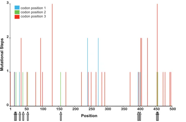
To gain further insight into the distribution of recurrent mutations we used parsimony to estimate the minimum number of mutational steps at each site, based on the inferred phylogenetic tree. Consistent with our maximum likelihood analysis, we found a large number of invariant sites, in combination with highly variable sites (see Figure 2). Interestingly, the highly mutable sites were distributed over the entire sequence and mainly affected the third codon position. Four amino acid replacements were inferred to be recurrent mutations (see Figure 2). While these data suggest that the divergence between the sequenced DCV isolate and our isolates might be due to a high rate of homoplasy not adequately recovered by maximum likelihood, we note that recombination, which is a well-described phenomenon among RNA viruses [29], also could have generated this pattern. To test this possibility we performed a PHI-Test (p.0.05) and GARD analysis. Both tests did not provide any evidence for recombination. Thus, within the limitations of the statistical power of tests for recombination, our analyses suggest that homoplasies found in our dataset result from recurrent mutations.
To investigate whether the presence of different isolates in a multiply infected host strain is changing over time, we propagated two Drosophila lines (D. melanogaster, D. mauritiana) for three months
and compared the DCV sequences at the beginning and the end of this period. For both lines, we observed identical sequences.
Since it is possible that DCV infection is mainly a laboratory phenomenon, we also inspected offspring of freshly collected wild-type D. melanogaster lines that have never been in contact with the laboratory environment. Out of 420 D. melanogaster lines, we selected two that showed clear DCV infection symptoms (see Material and Methods). RT-PCR confirmed infection of these lines with DCV. We found that one isolate represents a new haplotype, whereas the other was identical to one of our laboratory isolates. However, as a cautionary note, it must be mentioned that the flies were collected by a researcher with continuous exposure to laboratory D. melanogaster.
In summary, our data show that DCV can infect a much broader range of Drosophila species than previously reported. Due to the total absence of spatial structure and host specificity in our data, we conclude that there is a high degree of cross-infection with DCV among laboratory strains of the genus Drosophila. Since DCV is transmitted horizontally [1,30,31], laboratory mainte-nance might in fact facilitate cross-infections among different Drosophila lines and species that are kept in culture. Although this might be a potential nuisance for researchers working with multiple species, it also raises interesting questions. One such question is whether distinct host species co-evolve differently with DCV. For example, since the disease symptoms of DCV infection have so far only been characterized in D. melanogaster, it would be
Figure 1. Genealogy of DCV isolates. Unrooted maximum likelihood tree showing all virus samples identified in our dataset (see File S1), including host identity (ID), host species, last known origin of the sample, and duration of laboratory maintenance in our stock collection until the time of RNA extraction. Host lines ‘‘never in contact’’ are wild caught lines which have never been in physical contact with lab stocks; all other lines are lab stocks. Host ID contains information about whether strains were extracted from multiply infected samples (L: low peaks; H: high peaks; see Materials and Methods and File S2).
interesting to study in future work the pathophysiology of this virus in other Drosophila species that we have identified here as natural hosts. Moreover, since DCV is known to influence a number of physiological and life history traits (e.g., mortality, fecundity and body size) in D. melanogaster [30,32–34], future studies should take into account that DCV might have large and potentially confounding effects on such traits in a wide range of host species.
Supporting Information
File S1 Raw electropherograms of sequenced DCV isolates. Found at: doi:10.1371/journal.pone.0012421.s001 (1.72 MB ZIP)
File S2 FASTA alignment of consensus sequences from DCV isolates.
Found at: doi:10.1371/journal.pone.0012421.s002 (0.02 MB TXT)
Figure S1 Electropherograms from sample Bam73, which contains more than one virus isolate (Top row: forward sequence;
bottom row: reverse sequence). The arrows indicate three positions with polymorphisms. The different signal intensity of the variable sites allowed us to infer the two haplotypes. We determined C,T and T to belong to the high frequency haplotype and T,C and C to the low frequency haplotype.
Found at: doi:10.1371/journal.pone.0012421.s003 (0.83 MB EPS)
Acknowledgments
We thank Carolin Kosiol for help and comments on the phylogenetic analysis of the data; the members of the Institute of Population Genetics for fruitful discussions; Brian McAllister for providing D. americana flies; and two anonymous reviewers for helpful comments on an earlier version of the manuscript.
Author Contributions
Conceived and designed the experiments: MJK VN CS. Performed the experiments: MJK VN. Analyzed the data: MJK VN. Wrote the paper: MJK TF CS.
References
1. Jousset F-X, Plus N, Croizier G, Thomas M (1972) Existence chez Drosophila de deux groupes de Picornavirus de proprie´te´s se´rologiques et biologiques diffe´rentes. Comptes Rendus Hebdomadaire des Se´ances de l9Acade´mie des Sciences Series D. pp 3043–3046.
2. Huszar T, Imler J (2008) Chapter 6 Drosophila Viruses and the Study of Antiviral Host-Defense. Advances in Virus Research 72: 227–265.
3. Christian PD, Scotti PD (1998) The Picorna-like viruses of insects. The insect viruses. New York: Springer. pp 301–329.
4. Plus N, Croizier G, Reinganum C, Scotti PD (1978) Cricket paralysis virus and Drosophila C virus - Serological Analysis and Comparison of Capsid Polypeptides and Host Range. Journal of Invertebrate Pathology 31: 296–302.
5. Brun G, Plus N (1980) The viruses of Drosophila. In: Ashburner M, Wright TRF, eds. The Biology and Genetics of Drosophila. New York: Academic Press. pp 625–702.
6. Plus N, Croizier G, Jousset FX, David J (1975) Picornaviruses of laboratory and wild Drosophila melanogaster - Geographical distribution and serotypic composition. Annales de Microbiologie (Institut Pasteur) A126: 107–117.
7. Comendador MA, Plus N, Louis C, Lopezferber M (1986) Endemic microor-ganisms of a Drosophila simulans strain and their relationships with the non-mendelian transmission of a character. Genetics Selection Evolution 18: 131–143. 8. King LA, Possee RD, Hughes DS, Atkinson AE, Palmer CP, et al. (1994) Advances in Insect Physiology. In: Evans PD, ed. Advances in Insect Physiology, Volume 25. London: Academic Press Limited. 386 p.
9. Moore NF (1991) Identification, pathology, structure and replication of insect picornaviruses. In: Edouard K, ed. Viruses of invertebrates. New York: Dekker. 351 p.
10. Plus N, Croizier G, Reinganum C, Scott PD (1978) Cricket paralysis virus and Drosophila C virus: serological analysis and comparison of capsid polypeptides and host range. Journal of Invertebrate Pathology 31: 296–302.
11. Jousset FX (1976) Host range of Drosophila melanogaster C virus among Diptera and Lepidoptera. Annales de Microbiologie (Institut Pasteur) 127: 529–544. 12. Tamura K, Subramanian S, Kumar S (2004) Temporal patterns of fruit fly
(Drosophila) evolution revealed by mutation clocks. Molecular Biology and Evolution 21: 36–44.
Figure 2. Minimum mutational steps. Minimum number of mutational steps per site estimated by MacClade. The arrows indicate mutations which led to amino acid changes.
doi:10.1371/journal.pone.0012421.g002
13. Ashburner M, Roote J (2007) Maintenance of a Drosophila Laboratory: General Procedures. Cold Spring Harbor Protocols.
14. Tully T (2008) DCV Protocols: http://www.helicontherapeutics.com/DART/ DCV/Protocols.html.
15. Posada D (2008) jModelTest: Phylogenetic Model Averaging. Molecular Biology and Evolution 25: 1253–1256.
16. Tavare´ S (1986) Some probabilistic and statistical problems in the analysis of DNA sequences. Lectures on Mathematics in the Life Sciences: American Mathematical Society. pp 57–86.
17. Guindon S, Gascuel O, test (2003) PhyML: A simple, fast and accurate algorithm to estimate large phylogenies by maximum likelihood. Systematic Biology 52: 696–704.
18. Maddison DR, Maddison WP (2003) MacClade 4: Analysis of phylogeny and character evolution. 4.06 ed. Sunderland, Massachusetts: Sinauer Associates. 19. Bruen TC, Philippe H, Bryant D (2006) A simple and robust statistical test for
detecting the presence of recombination. Genetics 172: 2665–2681. 20. Pond SLK, Posada D, Gravenor MB, Woelk CH, Frost SDW (2006) Automated
phylogenetic detection of recombination using a genetic algorithm. Molecular Biology and Evolution 23: 1891–1901.
21. Clewley J, Pullin J, Avery R, Moore N (1983) Oligonucleotide fingerprinting of the RNA species obtained from six Drosophila C virus isolates. Journal of General Virology 64: 503.
22. Johnson KN, Christian PD (1999) Molecular characterization of Drosophila C virus isolates. Journal of Invertebrate Pathology 73: 248–254.
23. Johnson KN, Christian PD (1998) The novel genome organization of the insect picorna-like virus Drosophila C virus suggests this virus belongs to a previously undescribed virus family. Journal of General Virology 79. pp 191–203.
24. Drake JW, Holland JJ (1999) Mutation rates among RNA viruses. Proceedings of the National Academy of Sciences 96: 13910–13913.
25. Jenkins GM, Rambaut A, Pybus OG, Holmes EC (2002) Rates of molecular evolution in RNA viruses: A quantitative phylogenetic analysis. Journal of Molecular Evolution 54: 156–165.
26. Carpenter J, Obbard D, Maside X, Jiggins F (2007) The recent spread of a vertically transmitted virus through populations of Drosophila melanogaster. Molecular Ecology, 16: 3947–3954.
27. Nobusawa E, Sato K (2006) Comparison of the mutation rates of human Influenza A and B viruses. Journal of Virology 80: 3675–3678.
28. Yang ZH (1996) Among-site rate variation and its impact on phylogenetic analyses. Trends in Ecology & Evolution 11: 367–372.
29. Worobey M, Holmes EC (1999) Evolutionary aspects of recombination in RNA viruses. Journal of General Virology 80: 2535–2543.
30. GomarizZilber E, Poras M, ThomasOrillard M (1995) Drosophila C virus -Experimental study of infectious yields and underlying pathology in Drosophila melanogaster laboratory populations. Journal of Invertebrate Pathology 65: 243–247.
31. Filipe D, Thomas-Orillard M (1998) Experimental study of a Drosophila melanogaster laboratory population infected by food contamination. Endocyto-biosis & Cell Research 12: 163–176.
32. Gravot E, Thomas-Orillard M, Jeune B (2000) Virulence variability of the Drosophila C virus and effects of the microparasite on demographic parameters of the host (Drosophila melanogaster). Journal of Invertebrate Pathology 75: 144–151.
33. Lautie´-Harivel N, Thomas-Orillard M (1990) Location of Drosophila C virus target organs in Drosophila host population by an immunofluorescence technique. Biology of the Cell 69: 35–39.
34. Thomas-Orillard M (1984) Modifications of mean ovariole number, fresh weight of adult females and developmental time in Drosophila melanogaster induced by Drosophila C virus. Genetics 107: 635–644.