ORIGINAL ARTICLE
Effect of pulverized natural bone mineral on regeneration
of three-wall intrabony defects. A preclinical study
A. Ivanovic&D. D. Bosshardt&I. Mihatovic&F. Schwarz& R. Gruber&A. Sculean
Received: 28 February 2013 / Accepted: 11 August 2013 / Published online: 25 August 2013 # Springer-Verlag Berlin Heidelberg 2013
Abstract
Aims The objective of this study is to evaluate the effects of a paste-like bone substitute material with easy handling proper-ties and improved mechanical stability on periodontal regen-eration of intrabony defects in dogs.
Materials and methods Mandibular and maxillary first and third premolars were extracted, and three-wall intrabony de-fects were created on second and fourth premolars. After a healing period of 3 months, acute type defects were filled with a paste-like formulation of deproteinized bovine bone mineral (DBBM) (particle size, 0.125–0.25 mm) in a collagenous carrier matrix (T1), pulverized DBBM (particle size, 0.125– 0.25 mm) without the carrier (T2), or Bio-Oss® granules (particle size, 0.25–1.00 mm) as control (C). All defects were covered with a Bio-Gide® membrane. The dogs were sacrificed after 12 weeks, and the specimens were analyzed histologically and histometrically.
Results Postoperative healing of all defects was uneventful, and no histological signs of inflammation were observed in
the augmented and gingival regions. New cementum, new periodontal ligament, and new bone were observed in all three groups. The mean vertical bone gain was 3.26 mm (T1), 3.60 mm (T2), and 3.81 mm (C). That of new cementum was 2.25 mm (T1), 3.88 mm (T2), and 3.53 mm (C). The differences did not reach statistical significance. The DBBM particles were both incorporated in new bone and embedded in immature bone marrow.
Conclusions The results of this preclinical study showed that the 0.125–0.25-mm DBBM particles in a powder or paste formulation resulted in periodontal regeneration comparable to the commercially available DBBM. Osteoconductivity, in particular, was not affected by DBBM size or paste formulation.
Clinical relevance The improved handling properties of the paste-like bone substitute consisting of small DBBM particles embedded in a collagen-based carrier hold promise for clinical applications.
Keywords Natural bone mineral . Intrabony defects . Regeneration . Periodontal ligament . Cementum . Bone
Introduction
Regenerative periodontal therapy aims at restoring the struc-ture and function of the periodontium (i.e., periodontal liga-ment, cementum with inserting connective tissue fibers, and bone), which have been lost following injury or periodontal disease [1]. Grafting materials can support bone regeneration in non-contained type periodontal defects (i.e., supra-alveolar and two-wall intrabony with a missing buccal bone wall), particularly when combined with a barrier membrane [2]. Deproteinized bovine bone mineral (DBBM; Bio-Oss®, Geistlich Pharma, Wolhusen, Switzerland) combined with a biodegradable collagen barrier membrane (Bio-Gide®, Geistlich Pharma) is widely used in regenerative periodontal
A. Ivanovic
:
D. D. Bosshardt:
R. Gruber:
A. Sculean (*) Department of Periodontology, School of Dental Medicine, University of Bern, Freiburgstrasse 7, 3010 Bern, Switzerland e-mail: [email protected]D. D. Bosshardt
:
R. GruberDepartment of Oral Surgery and Stomatology, School of Dental Medicine, University of Bern, Bern, Switzerland
D. D. Bosshardt
Robert K. Schenk Laboratory of Oral Histology, School of Dental University of Bern, Bern, Switzerland
I. Mihatovic
:
F. SchwarzDepartment of Oral Surgery, Heinrich Heine University, Düsseldorf, Germany
R. Gruber
Laboratory of Oral Cell Biology, School of Dental University of Bern, Bern, Switzerland
therapy [3–9]. However, in some clinical situations, for ex-ample in non-contained type defects, the DBBM particles are difficult to apply and to keep in place. This may lead to mobilization and displacement of the graft particles, which in turn might negatively affect the stability of the blood clot and the regenerative process. Several studies have shown the importance of blood clot stability to prevent apical growth of the junctional epithelium and enhancement of connective tissue attachment to the treated root surface (for a review, see Wikesjo et al. [10]). Thus, there is a demand for DBBM with improved handling and biological properties.
One possibility to enhance the handling properties and to immobilize graft particles is to embed them in a carrier matrix and to apply this premixed composite or paste-like material into the periodontal defect. While one such product (Ostim®), a synthetic, nanocrystalline, phase-pure hydroxyapatite in an aqueous paste, showed a sig-nificant improvement in clinical parameters [11–13], his-tological evaluation revealed only limited potential to promote true periodontal regeneration [14]. Thus, histo-logical studies are indispensable for the evaluation of the regenerative potential of new biomaterials. Variables that may affect the histological and clinical outcomes are chemical composition of the carrier material as well as chemical composition and particle size of the bone sub-stitute material.
The size and geometry of a bone substitute material is important, since ingrowth of blood vessels and accompanying tissues should not be obstructed [15]. Thus, particle size may influence periodontal regeneration including the amount of new bone. There are different views on the ideal particle size. Larger particles may leave more space for vascular ingrowth, whereas small particles may facilitate their use as injectable paste-like formulations. No differences were observed for small and large DBBM granules with regard to bone forma-tion in bone defects [16,17] and after sinus floor elevation [18]. When a synthetic biomaterial was used in bone defects, more bone formation was observed in association with small particles [19]. There are, however, no data available at present on the use of a paste-like formulation consisting of very small DBBM particles and a new carrier material for periodontal applications.
Therefore, the aim of this preclinical study was to evaluate histologically and histometrically the effect of a paste-like formulation of pulverized (0.125–0.25 mm granule size) DBBM embedded in a carrier in comparison to pulverized (0.125–025 mm) and medium-sized (0.25–1.00 mm) DBBM granules without carrier on periodontal tissue healing in the treatment of experimentally created and self-contained (three-wall) intrabony defects in dogs.
The null hypothesis is that the paste-like and pulverized DBBM are as effective as the commercially available DBBM for the regeneration of three-wall intrabony defects.
Materials and methods Animal model
Five male Beagle dogs, approximately 15 months old, weighing 10–15 kg, and bred exclusively for biomedical research purposes, were used. The animals had an intact dentition with a healthy periodontium. Animal selection and management, surgical protocol, and preparation followed rou-tines were approved by the Institutional Biomatech, Chasse-sur-Rhône, France. The animals had ad libitum access to water and a pelleted laboratory diet with the exception of 1 week immediately post-surgery when they were fed a canned soft dog food diet. Food was withheld the night preceding surgery. The surgical procedures were performed under general anes-thesia according to an already established protocol. The man-dibular and maxillary first and third premolars were extracted, and three-wall intrabony defects (5×5×5 mm) were created on the second and fourth premolars. The defects for the fourth premolars were always localized mesially of the tooth. The defects for the second premolars were always localized distal-ly of the tooth, except at three sites where the defects were made mesially. The extraction and intrabony defect sites were allowed to heal for 12 weeks. The remaining dentition re-ceived oral prophylaxis in conjunction with the extractions. Experimental approach
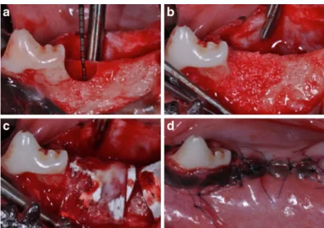
Under general anesthesia, vestibular and oral mucoperiosteal flaps were elevated. The three-wall intrabony defects were refreshed because original defects showed almost complete spontaneous healing [20,21] (Fig.1a). Following root plan-ing, a reference notch was made into the root surface at the base of each defect using a small round bur. Thus, de novo periodontal ligament tissue can be detected in the histological sections. The intrabony defects were then randomly assigned to one of the following treatment groups (14 sites for T1, 14 sites for T2, and 12 sites for C): (T1) a paste-like formulation of pulverized DBBM with 0.125–0.25 mm particles embed-ded in a carrier containing collagen types I and III, (T2) a pulverized formulation of DBBM with 0.125–0.25 mm parti-cles but without a carrier, and (C) Bio-Oss® granules (0.25– 1.00 mm) (Geistlich Pharma, Wolhusen, Switzerland) (Fig. 1b). Defects were covered with a resorbable collagen membrane (Bio-Gide®, Geistlich) with the double-layer tech-nique (Fig.1c). The flaps were positioned and sutured in a coronally displaced position with double-sling sutures (Seralon® Wiessner) (Fig.1d). Animals received spiramycin 750,000 IU and metronidazole 125 mg (BUCCOVAL®, Laboratoire Sogeval), per os, beginning the day before surgery and continuing for at least 14 days after surgery. Penicillin 150,000 UI procaine and benzathine (Bicillin® C-R, Pfizer) were injected intramuscularly the day before defect creation.
The sutures were removed after 14 days. Hygiene procedures were performed daily including tooth brushing and topical application of 0.2 % chlorhexidine until suture removal. Oral hygiene measures were performed twice weekly. Observa-tions of experimental sites with regards to gingival health, maintenance of suture line closure, edema, and evidence of tissue necrosis or infection were made daily until suture re-moval and at least twice weekly thereafter.
Retrieval of specimens
After 3 months, the animals were sacrificed with an overdose of pentobarbital sodium. The oral tissues were fixed by per-fusion with 10 % buffered formalin administered through the carotid arteries. Jaw segments with surrounding soft tissues were removed and placed in formalin. The specimens con-taining the experimental teeth were further subdivided using a diamond-coated band saw. From the 40 defects, 28 were left undecalcified and embedded in methyl methacrylate. The remaining 12 defects were decalcified in 10 % ethylene-diaminetetraacetic acid and processed for embedding in par-affin. Mesiodistal serial paraffin sections were cut parallel to the long axis of the teeth with the microtome set at 8μm and stained with hematoxylin and eosin. The reason for using paraffin histology in addition to undecalcified ground sections was to have some samples available for a possible future immunohistochemical evaluation, if required. The undecal-cified samples embedded in methyl methacrylate were cut in the same direction into∼500-μm-thick ground sections. They were then mounted on Plexiglas slabs and ground to a final thickness of∼80 μm. Finally, the sections were superficially stained with toluidine blue and basic fuchsin.
Data analysis Qualitative analysis
All histological sections were observed in Zeiss Axioplan microscope (Carl Zeiss, Göttingen, Germany), and digital photography of the defects was performed using a ProgRes® C5 digital camera (Jenoptik Laser, Optik, Systeme GmbH, Jena, Germany). Particular attention was paid to the quality and maturity of newly formed bone, histological signs of inflammation and foreign body reac-tion, and the presence of multinucleated giant cells on the bone substitute materials.
Histomorphometric analysis
Both the principal investigator (A.I.) and a very experienced researcher working for many years in the field of periodontal histology (D.B.) inspected all histological sections directly in the microscope at a maximum magnification/resolution. Only after both examiners agreed on the position of each reference point, these points were marked and the examiner (A.I.) started to perform the measurements.
For descriptive and histomorphometric analyses, sections with the largest defect area were selected, since this was indicative of the center of the defect. The following histolog-ical linear measurements were performed by one calibrated and masked investigator using the software included in the ProgRes® C5 digital camera as follows:
– Defect height: distance between apical extension of the root planing (notch) and coronal extension of gingival margin (in millimeter)
Fig. 1 Views after a surgical creation of an intrabony periodontal defect at the distal aspect of the PM2, b application of pulverized DBBM filler, c coverage of the filled defect with a collagen barrier membrane, and d flap repositioning and suturing for primary intention healing
– Junctional epithelium: distance from coronal to apical extent of a junctional epithelium along the root surface (in millimeter)
– Sulcus depth: distance from coronal to apical extent of sulcus depth along the root surface (in millimeter) – Cementum regeneration (height): distance between
api-cal extension of the root planing (notch) and the coronal extension of a continuous layer of new cementum or cementum-like deposit on the planed root (in millimeter) – Bone regeneration (height): distance between apical ex-tension of root planing (notch) and the coronal exex-tension of regenerated alveolar bone along the planed root (in millimeter)
Mean and standard deviation were calculated for all parameters.
Statistical analysis
The number of defects per group was 14 for group T1, 14 for group T2, and 12 for group C. The differences in means between the three groups were analyzed using nonpara-metric analysis of variance (Kruskal–Wallis test, PROC NPAR1WAY, SAS® version 9.2, SAS Institute Inc., Cary, NC, USA). No sample size/power calculation was performed. A post hoc power calculation indicated that
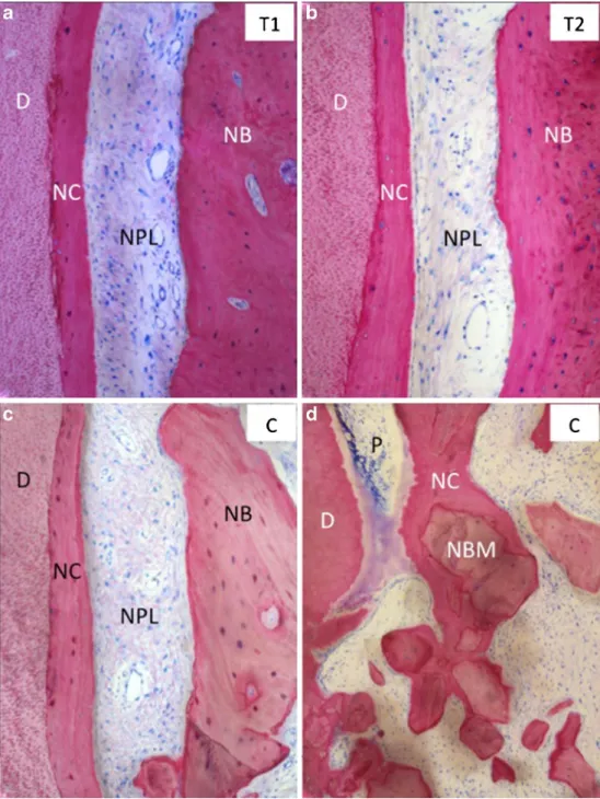
Fig. 2 Undecalcified ground sections from defects filled with a paste-like DBBM particles (T1), b pulverized DBBM particles (T2), and c, d control 0.25–1.0 mm Bio-Oss particles (C). The arrows demarcate the notch at the apical end of the defects. GM demarcates the gingival margin, aJE demarcates the most apical extension of the junctional epithelium, and C and B demarcate the most coronal extension of cementum and bone. Filling of the defect area with new bone can be seen in all three groups. Attachment of some DBBM particles to the root surface via new cementum was observed when the dental pulp was opened at the time of defect creation
16 subjects are necessary in each group to reach an alpha of 5 % and a power of 80.
Results
Histological analysis
Postoperative healing of all defects was uneventful. Five out of 14 defects in T1 were suitable for histomorphometric analysis. For T2, 11 out of 14 defects and for C 8 out of 12 defects were included in the histomorphometric evaluation.
Reasons for exclusion were as follows: (1) no removal of cementum and no notch were detected on the root surface in the histological sections, (2) opening of the dental pulp due to excessive removal of hard tissues from the root surface, and (3) technical difficulties during the preparation of histological samples. The notch and the periodontal defect region were clearly recognizable (Fig.2a–d). In a few defects, two notches were observed (Fig.2b), likely due to the creation of a second defect. An opening of the dental pulp was observed in four defects. The DBBM particles were integrated in new bone in the defect region adjacent to the root surface (Fig.2a–d). In five defects, some DBBM particles were connected to the root
Fig. 3 Representative
photomicrographs demonstrating the formation of new cementum (NC) on root dentin (D), new periodontal ligament (NPL), and new bone (NB) in a group T1 (paste-like DBBM), b group T2 (pulverized DBBM), and c group C (control DBBM). d New cementum formation between the root and deproteinized bovine bone mineral (DBBM) particles, when the dental pulp (P) was opened during defect creation
surface via new cementum (Fig.2d). Number and distribution of the DBBM particles in the augmented region varied greatly between defects, but did not reveal any obvious differences between groups. In all defects, no adverse inflammatory reac-tions were observed in both the augmented region and the gingival seal. Some multinucleated osteoclast-like cells were seen on some DBBM particles in all three groups.
New cementum, new periodontal ligament, and new bone were observed in all groups (Fig.3a–c). The cementum was cellular, quite uniform in thickness, and inserting collagen fibers were seen in places. In the coronal portion, the cemen-tum layer was often thinner than in apical locations. The newly formed periodontal ligament had a width correspond-ing to that of the original periodontal ligament and contained many blood vessels, fibroblasts, and collagen fibers. New cementum connecting DBBM particles with the root surface and discontinuity of the periodontal ligament was observed in five defects (one defect in group T1, two defects in group T2, and two defects in group C). In four of these five defects, the dental pulp was opened (Fig.3d). For all three groups, regions were found, where the DBBM particles were integrated in dense mature bone (Fig.4a–c). In the majority of defects, this occurred in the more apical region of the defects. Likewise, all three groups showed regions where DBBM particles were surrounded and connected to one another by a thin layer of
new bone (Fig.4d–f). The so formed bone–DBBM composite was typically localized in more marginal defect regions. DBBM particles were also found surrounded by soft tissue and without bone being formed on their surface (Fig.5a–c). The soft tissue found at these sites resembled either an imma-ture bone marrow (Fig.5b) or a soft connective tissue with a high number of fibroblast-like cells (Fig.5a, c). In all defects, remnants of the collagen membrane were clearly recognizable (Fig. 5d, e). The residual collagen membrane material consisted of wave-shaped fragments that were intensely stained in red. Morphology and staining of these fragments allowed differentiation between residual collagen membrane and host collagen.
Histomorphometric analysis
The histometric results are shown in Tables1,2,3, and4. The defect height from the apical notch to the gingival margin was on average 6.43 mm (SD 1.46). The mean scores of all defects (24 defects) were calculated and presented in Table 1. The mean length of cementum and bone was 3.42 mm (SD 1.99) and 3.60 mm (SD 1.39), respectively, and that of the connec-tive tissue, junctional epithelium, and sulcus depth averaged 0.88 (SD 1.00), 1.45 (SD 0.91), and 0.67 mm (SD 0.47), respectively.
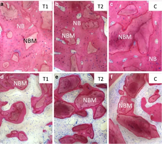
Fig. 4 Representative photomicrographs showing integration of DBBM particles in a–c dense and d–f loose new bone (NB) for the groups a, d T1 (paste-like DBBM), b, e T2 (pulverized DBBM), and c, f the control group C (control DBBM)
The mean defect height was very similar in all groups (6.16 mm (SD 1.94) for T1, 6.55 mm (SD 1.4) for T2, and 6.44 mm (SD 1.36) for C) and did not reach statistical signif-icance (P > 0.05). All groups showed an increase in new cementum measuring on average 2.25 (SD 2.24), 3.88 (SD 2.00), and 3.53 mm (SD 1.78) for T1, T2, and C, respectively. The differences between groups did not reach statistical sig-nificance (P >0.05) (Tables2,3, and4). All groups showed an average increase in new bone of 3.26 (SD 1.03), 3.60 (SD 1.66), and 3.81 mm (SD 1.30), respectively. The differences among the groups did not reach statistical significance (P > 0.05) (Tables2,3, and4). The connective tissue showed a mean value of 1.45 (SD 1.71), 0.60 (SD 0.55), and 0.92 mm
(SD 0.90) for T1, T2, and C, respectively. The differences among the groups did not reach statistical significance (P > 0.05) (Tables2,3, and4). The junctional epithelium measured 1.78 (SD 0.81), 1.53 (SD 1.12), and 1.16 mm (SD 0.63) for T1, T2, and C, respectively. The differences among the groups did not reach statistical significance (P >0.05) (Tables 2,3, and4). The sulcus depth measured 0.68 (SD 0.35), 0.55 (SD 0.32), and 0.83 mm (SD 0.69) for T1, T2, and C, respectively. The differences among the groups did not reach statistical significance (P >0.05) (Tables2,3, and4).
Fig. 5 Representative photomicrographs illustrating embedding of DBBM particles in soft tissue without the formation of new bone for a group T1 (paste-like DBBM), b group T2 (pulverized DBBM), and c group C (control DBBM). Remnants of the collagen barrier membrane (BM) were found in all defects adjacent to d new bone (NB) and e DBBM particles. The residual collagen membrane material consisted of wave-shaped fragments that were intensely stained in red (arrows). These characteristic features allowed differentiating of residual membrane material from host collagen
Table 1 Histomorphometric analysis of mean scores of all defects (T1, T2, and C) for cementum, bone, connective tissue, junctional epithelium, sulcus depth, and defect height (means (millimeter), standard deviations, standard error, minimum, maximum, median)
Variable Number Mean Std dev
Std error
Minimum Maximum Median
C 24 3.42 1.99 0.41 0.18 7.65 3.64 B 24 3.60 1.39 0.28 0.75 5.72 3.56 CT 24 0.88 1.00 0.20 0.00 4.21 0.60 JE 24 1.45 0.91 0.19 0.46 4.44 1.31 SD 24 0.67 0.47 0.10 0.15 2.33 0.59 DH 24 6.43 1.46 0.30 3.53 9.01 6.35 C cementum, B bone, CT connective tissue, JE junctional epithelium, SD sulcus depth, DH defect height
Table 2 Histomorphometric analysis of mean scores of T1 (paste-like DBBM) for cementum, bone, connective tissue, junctional epithelium, sulcus depth, and defect height (means (mm), standard deviations, stan-dard error, minimum, maximum, median)
Variable Number Mean Std dev
Std error
Minimum Maximum Median
C 5 2.25 2.24 1.00 0.18 5.48 1.31 B 5 3.26 1.03 0.46 1.85 4.76 3.25 CT 5 1.45 1.71 0.76 0.00 4.21 1.21 JE 5 1.78 0.81 0.36 0.80 3.03 1.62 SD 5 0.68 0.35 0.16 0.23 1.16 0.72 DH 5 6.16 1.94 0.87 3.72 9.01 6.24 No statistically significant differences between the paste-like, pulverized, and control DBBM for all parameters; P >0.05
C cementum, B bone, CT connective tissue, JE junctional epithelium, SD sulcus depth, DH defect height
Discussion
This study investigated healing and periodontal regeneration of three-wall intrabony defects in dogs filled with a paste-like bone filler consisting of 0.125–0.25-mm DBBM particles embedded in a collagenous carrier in comparison to 0.125– 0.25 mm large and to commercially available 0.25–1.00 mm large DBBM particles without the addition of the carrier material. Unexpectedly, complete bone fill was observed 3 months after the first defect creation. This shows that such defects in dogs have a high spontaneous healing potential. Nevertheless, the similar histological and histomorphometrical outcome for the three biomaterials tested after the second surgical intervention clearly shows that the two new formula-tions did not negatively interfere with periodontal healing and regeneration when compared with the commercially available and well-documented DBBM. Furthermore, it should be
clearly pointed out that the aim of this study was to evaluate the tissue responses to the three tested biomaterials. Therefore, in order to minimize/eliminate confounding factors like bac-terial challenge through plaque microorganisms as seen in chronic defects, acute type three-wall defects were used. After the positive results of the present study, the next experiments can be performed in a clinically more relevant but also more challenging situation like a chronic defect and in a larger sample number allowing proper statistical analysis.
All three groups showed no inflammatory reactions and resulted in comparable amounts of new cementum, new peri-odontal ligament, and new bone. Furthermore, integration of the DBBM particles in new bone was observed for all three groups. These results are consistent with studies using a combination of commercially available DBBM (Bio-Oss®) and a collagen barrier membrane (Bio-Gide®) in two-wall intrabony [22], one-wall intrabony [23], and fenestration de-fects [24] in canines.
The barrier membrane
The need for the use of a barrier membrane [23,24] and the beneficial effect of the placement of DBBM underneath the membrane [22] to achieve periodontal regeneration has clearly been demonstrated. In the present study, the collagen barrier membrane was placed in a double-layer technique. Our ob-servation that residual membrane material was found in all defects after 3 months indicates a long-lasting barrier function, as was shown for the double-layer technique in other defect models [25,26]. Thus, it may be concluded that for all groups in the present study, comparable conditions in terms of barrier function existed, which lowered the chance for confounding factors interfering with the regenerative process.
Granule size
Bone substitute materials are available in different granule sizes. A DBBM granule size of 0.25–1.00 mm is recommend-ed for periodontal applications. Using particle sizes smaller than 0.25–1.00 mm may be beneficial for applications in small or narrow periodontal bone defects and may allow their pas-sage through the tip of a syringe when used as an injectable bone substitute material. To the best of our knowledge, this is the first study where 0.125–0.25 mm granules with or without a carrier material were tested in periodontal defects. Granules with a small particle size may have an influence on their degradation/resorption rate, on the speed and amount of bone formation, and on ankylosis.
Ankylosis was not observed in the present study, yet a connection of some DBBM particles with the root surface via cementum was seen in five defects. DBBM connection via hard tissue to the root surface has occasionally been observed in other studies [27,28]. Since this phenomenon
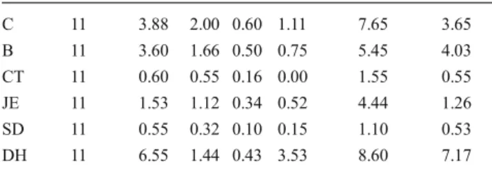
Table 3 Histomorphometric analysis of mean scores of T2 (pulverized DBBM) group for cementum, bone, connective tissue, junctional epithe-lium, sulcus depth, and defect height (means (millimeter), standard devi-ations, standard error, minimum, maximum, median)
Variable Number Mean Std dev
Std error
Minimum Maximum Median
C 11 3.88 2.00 0.60 1.11 7.65 3.65 B 11 3.60 1.66 0.50 0.75 5.45 4.03 CT 11 0.60 0.55 0.16 0.00 1.55 0.55 JE 11 1.53 1.12 0.34 0.52 4.44 1.26 SD 11 0.55 0.32 0.10 0.15 1.10 0.53 DH 11 6.55 1.44 0.43 3.53 8.60 7.17 No statistically significant differences between the paste-like, pulverized, and control DBBM for all parameters; P >0.05
C cementum, B bone, CT connective tissue, JE junctional epithelium, SD sulcus depth, DH defect height
Table 4 Histomorphometric analysis of mean scores of C (control DBBM) for cementum, bone, connective tissue, junctional epithelium, sulcus depth, and defect height (mean (millimeter), standard deviations, standard error, minimum, maximum, median)
Variable Number Mean Std dev
Std error
Minimum Maximum Median
C 8 3.53 1.78 0.63 1.22 6.51 3.55 B 8 3.81 1.30 0.46 2.14 5.72 3.55 CT 8 0.92 0.90 0.32 0.00 2.31 0.56 JE 8 1.16 0.63 0.22 0.46 2.35 1.21 SD 8 0.83 0.69 0.24 0.15 2.33 0.65 DH 8 6.44 1.36 0.48 4.84 8.68 6.25 No statistically significant differences between the paste-like, pulverized, and control DBBM for all parameters; P >0.05
C cementum, B bone, CT connective tissue, JE junctional epithelium, SD sulcus depth, DH defect height
was observed in one defect of group T1, two defects of group T2, and two defects of group C, the particle size is unlikely responsible for this. In four of these five defects, the pulp was opened. Thus, it is likely that pulp opening caused an inter-ruption of periodontal ligament expansion leading to this phenomenon. Pulp opening likely occurred because defect creation occurred twice.
Although multinucleated osteoclast-like cells are normally found on DBBM particles and other calcium phosphate bio-materials [29], the degradation rate of DBBM is very low [30–32]. In the present study, osteoclast-like cells were seen on some DBBM particle in all three groups, and no pro-nounced signs of resorption such as deep Howship’s lacunae or extremely small DBBM particles, as demonstrated in an-other studies [16, 22–24, 28], were observed. Thus, it is concluded that neither the 0.125–0.25 mm DBBM particles nor the carrier enhanced the resorptive activity.
Concerning the amount of augmented bone and integration of DBBM particles in bone, no qualitative differences be-tween small and large DBBM particles were observed in the present study. Similar conclusions were made for sinus floor elevation [18] and the regeneration of calvarial defects [17]. In contrast, a higher bone density was observed with small synthetic bone substitute granules (0.3–0.5 mm) compared with larger (0.85–1.00 mm) granules in another bone defect model [19]. Thus, our data suggest that the osteoconductive properties of the small 0.125–0.25 mm DBBM particles are comparable to those of the larger 0.25–1.00 mm DBBM particles. However, one drawback of the present study was that we had to refrain from a histometrical evaluation of area fractions of new bone and DBBM. Due to the fact that defect morphology was so heterogeneous, and since the distribution of the DBBM particles and the density of both DBBM and new bone were so variable from defect to defect, any possible material-related differences may have been overruled by this heterogeneity. Reasons for this heterogeneity may be related to the fact that defect creation was performed twice due to spontaneous healing of the first defect and defect creation on different tooth types with different root morphologies and both in the lower and upper jaws. There was a big variation of the scores within the groups of all parameters (cementum, bone, connective tissue, junctional epithelium, sulcus depth, and defect height).
The carrier/scaffold
A carrier may influence the osteoconductivity of a bone substitute material, alter the migration behavior of the parti-cles, and/or elicit unwanted tissue responses like enhanced inflammation. Our data suggest that the presence of the carrier did not negatively influence bone formation and did not result in adverse tissue reactions. A similar conclusion was drawn
from a bone defect study, where a hydrogen carrier consisting of carboxymethyl cellulose and collagen was used [17].
Clinical relevance and conclusions
From a clinical standpoint, a paste-like bone substitute mate-rial, which can easily be injected in a periodontal defect, may enhance handling and mechanical stability. The biomaterials tested in the present study did not differ in regard to both their periodontal regenerative capacity and osteoconductivity. It may thus be concluded that the 0.125–0.25-mm DBBM par-ticles combined with the collagen-based carrier may be suit-able for clinical applications.
Acknowledgments The authors wish to thank Ms. Monika Aeberhard, Ms. Thuy-Trang Nguyen, and Mr. David Reist for the histological prep-aration of the specimens and Mr. Walter Bürgin for performing the statistical analyses. The study was funded by Geistlich Pharma, Wolhusen, Switzerland.
Conflict of interest The authors report no conflicts of interest related to this study.
References
1. Karring T, Lindhe J, Cortellini P (2003) Regenerative periodontal therapy. In: Lindhe J, Karring T, Lang NP (eds) Clinical periodon-tology and implant dentistry, 4th edn. Blackwell-Munksgard, Copenhagen, pp 650–704
2. Sculean A, Nikolidakis D, Schwarz F (2008) Regeneration of peri-odontal tissues: combinations of barrier membranes and grafting materials—biological foundation and preclinical evidence: a system-atic review. J Clin Periodontol 35(8 Suppl):106–116. doi:10.1111/j. 1600-051X.2008.01263.x
3. Camelo M, Nevins ML, Schenk RK, Simion M, Rasperini G, Lynch SE, Nevins M (1998) Clinical, radiographic, and histologic evalua-tion of human periodontal defects treated with Bio-Oss and Bio-Gide. Int J Periodontics Restor Dent 18(4):321–331
4. Mellonig JT (2000) Human histologic evaluation of a bovine-derived bone xenograft in the treatment of periodontal osseous defects. Int J Periodontics Restor Dent 20(1):19–29
5. Sculean A, Berakdar M, Chiantella GC, Donos N, Arweiler NB, Brecx M (2003) Healing of intrabony defects following treatment with a bovine-derived xenograft and collagen membrane. A con-trolled clinical study. J Clin Periodontol 30(1):73–80
6. Sculean A, Stavropoulos A, Windisch P, Keglevich T, Karring T, Gera I (2004) Healing of human intrabony defects following regen-erative periodontal therapy with a bovine-derived xenograft and guided tissue regeneration. Clin Oral Investig 8(2):70–74. doi:10. 1007/s00784-004-0254-7
7. Sculean A, Chiantella GC, Windisch P, Arweiler NB, Brecx M, Gera I (2005) Healing of intrabony defects following treatment with a composite bovine-derived xenograft (Bio-Oss Collagen) in combi-nation with a collagen membrane (Bio-Gide PERIO). J Clin Periodontol 32(7):720–724. doi:10.1111/j.1600-051X.2005.00758.x
8. Sculean A, Schwarz F, Chiantella GC, Donos N, Arweiler NB, Brecx M, Becker J (2007) Five-year results of a prospective, randomized, controlled study evaluating treatment of intrabony defects with a
natural bone mineral and GTR. J Clin Periodontol 34(1):72–77. doi:10.1111/j.1600-051X.2006.01007.x
9. Tonetti MS, Cortellini P, Lang NP, Suvan JE, Adriaens P, Dubravec D, Fonzar A, Fourmousis I, Rasperini G, Rossi R, Silvestri M, Topoll H, Wallkamm B, Zybutz M (2004) Clinical outcomes following treatment of human intrabony defects with GTR/bone replacement material or access flap alone. A multicenter randomized controlled clinical trial. J Clin Periodontol 31(9):770–776. doi: 10.1111/j.1600-051X.2004.00562.x
10. Wikesjo UM, Selvig KA (1999) Periodontal wound healing and regeneration. Periodontol 2000 19:21–39
11. Schwarz F, Bieling K, Latz T, Nuesry E, Becker J (2006) Healing of intrabony peri-implantitis defects following application of a nano-crystalline hydroxyapatite (Ostim) or a bovine-derived xenograft (Bio-Oss) in combination with a collagen membrane (Bio-Gide): a case series. J Clin Periodontol 33(7):491–499. doi: 10.1111/j.1600-051X.2006.00936.x
12. Kasaj A, Willershausen B, Reichert C, Rohrig B, Smeets R, Schmidt M (2008) Ability of nanocrystalline hydroxyapatite paste to promote human periodontal ligament cell proliferation. J Oral Sci 50(3):279–285
13. Chitsazi MT, Shirmohammadi A, Faramarzie M, Pourabbas R, Rostamzadeh A (2011) A clinical comparison of nano-crystalline hydroxyapatite (Ostim) and autogenous bone graft in the treatment of periodontal intrabony defects. Med Oral Patol Oral Cir Bucal 16(3):e448–e453
14. Horvath A, Stavropoulos A, Windisch P, Lukacs L, Gera I, Sculean A (2012) Histological evaluation of human intrabony periodontal de-fects treated with an unsintered nanocrystalline hydroxyapatite paste. Clin Oral Investig 17(2):423–30. doi:10.1007/s00784-012-0739-8
15. Carano RA, Filvaroff EH (2003) Angiogenesis and bone repair. Drug Discov Today 8(21):980–989
16. Busenlechner D, Tangl S, Arnhart C, Redl H, Schuh C, Watzek G, Gruber R (2012) Resorption of deproteinized bovine bone mineral in a porcine calvaria augmentation model. Clin Oral Implants Res 23(1):95–99. doi:10.1111/j.1600-0501.2011.02198.x
17. Busenlechner D, Tangl S, Fitzl C, Bernhart T, Gruber R, Watzek G (2009) Paste-like inorganic bone matrix: preclinical testing of a prototype preparation in the porcine calvaria. Clin Oral Implants Res 20(10):1099–1104. doi:10.1111/j.1600-0501.2009.01743.x
18. Chackartchi T, Iezzi G, Goldstein M, Klinger A, Soskolne A, Piattelli A, Shapira L (2011) Sinus floor augmentation using large (1–2 mm) or small (0.25–1 mm) bovine bone mineral particles: a prospective, intra-individual controlled clinical, micro-computerized tomography, and histomorphometric study. Clin Oral Implants Res 22(5):473– 480. doi:10.1111/j.1600-0501.2010.02032.x
19. Zhou X, Zhang Z, Li S, Bai Y, Xu H (2011) Osteoconduction of different sizes of anorganic bone particles in a model of guided bone regeneration. Br J Oral Maxillofac Surg 49(1):37–41. doi:10.1016/j. bjoms.2010.01.001
20. Kim CS, Choi SH, Chai JK, Cho KS, Moon IS, Wikesjo UM, Kim CK (2004) Periodontal repair in surgically created intrabony defects in dogs: influence of the number of bone walls on healing response. J Periodontol 75(2):229–235. doi:10.1902/jop.2004.75.2.229
21. Kim CS, Choi SH, Cho KS, Chai JK, Wikesjo UM, Kim CK (2005) Periodontal healing in one-wall intrabony defects in dogs following
implantation of autogenous bone or a coral-derived biomaterial. J Clin Periodontol 32(6):583–589. doi:10.1111/j.1600-051X.2005. 00729.x
22. Yamada S, Shima N, Kitamura H, Sugito H (2002) Effect of porous xenographic bone graft with collagen barrier membrane on periodontal regeneration. Int J Periodontics Restor Dent 22(4):389–397
23. Sakata J, Abe H, Ohazama A, Okubo K, Nagashima C, Suzuki M, Hasegawa K (2006) Effects of combined treatment with porous bovine inorganic bone grafts and bilayer porcine collagen membrane on refractory one-wall intrabony defects. Int J Periodontics Restor Dent 26(2):161–169
24. Tal H, Artzi Z, Moses O, Nemcovsky C, Kozlovsky A (2005) Guided periodontal regeneration using bilayered collagen membranes and bovine bone mineral in fenestration defects in the canine. Int J Periodontics Restor Dent 25(5):509–518
25. Kim SH, Kim DY, Kim KH, Ku Y, Rhyu IC, Lee YM (2009) The efficacy of a double-layer collagen membrane technique for overlay-ing block grafts in a rabbit calvarium model. Clin Oral Implants Res 20(10):1124–1132. doi:10.1111/j.1600-0501.2009.01744.x
26. Kozlovsky A, Aboodi G, Moses O, Tal H, Artzi Z, Weinreb M, Nemcovsky CE (2009) Biodegradation of a resorbable collagen membrane (Bio-Gide) applied in a double-layer technique in rats. Clin Oral Implants Res 20(10):1116–1123. doi:10.1111/j.1600-0501. 2009.01740.x
27. Sculean A, Chiantella GC, Arweiler NB, Becker J, Schwarz F, Stavropoulos A (2008) Five-year clinical and histologic results fol-lowing treatment of human intrabony defects with an enamel matrix derivative combined with a natural bone mineral. Int J Periodontics Restor Dent 28(2):153–161
28. Stavropoulos A, Wikesjo UM (2010) Influence of defect dimen-sions on periodontal wound healing/regeneration in intrabony defects following implantation of a bovine bone biomaterial and provisions for guided tissue regeneration: an experimental study in the dog. J Clin Periodontol 37(6):534–543. doi:10.1111/ j.1600-051X.2010.01566.x
29. Jensen B, Buser D (2009) Bone grafts and bone substitute materials for GBR procedures. In: Buser D (ed) 20 years of guided bone regeneration in implant dentistry, 2nd edn. Quintessence, Chicago, pp 71–96
30. Jensen SS, Broggini N, Hjorting-Hansen E, Schenk R, Buser D (2006) Bone healing and graft resorption of autograft, anorganic bovine bone, and beta-tricalcium phosphate. A histologic and histomorphometric study in the mandibles of minipigs. Clin Oral Implants Res 17(3):237–243. doi:10.1111/j.1600-0501.2005. 01257.x
31. Jensen SS, Yeo A, Dard M, Hunziker E, Schenk R, Buser D (2007) Evaluation of a novel biphasic calcium phosphate in standardized bone defects: a histologic and histomorphometric study in the man-dibles of minipigs. Clin Oral Implants Res 18(6):752–760. doi:10. 1111/j.1600-0501.2007.01417.x
32. Jensen SS, Bornstein MM, Dard M, Bosshardt DD, Buser D (2009) Comparative study of biphasic calcium phosphates with different HA/TCP ratios in mandibular bone defects. A long-term histomor-phometric study in minipigs. J Biomed Mater Res B Appl Biomater 90(1):171–181. doi:10.1002/jbm.b.31271