Diagnosing breast cancer using diffuse reflectance
spectroscopy and intrinsic fluorescence spectroscopy
The MIT Faculty has made this article openly available.
Please share
how this access benefits you. Your story matters.
Citation
Volynskaya, Zoya et al. “Diagnosing Breast Cancer Using Diffuse
Reflectance Spectroscopy and Intrinsic Fluorescence Spectroscopy.”
Journal of Biomedical Optics 13.2 (2008): 024012. Web. 16 Feb. 2012.
© 2008 SPIE - International Society for Optical Engineering
As Published
http://dx.doi.org/10.1117/1.2909672
Publisher
SPIE - International Society for Optical Engineering
Version
Final published version
Citable link
http://hdl.handle.net/1721.1/69126
Terms of Use
Article is made available in accordance with the publisher's
policy and may be subject to US copyright law. Please refer to the
publisher's site for terms of use.
Maryann Fitzmaurice Robert Shenk Nancy Wang
University Hospitals–Case Medical Center and
Case Western Reserve University Cleveland, Ohio 44106
Jon Nazemi
Ramachandra R. Dasari Michael S. Feld
Massachusetts Institute of Technology Laser Biomedical Research Center G. R. Harrison Spectroscopy Laboratory 77 Massachusetts Avenue, 6-218M Cambridge, Massachusetts 02139
lected via a portable clinical spectrometer and specially designed op-tical fiber probe. The diffuse reflectance spectra are fit using modified diffusion theory to extract absorption and scattering tissue parameters. Intrinsic fluorescence spectra are extracted from the combined fluo-rescence and diffuse reflectance spectra and analyzed using multivari-ate curve resolution. Spectroscopy results are compared to pathology diagnoses, and diagnostic algorithms are developed based on param-eters obtained via logistic regression with cross-validation. The sensi-tivity, specificity, positive predictive value, negative predictive value, and overall diagnostic accuracy共total efficiency兲 of the algorithm are 100, 96, 69, 100, and 91%, respectively. All invasive breast cancer specimens are correctly diagnosed. The combination of diffuse reflec-tance spectroscopy and intrinsic fluorescence spectroscopy yields promising results for discrimination of breast cancer from benign breast lesions and warrants a prospective clinical study. © 2008 Society of
Photo-Optical Instrumentation Engineers. 关DOI: 10.1117/1.2909672兴
Keywords: cancer; breast; diagnosis; reflectance; fluorescence; spectroscopy.
Paper 07381R received Sep. 14, 2007; revised manuscript received Dec. 11, 2007; accepted for publication Dec. 12, 2007; published online Apr. 30, 2008.
1 Introduction
Screening procedures such as x-ray mammography, ultra-sound, and/or palpation are the first step in diagnosing breast cancer. Screening procedures do not accurately identify ma-lignant tissue. Therefore, to establish a diagnosis, the patient must undergo breast biopsy followed by histopathological di-agnosis. Due to the lack of specificity of mammography, a high percentage of suspected abnormal lesions 共70 to 90%兲 are found to be benign or normal upon biopsy,1resulting in unnecessary cost, patient trauma, and time delay to obtain histopathological diagnosis. Imaging and spectroscopic opti-cal diagnostic techniques are currently being explored to re-duce the number of unnecessary breast biopsies in a mini-mally invasive manner.
Diffuse optical tomography 共DOT兲, an optical imaging technique to identify suspect lesions, is currently under study.2 Similar to current screening techniques, it is noninvasive and can detect lesions deep within the breast. It employs light of a number of near-IR wavelengths. Contrast, provided by scat-tering and absorption, measures relative concentrations of oxy- and deoxyhemoglobin, as well as the presence of lipids
and water content.3 However, this technique has a low resolution,4,5due to which lesions of small size go undetected; a resolution of 4 to13 mm has been reported, and the sources of contrast are limited.3,2,6Despite these challenges, DOT has been utilized in several reported breast cancer studies.7,8
A number of promising optical spectroscopy techniques for diagnosing breast cancer are also under study. These tech-niques currently employ point contact probes, but have the potential to be extended to imaging modes.9 The most fre-quently utilized methods are diffuse reflectance spectroscopy10–12 共DRS兲 and fluorescence spectroscopy.13–15 As of now, intrinsic fluorescence spectroscopy16共IFS兲, a tech-nique that uses diffuse reflectance to suppress the effects of scattering and absorption in fluorescence, has not been imple-mented. The objective of this study is to explore the use of DRS and IFS, in combination, to identify and diagnose sus-pect lesions.
Briefly, DRS measures tissue scattering and absorption properties, which provide information about the morphology and biochemistry of the breast tissue epithelium and stroma. DRS typically utilizes broadband UV-visible excitation in the wavelength range 300 to800 nm, spanning the prominent he-moglobin absorption features. Fluorescence spectroscopy
pro-1083-3668/2008/13共2兲/024012/9/$25.00 © 2008 SPIE Address all correspondence to Zoya Volynskaya, MS, Laser Biomedical
Re-search Center, G.R. Harrison Spectroscopy Laboratory, Massachusetts Institute of Technology, 77 Massachusetts Ave, 6-218M, Cambridge, MA 02139; Tel: 共617兲 253-5077; Fax: 共617兲 253-4513; E-mail: zivolyns@mit.edu
vides information about the endogenous tissue fluorophores excited with UV-visible light in the same wavelength range 300 to 800 nm. Fluorophores excited in this range have the most diagnostic significance. However, fluorescence spectra recorded from tissue are distorted by absorption and scatter-ing, thereby limiting the accuracy of the extracted informa-tion. To overcome this problem, DRS can be used in conjunc-tion with the measured fluorescence spectrum to extract the intrinsic共undistorted兲 fluorescence.17,16This process is called intrinsic fluorescence spectroscopy共IFS兲.
The DRS and fluorescence studies published up to now have focused on differentiating malignant from nonmalignant tissues. However, it is also clinically important to discriminate benign lesions that may carry a risk of breast cancer or that need to be surgically excised from normal breast tissue. For example, fibrocystic change is a benign condition that does not require excision, whereas fibroadenoma proliferates rap-idly and must be excised. As our data will show, DRS and fluorescence spectroscopy have the potential to identify these lesions in real time at the bedside when performed percutane-ously using a fiber optic probe inserted through a breast bi-opsy needle.
Furthermore, previously used diagnostic algorithms have not been based on parameters that can be identified as patho-physiological features of disease. While doing so is not a re-quirement for success, the use of parameters that may be di-rectly compared to histopathology is likely to lead to more robust algorithms and to aid clinicians in interpreting spectro-scopic results. It also represents a step toward quantifiable, objective tissue diagnosis, as compared to subjective histopa-thology analysis.
We report an ex vivo spectroscopic study of freshly excised human breast tissue. We use DRS and fluorescence, combined in a particular manner, to discriminate breast cancer from both benign breast lesions and normal breast tissue. Our technique, which we refer to subsequently as DRS-IFS, provides a more complete biochemical, morphological, and metabolical tissue profile than either modality alone. Our analysis extracts pa-rameters that optimize classification and, at the same time, provide physical insight. This added information should en-able the development of a more robust diagnostic algorithm.
2 Instrumentation
A clinical instrument for DRS-IFS studies, the FastEEM关Fig. 1共a兲兴, was developed at the G.R. Harrison Spectroscopy Labo-ratory at the Massachusetts Institute of Technology18 共MIT兲. This instrument collects white light reflectance and fluores-cence excitation-emission matrices共EEMs兲 in a fraction of a second. It delivers a sequence of 10 excitation laser pulses 共308 to 480 nm兲 and two white light pulses 共300 to 800 nm兲 to the tissue via an optical fiber probe, which is in the form of a flexible catheter with an overall length of3 m and a diam-eter of approximately1.2 mm关Fig.1共b兲兴. The distal tip con-sists of a single delivery fiber surrounded by six collection fibers, all with core diameters of 200m, and terminated with a transparent, protective optical shield.18 The tip is brought into gentle contact with the tissue when data are taken. The diameter of tissue sampled is approximately 600m, with depth sensing of approximately 100m. The same probe delivers the excitation light and collects the dif-fusely reflected white light and fluorescence. At the proximal end of the probe, the collection fibers are confined in a single line and imaged onto the slit of an intensified CCD detector. Ten laser-induced fluorescence emission spectra and the two white light reflectance spectra are collected in approximately 0.3 s. Sufficient SNR is obtained by averaging five measure-ments, resulting in a typical acquisition time of 1.5 s. The SNR, determined as the ratio of the peak of the signal to the standard deviation of the noise, was found to be 100.
The instrument was calibrated daily prior to data collec-tion. Calibration consisted of four steps: 共1兲 a background spectrum of water was collected and subtracted from all sub-sequent measurements to correct for dark current and stray light;共2兲 the excitation intensities at each of 10 wavelengths were then normalized by measuring a rhodamine B dye mix-ture;共3兲 a Spectralon standard with 20% reflectance was then measured to account for the wavelength-dependent system re-sponse; 共4兲 and finally, a mercury spectrum was acquired to calibrate wavelength.
3 Patients and Breast Tissue Handling
This ex vivo study was conducted at the University Hospitals-Case Medical Center 共UHC兲 under approval from the UHC
Fig. 1 共a兲 FastEEM clinical spectrophotometer and 共b兲 schematic diagram of the distal tip of the optical fiber probe. Volynskaya et al.: Diagnosing breast cancer using diffuse reflectance spectroscopy…
that no artifacts were introduced in the data as a result of this processing step.
Some of the data collected were subsequently excluded from the analysis. Specifically, DRS data with overall reflec-tance of less than 1% were excluded because of the inability to use reflectance information of that data to process the fluo-rescence data to obtain the intrinsic fluofluo-rescence. Sites whose spectra failed this criterion共11 sites兲 were found to be con-taminated by the presence of surface blood that absorbed too much of the reflected light. Additionally, specimens from pa-tients with preoperative chemotherapy or who underwent re-peat excisional biopsy were excluded from analysis共7 speci-mens兲. Sites exhibiting other lesions 关ductal epithelial hyperplasia, n= 2; fat necrosis, n = 1; and ductal carcinoma
in situ 共DCIS兲, n=6兴 were also excluded, as their limited
numbers precluded development of a robust diagnostic algo-rithm. Following these exclusions, 202 IFS and DRS spectra acquired from a total of 104 breast tissue sites共1 and often 2 spectra per site兲 in specimens from 17 consecutive patients remained and were used for further analysis.
After acquisition of the spectra, the exact site of probe placement was marked with colloidal ink for registration with histopathology. The breast specimens were then fixed in for-malin and submitted for routine pathology evaluation, per-formed by an experienced breast pathologist blinded to the spectroscopy results. The histopathology diagnoses were1931 normal, 55 fibrocystic change, 9 fibroadenoma, and 9 invasive carcinoma 共all infiltrating ductal carcinoma兲. The relative number of each diagnosis in this data set closely resembles the typical prevalence for diseases of breast tissue.1
4 Data Analysis
We analyzed the DRS spectrum from each breast tissue site using a mathematical model based on the diffusion approxi-mation of light propagation in tissue to determine the values of the absorption coefficienta共兲 and the reduced scattering coefficients
⬘
共兲, previously described in detail.17The model requires index of refraction n, which for soft biological tissue has a typical value20 of 1.35 to 1.45. Two absorbers, oxyhe-moglobin and-carotene,21were used to model the extracted absorption coefficient a. During our study, the tissue was exposed to room air and hemoglobin was oxygenated; there-fore, deoxyhemoglobin was not included. For the reduced scattering coefficient s⬘
共兲, wavelength dependence of the form A−Bwas employed.22IFS spectra were extracted from the combined fluorescence and DRS spectra, as previously described.23An IFS spectrum
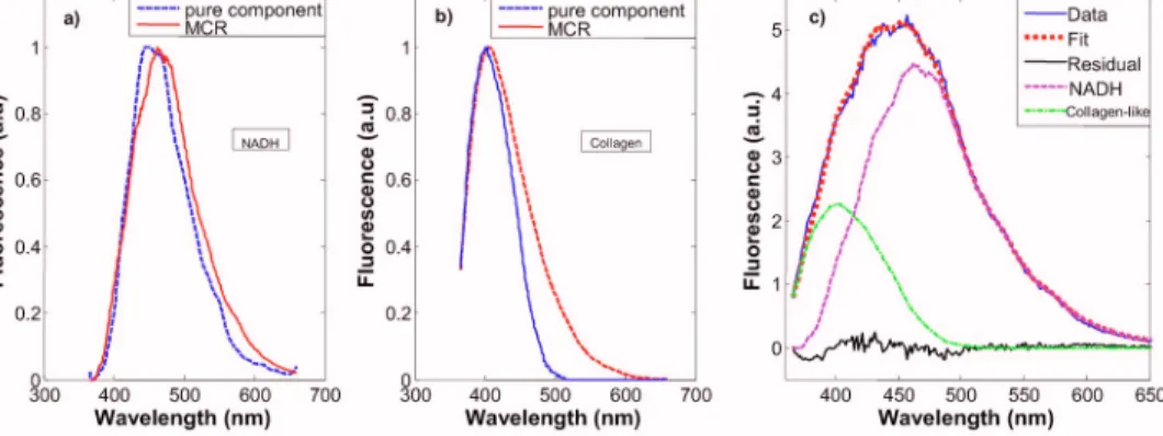
is composed of a linear combination of the spectra of the individual fluorophores present in the tissue. However, differ-ences in local chemical environments, such as in tissue, may alter the shape of a fluorophore spectrum from that of its pure component.24Therefore, pure component spectra were not di-rectly fit to a given IFS spectrum. Instead, the IFS spectra were analyzed using multivariate curve resolution 共MCR兲 with nonnegativity constraints, a standard chemometric method.25MCR generates spectral components by adjusting the spectral shape of initial input parameters to minimize the fitting error to a given tissue spectrum. The initial input spec-tra in our case were the pure component specspec-tra of fluoro-phores known to be present in breast tissue24共Table 1兲. The maximum number of the initial input spectra was not con-strained; however, on analysis, it was found that only two spectra were required to produce good fits to the data. The resulting MCR-generated spectral components at340 nm are shown in Figs.2共a兲and2共b兲. The combined basis spectra and fit to a representative IFS spectrum are shown in Fig.2共c兲. The MCR-generated spectral components are similar to the spectra of NADH and collagen-like, respectively, but are broader and red-shifted, as expected of fluorophores in a tis-sue environment.26
As stated previously, the FastEEM instrument provides emission spectra at 10 different excitation wavelengths. Col-lagen, NADH, elastin, tryptophan, FAD, and porphyrins, the native fluorophores of breast tissue, have their maximum emission at 308, 340, 360, and 425 nm excitation wavelengths26共Table 1兲. Thus, those four fluorescence exci-tation wavelengths were employed in the initial analysis. However, it was found that not all of these fluorophores could be detected, presumably because they were not present in our specimens at high enough levels. These include elastin excited at 340 nm and FAD and porphyrins excited at 360 nm. The absence of these fluorophores was confirmed by attempting to extract their basis spectra using MCR and finding that the contributions were negligible.
Of the measured fluorophores, only NADH and collagen-like excited at 340 and360 nm were required to obtain good fits to the IFS spectra. However, no substantial differences in the fluorophore contributions at those two wavelengths were observed. Due to the proximity in wavelength of 340 and 360 nm, the difference in the penetration depth of the light 共⬍5m兲 evidently was not sufficient to reveal these differ-ences. Averaging the information at these two wavelengths
did not improve the fits. To reduce complexity, we opted to utilize only one excitation wavelength, with the added benefit that as a result only one UV source will be required in future clinical instruments. Therefore, contributions of NADH and collagen-like excited at 340 nm were chosen for algorithm
development, as this is the wavelength for maximum emission of NADH. Representative DRS and IFS spectra collected with 340-nm excitation are shown in Fig.3.
Thus, the data analysis of each tissue site was performed using two spectra共one DRS and one IFS兲. Spectra were
fur-Fig. 3 Representative spectra of DRS and IFS excited at 340 nm for all pathologies:共a兲 normal tissue, 共b兲 fibrocystic change lesion, 共c兲 fibroad-enoma lesion, and共d兲 infiltrating ductal carcinoma lesion 关data 共blue, solid line兲; fit 共red, dashed line兲; residual 共black, dash-dotted line兲兴. 共Color online only兲
Fig. 2 共a兲 and 共b兲 Comparison of pure component spectra 共blue, dashed lines兲 and MCR basis spectra 共red solid lines兲 excited at 340 nm for 共a兲
NADH and共b兲 collagen; 共c兲 representative spectrum fit with a linear combination of NADH 共magenta, dotted line兲 and collagen-like basis spectra 共green, dash-dotted line兲 关data 共blue, solid line兲; fit 共red, dotted line兲; residual 共black兲兴. 共Color online only兲
ther characterized by six parameters: A and B—scattering pa-rameters, fit coefficients of oxyhemoglobin and-carotene, as well as the fit coefficients of collagen-like and NADH.
5 Algorithm Development
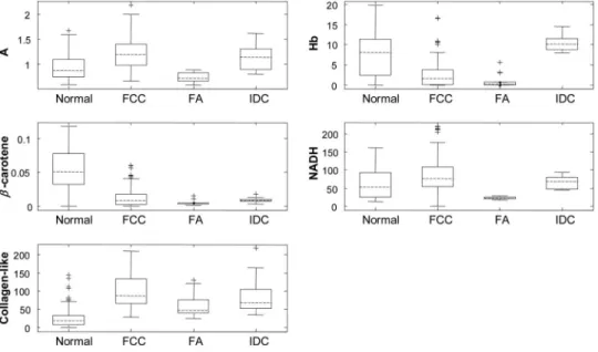
Following data analysis, all of the fit parameters, except B, were found to be diagnostically relevant. Boxplots for these parameters are shown in Fig. 4. 共A boxplot is a graphical representation of data spread, with the first, second, and third quartiles “contained” in the box; the median for the data set is indicated by the black center line. The interquartile range is defined as the distance between the first and third quartiles. The extreme values are defined as 1.5 times the interquartile range, measured from the median. Data points outside of this range, indicated by crosses, are defined as outliers.兲 This form of data depiction is useful for visually examining the diagnos-tic capability of each parameter. For example, the boxplot for collagen-like indicates that normal breast sites may be par-tially separated from other sites using this parameter. Note that diagnostic parameters in a particular tissue type may be correlated. Hence, one parameter may provide only partial separation, but the use of a second parameter in combination with it may provide full separation. Visualizing this behavior requires a 2-D scatter plot and is not revealed by the boxplots. The desired algorithm must be able to classify invasive breast cancer共in this case, infiltrating ductal carcinoma兲, be-nign breast lesions such as fibroadenoma and fibrocystic change, and normal breast tissue. Because each of these tissue types can likely be characterized using different diagnostic parameters, we performed the analysis in a stepwise fashion, as shown in Fig.5. Initially, normal specimens are separated from the rest of the specimens共step 1兲, followed by the sepa-ration of fibroadenoma from fibrocystic change and infiltrat-ing ductal carcinoma 共step 2兲, and finally, the separation of infiltrating ductal carcinoma from fibrocystic change lesions 共step 3兲. The diagnostic algorithm was developed using
logis-tic regression and leave-one-out cross-validation. Histopathol-ogy diagnoses were used as the standard against which the spectroscopic diagnoses were compared. By maximizing the sensitivity of each stage of the algorithm we were able to identify the diagnostic parameters that can best discriminate pathologies of breast tissue.
The scatter plots and decision lines for each step of the diagnostic algorithm are depicted in Fig.6. In step 1, normal sites are classified using the collagen-like and-carotene fit coefficients extracted from IFS at 340-nm excitation and DRS, respectively 关Fig. 6共a兲兴. In step 2, fibroadenoma sites are discriminated from fibrocystic change and infiltrating duc-tal carcinoma using the DRS scattering parameter A and the IFS NADH fit coefficient关Fig.6共b兲兴. Finally, in step 3, infil-trating ductal carcinoma sites are distinguished from fibrocys-tic change using the DRS oxyhemoglobin and a weaker de-pendence of IFS collagen-like 共excited at 340 nm兲 fit coefficients关Fig.6共c兲兴.
Fig. 4 Boxplots of normal tissue共Normal兲, fibrocystic change 共FCC兲, fibroadenoma 共FA兲, and infiltrating ductal carcinoma 共IDC兲 for diagnostically relevant parameters: A, scattering parameter extracted from DRS; Hb共oxyhemoglobin兲 and-carotene, absorption fit coefficients extracted from DRS; and NADH and collagen-like, fit coefficients from IFS spectra excited at 340 nm.
6 Results and Discussion
Table 2 compares the DRS-IFS spectral diagnoses and the histopathology diagnoses. The diagnostic algorithm achieves the goal of distinguishing normal breast, fibrocystic change, fibroadenoma, and infiltrating ductal carcinoma using physi-cally meaningful parameters extracted from DRS and IFS. All of the invasive carcinomas are correctly classified and only three normal specimens and one fibrocystic change lesion are misclassified as infiltrating ductal carcinoma. The total effi-ciency共overall accuracy兲 is 91% 共95/104兲. The sensitivity and specificity are 100 and 96%, respectively, with sensitivity de-fined as the fraction of cancers correctly identified by the algorithm, and specificity defined as the fraction of correctly identified noncancerous lesions. The positive and negative predictive values are 69 and 100%, respectively. The positive predictive value共PPV兲 is the probability that a positive result is accurate, i.e., that the patient actually has the specified dis-ease. The negative predictive value共NPV兲 is the probability that a negative result is accurate.27NPV is the most important value for the clinical test we envision because it is less ad-verse to the health of a patient to excise normal or benign lesions共owing to a low PPV for a given technique兲 than to leave a cancerous lesion in place 共owing to a low NPV兲.
Therefore, it is important that a technique have a high NPV to be effective in clinical practice. Note that, unlike sensitivity and specificity, predictive values depend on the characteristics of the population and, in particular, the prevalence of disease. In our study, the disease prevalence of the data set is similar to the usual prevalence encountered in breast cancer; predic-tive values achieved by our technique are thus meaningful.
Our use of multiple parameters from multiple spectro-scopic modalities enables discrimination of benign lesions from normal and malignant tissues. Furthermore, it allows for discrimination among different types of benign lesions in the majority of cases. This is difficult to accomplish using only one or two parameters. Gupta et al. utilized differences in the integrated fluorescence intensities to separately discriminate cancerous from fibroadenoma and cancerous from normal tis-sue types in an ex vivo study. They were unsuccessful in their attempt to discriminate fibroadenoma from normal tissue ow-ing to similar fluorescence intensities.14Palmer et al. exam-ined both fluorescence and diffuse reflectance spectroscopy during an ex vivo study.28Their study utilized principal com-ponent analysis followed by a support vector machine to dis-cern differences in spectral components of the pathology groupings. It was found that multiexcitation fluorescence
Fig. 6 Discrimination of pathologies using the stepwise algorithm. The three steps are indicated.
Table 2 Comparison of DRS-IFS and histopathologic classifications, where DRS-IFS diagnostic algorithm has an overall accuracy of 91%共95/104兲. Pathology
DRS–IFS
Normal
共31 Samples兲 Fibrocystic Change共55 Samples兲 Fibroadenoma共9 Samples兲 Infiltrating Ductal Carcinoma共9 Samples兲
Normal 26 3 0 0
Fibrocystic change 2 51 0 0
Fibroadenoma 0 0 9 0
Infiltrating ductal carcinoma 3 1 0 9
spectroscopy was successful at discriminating malignant and nonmalignant tissues with a sensitivity and specificity of 70 and 92%, respectively. However, the sensitivity 共30%兲 and specificity 共78%兲 of diffuse reflectance spectroscopy alone were significantly lower. They found that the combination of fluorescence and diffuse reflectance spectroscopy did not im-prove the classification accuracy of an algorithm based on fluorescence spectra alone. Breslin et al.11 also explored the combination of fluorescence and diffuse reflectance spectros-copy in an ex vivo study. Using principle component analysis to discriminate malignant from nonmalignant tissue, they were unable to improve sensitivity and specificity with the combination of techniques from that achieved using only fluo-rescence spectroscopy. Table 3 provides a synopsis of pub-lished results for the application of DRS and fluorescence modalities to the study of breast cancer.
In contrast to these studies, we find that the majority of our diagnostic accuracy in discriminating benign from malignant lesions arises from parameters extracted from DRS. For ex-ample, generating a classification scheme from DRS param-eters alone共scattering coefficient A and relative contributions of -carotene and oxyhemoglobin兲, provides a sensitivity, specificity, PPV, NPV, and total efficiencies of 100, 100, 100, 100, and 81%共85/104兲, respectively. That is, all sites are cor-rectly diagnosed as either benign or malignant. However, be-nign sites may be misclassified. A total of six normal and one fibroadenomas are misclassified as fibrocystic change, eight fibrocystic change lesions are misclassified as normal, and four fibrocystic change lesions are misclassified as fibroadenoma.
Somewhat surprisingly, an accurate classification scheme could not be generated from IFS parameters alone 共relative contributions of NADH and collagen-like兲. Results for the sensitivity, specificity, PPV, NPV and total efficiency are 89, 66, 20, 98, and 80%, respectively. Note that although the in-clusion of IFS parameters into the combined algorithm does
not improve the classification of benign versus malignant le-sions, IFS significantly improves the total efficiency共increase of 12% from the DRS algorithm alone兲. The total efficiency is the only metric that considers the accuracy of the technique in subclassifying the benign lesions; specifically: normal, fibro-cystic change, and fibroadenoma.
Although it is difficult to compare and contrast past studies with this current study given the wide range of experimental variables, differences in analysis methods, and differences in classification schemes and scope, we hypothesize that our model-based approach is the key advantage. We correct the observed fluorescence spectra using DRS and extract param-eters that represent NADH and collagen-like fluorescence sig-natures. We also fit the DRS spectra to extract multiple physi-ological parameters. Further, our analysis better accounts for physical changes in tissue composition that contribute to changes in histopathology. Normal breast tissue consists mostly of adipocytes 共fat兲, whereas most breast lesions are characterized by an increase in fibrous stroma and, as a result, an increase in the amount of collagen.1Therefore, in the first stage of our diagnostic algorithm, we expect that normal tis-sue can be separated from the three types of breast lesions by the relative contributions of collagen-like and -carotene, a lipid-soluble marker of adipocytes. In the second stage of the algorithm, fibroadenoma is separated from fibrocystic change and infiltrating ductal carcinoma with the aid of NADH and the scattering parameter A. By histopathology, fibrocystic change and infiltrating ductal carcinoma are both cellular pro-liferative lesions, accounting for their relatively large contri-butions of NADH, a product of cellular metabolism. The pa-rameter A is representative of the density of scatterers in the tissue.17 We expect infiltrating ductal carcinoma to have a relatively high value of the parameter A, as a result of frag-mentation and disorganization of the collagen fibers in the stroma of invasive cancers due to the action of matrix
Fluorescence Ex vivo 99.6 98.4 63 911 IDC versus fibroadenoma Gupta et al.
98.8 98.7 IDC versus normal
Ex vivo 70 92 32 56 Malignant versus nonmalignant Palmer et al.28a
Intrinsic fluorescence Ex vivo 89 66 17 104 Normal, FCC, fibroadenoma, IDC Volynskaya, 2008
DRS and fluorescence Ex vivo 70 91.7 32 56 Malignant versus nonmalignant Breslin et al.11a
DRS and instrinsic fluorescence Ex vivo 100 96 17 104 Normal, FCC, fibroadenoma, IDC Volynskaya, 2008
metalloproteinases.29 Finally, in the third stage of the algo-rithm, infiltrating ductal carcinoma is separated from fibrocys-tic change on the basis of collagen-like and oxyhemoglobin fit coefficients. As most malignant tumors, including infiltrating ductal carcinoma, are characterized by angiogenesis1 共in-creased vasculature兲, it is not surprising that oxyhemoglobin is useful in separating infiltrating carcinoma from its benign counterpart, fibrocystic change. Interestingly, oxyhemoglobin is also a diagnostic parameter in diffuse optical tomography imaging of breast cancer. It is not as clear why collagen-like is useful in distinguishing infiltrating ductal carcinoma from fi-brocystic disease, as both demonstrate stromal fibrosis. How-ever, since there are differences in the types of collagen present in the stroma of these two lesions,30it is possible that the MCR collagen-like basis spectrum better reflects the ty-peof collagen present in fibrocystic change.
The number of DCIS specimens is too small to develop a diagnostic algorithm. A larger clinical study is required to identify DCIS using our technique.
7 Conclusion
We demonstrated the potential of DRS-IFS as a clinical tool for breast cancer diagnosis. Our study is the first to show a marked benefit from the combined use of diffuse reflectance and fluorescence modalities. Our diagnostic algorithm is based on physically meaningful parameters, which include the scattering parameter A, the absorption of-carotene and oxy-hemoglobin, and the fluorescence of NADH and collagen-like excited at340 nm, extracted from their spectra using a math-ematical model based on the diffusion approximation of light propagation in tissue and multivariate curve resolution. Logis-tic regression and a leave-one-out cross-validation scheme were employed to determine the optimal diagnostic algorithm. The algorithm resulted in 100% sensitivity, 96% specificity, 69% positive predictive value, 100% negative predictive value and 91% total efficiency. These results are promising, and further study is warranted. As an important next step, our diagnostic algorithm should be prospectively tested in an ex
vivo or in vivo independent clinical study with a larger
num-ber of patients. As our diagnostic algorithm was constructed using an ex vivo data set that closely reflects disease preva-lence, we expect the performance of the algorithm to remain high in a larger study.
Recent work from our group in the application of Raman spectroscopy, a type of vibrational spectroscopy with chemi-cal specificity, to breast cancer diagnosis demonstrates com-parable results to the present DRS-IFS study.31By combining Raman spectroscopy with DRS-IFS, a multidimensional algo-rithm capable of discriminating an even broader range of breast lesions may be possible. In addition, other routinely encountered diagnoses, particularly ductal carcinoma in situ, should be included in future diagnostic algorithms. This may be accomplished by incorporating parameters from additional techniques, such as Raman spectroscopy.
Acknowledgments
This study was supported by National Center for Research Resources program Grant No. P41-RR-02594 and the Pathol-ogy Associates of University Hospitals.
References
1. R. Cotran, V. Kumar, and T. Collins, Robbins Pathologic Basis of
Disease, Philadelphia共1999兲.
2. J. C. Hebden, H. Veenstra, H. Dehghani, E. M. Hillman, M. Schweiger, S. R. Arridge, and D. T. Delpy, “Three-dimensional time-resolved optical tomography of a conical breast phantom,”Appl. Opt.
40共19兲, 3278–3287 共2001兲.
3. M. Guven, B. Yazici, X. Intes, and B. Chance, “Diffuse optical to-mography with a priori anatomical information,”Phys. Med. Biol.
50共12兲, 2837–2858 共2005兲.
4. J. Culver, V. Ntziachristos, M. Holboke and A. Yodh, “Optimization of optode arrangement for diffuse optical tomography: a singular-value analysis,” Opt. Lett. 26, 701–703共2001兲.
5. B. W. Pogue, T. O. McBride, J. Prewitt, U. L. Osterberg, and K. D. Paulsen, “Spatially variant regularization improves diffuse optical to-mography,” Appl. Opt. 38共13兲, 2950–2961 共1999兲.
6. V. Quaresima, S. J. Matcher, and M. Ferrari, “Identification and quan-tification of intrinsic optical contrast for near-infrared mammogra-phy,”Photochem. Photobiol.67共1兲, 4–14 共1998兲.
7. X. Cheng, J. M. Mao, R. Bush, D. B. Kopans, R. H. Moore, and M. Chorlton, “Breast cancer detection by mapping hemoglobin concen-tration and oxygen saturation,”Appl. Opt.42共31兲, 6412–6421 共2003兲.
8. G. Boverman, E. L. Miller, A. Li, Q. Zhang, T. Chaves, D. H. Brooks, and D. A. Boas, “Quantitative spectroscopic diffuse optical tomography of the breast guided by imperfect a priori structural in-formation,”Phys. Med. Biol.50共17兲, 3941–3956 共2005兲.
9. C. Yu, C. Lau, J. Mirkovic, S. A. McGee, G. O’Donoghue, K. Badizadegan, R. R. Dasari, and M. S. Feld, “Tri-modal spectroscopy imaging,” in Proc. Engineering Conferences, Int.—Advances in
Op-tics for Biotechnology, Medicine and Surgery, pp., Naples, FL共2007兲.
10. I. J. Bigio, S. G. Bown, G. Briggs, C. Kelley, S. Lakhani, D. Pickard, P. M. Ripley, I. G. Rose, and C. Saunders, “Diagnosis of breast can-cer using elastic-scattering spectroscopy: preliminary clinical re-sults,”J. Biomed. Opt.5共2兲, 221–228 共2000兲.
11. T. M. Breslin, F. Xu, G. M. Palmer, C. Zhu, K. W. Gilchrist, and N. Ramanujam, “Autofluorescence and diffuse reflectance properties of malignant and benign breast tissues,” Ann. Surg. Oncol. 11共1兲, 65–70 共2004兲.
12. C. Zhu, G. M. Palmer, T. M. Breslin, J. Harter, and N. Ramanujam, “Diagnosis of breast cancer using diffuse reflectance spectroscopy: comparison of a Monte Carlo versus partial least squares analysis based feature extraction technique,”Lasers Surg. Med.38共7兲, 714–
724共2006兲.
13. G. M. Palmer, C. Zhu, T. M. Breslin, F. Xu, K. W. Gilchrist, and N. Ramanujam, “Monte Carlo-based inverse model for calculating tissue optical properties. Part II: Application to breast cancer diagnosis,” Appl. Opt.45共5兲, 1072–1078 共2006兲.
14. P. K. Gupta, S. K. Majumder, and A. Uppal, “Breast cancer diagnosis using N2 laser excited autofluorescence spectroscopy,”Lasers Surg. Med.21共5兲, 417–422 共1997兲.
15. S. Majumder, P. Gupta, B. Jain, and A. Uppal, “UV excited autofluo-rescence spectroscopy of human breast tissues for discriminating can-cerous tissue from benign tumor and normal tissue,” Lasers Life Sci.
00, 1–16共1998兲.
16. I. Georgakoudi, B. C. Jacobson, M. G. Muller, E. E. Sheets, K. Badizadegan, D. L. Carr-Locke, C. P. Crum, C. W. Boone, R. R. Dasari, J. Van Dam, and M. S. Feld, “NAD共P兲H and collagen as in vivo quantitative fluorescent biomarkers of epithelial precancerous changes,” Cancer Res. 62共3兲, 682–687 共2002兲.
17. G. Zonios, L. Perelman, V. Backman, R. Manoharan, M. Fitzmaurice, J. Crowe, R. R. Dasari, and M. Feld, “Diffuse reflectance spectros-copy of human adenomatous colon polyps in vivo,” Appl. Opt., 38, 6628–6637共1999兲.
18. J. W. Tunnell, A. E. Desjardins, L. Galindo, I. Georgakoudi, S. A. McGee, J. Mirkovic, M. G. Mueller, J. Nazemi, F. T. Nguyen, A. Wax, Q. Zhang, R. R. Dasari, and M. S. Feld, “Instrumentation for multi-modal spectroscopic diagnosis of epithelial dysplasia,”
Tech-nol. Cancer Res. Treat. 2共6兲, 505–514 共2003兲.
19. Z. Volynskaya, “Multi-modal spectroscopy of breast tissue,” MS The-sis, MIT, p. 67共2005兲.
20. J. M. Schmitt and G. Kumar, “Optical scattering properties of soft tissue: a discrete particle model,” Appl. Opt. 37共13兲, 2788–2798 共1998兲.
lution,” Anal. Chem. 74共23兲, 6031–6039 共2002兲.
26. R. Richards-Kortum and E. Sevick-Muraca, “Quantitative optical
Wang, R. R. Dasari, and M. S. Feld, “Diagnosing breast cancer using raman spectroscopy: prospective analysis,” in preparation.


