HAL Id: inserm-00276420
https://www.hal.inserm.fr/inserm-00276420
Submitted on 29 Apr 2008HAL is a multi-disciplinary open access archive for the deposit and dissemination of sci-entific research documents, whether they are pub-lished or not. The documents may come from teaching and research institutions in France or abroad, or from public or private research centers.
L’archive ouverte pluridisciplinaire HAL, est destinée au dépôt et à la diffusion de documents scientifiques de niveau recherche, publiés ou non, émanant des établissements d’enseignement et de recherche français ou étrangers, des laboratoires publics ou privés.
the shell of the nucleus accumbens and in discrete
regions of the striatum.
Catalina Betancur, William Rostène, Anne Bérod
To cite this version:
Catalina Betancur, William Rostène, Anne Bérod. Chronic cocaine increases neurotensin gene expres-sion in the shell of the nucleus accumbens and in discrete regions of the striatum.. Molecular Brain Research, Elsevier, 1997, 44 (2), pp.334-40. �inserm-00276420�
Chronic cocaine increases neurotensin gene expression
in the shell of the nucleus accumbens and in discrete
regions of the striatum
Catalina Betancur *, William Rostène and Anne Bérod
INSERM U339, Hôpital Saint-Antoine, 184 rue du Faubourg Saint-Antoine, 75571 Paris Cédex 12, France
*Corresponding author.
Abstract
The effects of chronic cocaine administration on neurotensin (NT) mRNA expression were investigated in the rat brain using in situ hybridization. Adult Wistar rats were injected daily with cocaine (15 mg/kg i.p.) or saline for 10 days. One hour after the last injection, the brains were removed and coronal sections of the nucleus accumbens and striatum processed for in situ hybridization using a 35S-labeled NT mRNA oligonucleotide probe. Repeated administration of cocaine induced a specific increase in the expression of NT mRNA in the shell of the nucleus accumbens whereas no changes were observed in the core compartment. In addition, cocaine enhanced the expression of the NT gene in neurons confined to the posterior dorsomedial striatum, but did not alter this same region in the anterior striatum. A strong increase in NT mRNA expression was also observed in rats treated with cocaine in the ventrolateral region of the striatum, the fundus striati. No modifications were seen in the dorsolateral or ventromedial striatum, the lateral septum, or the olfactory tubercle. These findings demonstrate that cocaine affects NT mRNA expression in discrete populations of neurons confined to the shell of the nucleus accumbens and dorsomedial and ventrolateral striatum of the rat. The shell of the nucleus accumbens is a limbic area considered the locus of the reinforcing and locomotor activating properties of cocaine while the dorsal striatum is implicated in the regulation of motor output, and appears to be involved in the stereotypies induced by cocaine. The specific increases in NT gene expression induced by chronic cocaine suggest that these changes could be physiologically relevant for the behavioral effects of psychostimulant drugs.
Introduction
Cocaine is a potent psychostimulant drug which acts by blocking dopamine re-uptake, thus increasing dopamine neurotransmission. Considerable evidence suggests that the mesolimbic dopaminergic system is the major neural substrate of the reinforcing and locomotor-stimulating properties of cocaine and other drugs of abuse [26]. Although the effects of cocaine on central dopaminergic systems have been extensively investigated, only recent
studies have examined the possible influence of cocaine on other neurotransmitter systems in the brain, such as peptidergic neurons [9, 18, 19]. In addition, the possibility that neuropeptide systems might play a role in the behavioral effects of cocaine has received very little attention. One candidate among these alternative neuromodulators which could be involved in the effects of cocaine is neurotensin (NT).
NT is a neuropeptide widely distributed in the brain of several mammalian species, including human [20, 29], which interacts closely with dopaminergic systems. Numerous anatomical, behavioral, biochemical and electrophysiological studies indicate that NT modulates dopamine transmission in the nigrostriatal and mesolimbic pathways [22]. Reciprocally, dopamine also modulates NT systems. For instance, psychostimulants, such as cocaine and amphetamine, markedly increase NT peptide content in the rat neostriatum and nucleus accumbens [3, 15, 16, 28, 30]. Moreover, chronic cocaine modifies NT binding in regions associated with dopamine systems [39]. These prominent effects of cocaine on central NT systems suggest that endogenous NT may mediate, at least in part, some of the behavioral effects of cocaine, through its interactions with dopamine or other neurotransmitter systems.
A recent study examined the effects of psychostimulant drugs, including cocaine, on NT content in discrete regions of the striatum and nucleus accumbens, showing that NT systems do not respond in an homogeneous manner to cocaine [15]. These results underscore the complexity of the nucleus accumbens and striatum and the need to study specific subregions of these structures, which present clearly different neuroanatomical connections, neurochemistry and functions [12, 41]. In particular, it appears important to characterize the effects of cocaine on NT systems in the nucleus accumbens, the main projection area of the mesolimbic dopaminergic system, and which is considered the relevant site for the psychostimulant effects of cocaine [26]. The nucleus accumbens is composed of two anatomically and functionally different compartments, the shell, associated with the limbic system, and the core, related to the caudate-putamen [10, 41]. Based on their different patterns of anatomical connections, it has been proposed that the shell is involved in various aspects of motivated behavior and plays thus an important role in the reinforcing properties of psychostimulants, whereas the core is preferentially associated to motor behavior.
Thus, in the present study, we examined by in situ hybridization histochemistry the effects of repeated cocaine administration on NT gene expression in the shell and core subdivisions of the nucleus accumbens, as well as in different territories of the striatum. In situ hybridization histochemistry, with its high anatomical resolution, can help to identify distinct NT systems which could be regulated differentially by cocaine. A chronic treatment schedule was used, in order to examine the effects of cocaine on NT systems that may be related to the abuse of this drug.
Materials and Methods
Animals
Male Wistar rats (Janvier, Le Genest, France), weighing 200–220 g at the beginning of the
experiment, were housed five per cage and maintained in a temperature-controlled room (24°C) with a constant light/dark cycle (lights on from 08:00 to 20:00 h). The animals had free access to standard laboratory food and water. After 1 week of habituation, rats were injected i.p. with cocaine chlorhydrate (15 mg/kg/day, calculated as the free base; Coopération Pharmaceutique Française, Melun, France) for 10 days. Control animals were injected with saline in a volume of 1 ml/kg. Animals were killed by decapitation 1 h after the last injection, the brains were rapidly removed, frozen on dry ice and stored at -80°C.
In situ hybridization histochemistry
Coronal sections (20 μm thick) were cut on a cryostat, thaw-mounted on slides (Superfrost plus, Menzel-Glaser, Madison, WI, USA) and stored at -20°C until used. In situ hybridization histochemistry was performed as described previously [35], using an oligodeoxynucleotide complementary to the mRNA sequence (bases 548–595) encoding for rat NT [25]. The probe was labeled at the 3 end using terminal deoxynucleotidyl transferase (Boehringer-Mannheim) and [35S]ATP (3000 Ci/mmol; Isotopchim, France). The sections were warmed at room temperature, fixed with 4% paraformaldehyde in phosphate-buffered saline (PBS) pH 7.4, acetylated in a pH 8 solution of 4 SSC (1 SSC: 0.15 M NaCl and 0.015 M sodium citrate) containing 0.25% acetic anhydride and 0.1 M triethanolamine, dehydrated in ascending concentrations of ethanol, delipidated in chloroform and allowed to dry. The hybridization medium, containing 1 pmol/ml of the oligonucleotide probe, was applied onto each slide and sealed under a coverslip. Hybridization was allowed to proceed for 18 h at 42°C in humidified boxes. Sections were then washed in decreasing concentrations of SSC, dehydrated and dried. Film autoradiograms were obtained by apposition of radiolabeled sections to Hyperfilm β-max (Amersham, Les Ulis, France) for 3 weeks at room temperature. This time of exposure ensured that the film optical density corresponding to the hybridization signal was in a linear range. The films were developed in Kodak D-19 developer and fixed in Kodak Unifix. The sections were then dipped in nuclear emulsion (Ilford K5), exposed for 2 months, developed with Kodak D-19 developer, fixed in Kodak Unifix, counterstained with Toluidine blue, dehydrated and coverslipped.
Analysis of hybridization signal
Quantitative optical density measurements were carried out using a computer-based image analysis system, HISTO-RAG (Biocom, Les Ulis, France). The optical density of the background (measured in the parietal cortex, a region which does not contain NT mRNA) was determined in each tissue section and substracted from all values. Optical density values were expressed as nCi/mg protein using 14C standards (exposed together with the hybridized tissue), which use has been validated for 35S isotopes and Amersham Hyperfilm [34]. Brain structures were identified according to the atlas of Paxinos and Watson [36]. Hybridization signal was analyzed in the shell and core regions of the nucleus accumbens at two levels (1.7 and 1.2 mm anterior to bregma) and in different subdivisions of the striatum (dorsolateral, dorsomedial, ventrolateral and ventromedial) at two levels: rostral (1.2 mm anterior to bregma)
and caudal (-0.4 mm posterior to bregma). Measurements were also performed in other terminal areas of the mesolimbic dopamine system, the olfactory tubercle and the lateral septum (1.7 and 1.2 mm anterior to bregma, respectively). The analysis was performed bilaterally on six brain sections per animal at each level. Further analysis of the distribution of cells expressing NT mRNA was carried out at the level of the nucleus accumbens using emulsion-coated sections examined under dark- and bright-field illuminations with a high-power Leitz Laborlux S microscope.
Results
In control rats, NT mRNA expression was observed in the nucleus accumbens, mainly concentrated in the shell sector, whereas the expression in the core was minimal (Fig. 1A and Fig. 2A). The caudate-putamen exhibited very low levels of NT mRNA, predominantly localized to the dorsomedial level, beneath the corpus callosum (Fig. 1B and Fig. 2A,C). In contrast, a discrete region of the ventrolateral striatum, the fundus striati, showed considerable basal expression of NT mRNA (Fig. 1A and Fig. 2A). The lateral septum exhibited an intense hybridization signal (Fig. 1A and Fig. 2A), as did the olfactory tubercle (not shown). More caudally, in the medial preoptic area, a strong labeling was observed in the medial preoptic nucleus, which can be seen at its ventromedial origin and extending dorsolaterally (Fig. 1B and Fig. 2C). Analysis of emulsion-coated sections revealed that the hybridization signal at the level of the nucleus accumbens was mainly located in the ventromedial area of the shell while only few cells expressing NT mRNA were observed in the dorsal sector of the medial shell, called the cone (Fig. 4). This distribution of NT mRNA is in agreement with other studies examining mRNA expression in the rat forebrain [1, 8, 11, 32].
In rats injected with cocaine (15 mg/kg) for 10 days, quantitative analysis of films autoradiograms revealed that the intensity of the NT mRNA signal was selectively increased in the shell of the nucleus accumbens at the two levels studied (1.7 and 1.2 mm to bregma); only level 1.2 mm is shown in Fig. 2B and Fig. 3A (t-test, P<0.01). In contrast, no such increase was observed in the core of the accumbens. As can be seen in Fig. 4, microscopic analysis of emulsion-coated sections indicated that the enhanced expression of NT mRNA in the accumbens shell after chronic cocaine treatment appears to be mainly due to an increase in the number of labeled cells, particularly in the ventromedial subdivision of the shell.
Moreover, sections from cocaine-treated animals exhibited a significant increase (82%) in NT mRNA expression in the dorsomedial striatum (Fig. 2D and Fig. 3B). Interestingly, this increase was restricted to the caudal part of the striatum (Fig. 2D and Fig. 3B; P<0.01) whereas no significant changes were observed at the rostral level (Fig. 2B and Fig. 3B). In addition, a strong increase in the hybridization signal was observed in the ventrolateral striatum, mainly restricted to the fundus striati (Fig. 2B and Fig. 3B). No modification of NT mRNA levels was observed in the other striatal territories analyzed, the dorsolateral or ventromedial striatum, at rostral or caudal levels (data not shown). Finally, the expression of NT mRNA in the lateral septum and the olfactory tubercle was not affected by chronic cocaine (Fig. 3C).
The present study demonstrates that repeated administration of cocaine induces a specific up-regulation of NT mRNA in the shell of the nucleus accumbens and in two distinct territories of the caudate-putamen, the dorsomedial striatum at its caudal level and the ventrolateral region (fundus striati). These results suggest that the previously reported increases in NT peptide concentrations in the rat striatum and nucleus accumbens after cocaine administration [3, 15, 16] can be at least partly explained by increased synthesis of NT.
Interestingly, the specific increase we observed in NT gene expression in the dorsomedial sector of the posterior striatum after chronic cocaine was also described after a single injection of methamphetamine [8, 32]. Like cocaine, methamphetamine also induced an up-regulation of NT mRNA levels in the ventrolateral aspect of the striatum, the fundus striati [8]. The similarity between cocaine-induced NT gene expression in the dorsomedial and ventrolateral striatum observed in the present study and methamphetamine-induced NT mRNA expression [8, 32] suggests that induction of NT gene in these areas might be implicated in the effects of psychostimulants. Although the effects of methamphetamine on NT mRNA expression in the nucleus accumbens have not been studied, it has been shown previously that both cocaine and methamphetamine produce similar increases in NT immunoreactivity in this region [15, 16, 30]. Furthermore, Castel et al. [8] reported increases of NT mRNA in the olfactory tubercle after acute administration of methamphetamine while we did not observe any changes in this structure after chronic cocaine treatment. Whether this difference is drug-specific or secondary to an habituation phenomenon due to different treatment protocols (acute vs. chronic) is not clear. However, it has been shown that cocaine and amphetamine induce different patterns of c-fos gene expression in the rat forebrain [14]. In particular, amphetamine induced a high number of Fos-positive neurons in the olfactory tubercle while cocaine did not [14], suggesting the existence of regional differences in the populations of neurons activated by these two psychostimulants.
The pharmacological mechanisms through which cocaine influences NT neurons involve dopaminergic systems. Although cocaine blocks the re-uptake of several monoamines, including dopamine, serotonin and noradrenaline, its effects on NT gene expression appear to be mediated by interference with the dopamine transporter because specific dopamine re-uptake blockers, such as GBR 12909, but not serotonin re-re-uptake blockers, markedly increase NT immunoreactivity in the striatum and accumbens, similar to what is seen after cocaine [16]. Moreover, it has been shown previously that activation of D1 receptors underlies psychostimulant-induced increases in NT peptide content and gene expression. Specifically, the effects of cocaine and methamphetamine on NT concentration in striatal and accumbal systems are blocked by co-administration of the D1-selective antagonist, SCH 23390 [16, 27, 30]. In addition, SCH 23390 is able to suppress the striatal up-regulation of NT mRNA caused by methamphetamine [6].
It is interesting to note that blockade of D2 receptors by antipsychotic drugs also significantly increases NT immunoreactive content and NT mRNA expression in the rat striatum and accumbens [2, 15, 27, 30, 31, 32]. Thus, D1 and D2 receptors exert opposite
effects on NT activity: D1 receptors stimulate NT systems whereas D2 receptors exert a tonic inhibitory influence of NT systems [31]. Antipsychotics and psychostimulants induce NT gene expression in different subregions of the dorsal striatum. Antipsychotics, such as haloperidol, stimulate NT mRNA expression in the dorsal and lateral striatum in the area subjacent to the corpus callosum [32] whereas cocaine (present data) and methamphetamine [8, 32] induce an increase in the dorsomedial region of the striatum. Hence, the previously reported additive increase of striatal NT content [15, 28, 30] and NT mRNA levels [6] after concomitant administration of psychostimulant drugs and neuroleptics can be explained by differential targeting of neuronal populations of the dorsal striatum. These data, together with evidence indicating that D1 and D2 receptors in the striatum are differentially expressed in neurons projecting to the substantia nigra and to the globus pallidus, respectively [13], suggest that there are at least two populations of NT neurons in the caudate putamen. One population of NT neurons, projecting to the substantia nigra pars reticulata, is modulated by D1 receptors after stimulation of dopamine transmission by psychostimulants [5]. The second population is modulated by neuroleptics acting through D2 receptors, and may project to the globus pallidus, as the majority of striatal neurons expressing D2 receptors [7].
Recently, D3 receptors, which belong to the D2-like subfamily of dopamine receptors, have been implicated in the modulation of NT systems in the nucleus accumbens. Diaz and coworkers [11] showed that D3 receptor mRNA is abundantly expressed in the ventromedial subdivision of the shell, where it is co-localized with NT mRNA in a large fraction of neurons. In contrast, D2 receptor mRNA is present in the dorsal area of the shell, called the cone. Interestingly, D2-like receptor antagonists, such as haloperidol and sulpiride, increased NT mRNA expression in the D2 receptor-rich cone region but decreased it in the D3 receptor-rich area, the ventromedial shell, suggesting that D3 receptors exert a tonic stimulation of NT expression, as opposed to the inhibitory influence of D2 receptors [11]. Since it has been suggested that D3 receptors could play an important role in mediating the reinforcing actions of cocaine [4], we examined whether cocaine induced NT gene expression in the area of the nucleus accumbens shell that is rich in D3 receptors. At the level of the island of Calleja major, where the different subdivisions of the nucleus accumbens can be easily identified, the hybridization signal was mainly concentrated over the D3 receptor-rich ventromedial region of the shell while very low levels of NT mRNA were expressed dorsally in the cone. After cocaine, NT mRNA expression was increased particularly in the ventromedial subdivision. The use of highly selective D3 receptor antagonists, which were not available until recently, could contribute to the determination of the precise role of D3 receptors in the modulation of NT systems by cocaine.
Acute as well as chronic treatment with cocaine has been shown to increase mRNA expression of several other peptides in the striatum and accumbens, including enkephalin, dynorphin and substance P [9, 18, 19], suggesting that the effects of cocaine are probably modulated by a variety of neuropeptides located post-synaptically to dopamine neurons. However, the selective regional increases in NT gene expression in the nucleus accumbens
shell and in the dorsomedial striatum after long-term cocaine administration appear unique among neuropeptide neurons.
Although the functional consequences of the cocaine-induced increases in NT gene expression observed in the present study are not known, several lines of evidence suggest that they may be related to the behavioral effects of chronically administered cocaine. The selective increase in NT mRNA expression in the nucleus accumbens shell after cocaine may be important in view of the proposed role of this region in the reinforcement and locomotor activating effects of drugs of abuse [21, 26]. In support of this assumption, it has recently been shown that the increase in dopamine transmission in the nucleus accumbens associated with cocaine-induced behavioral sensitization is more marked in the shell than in the core [38]. Furthermore, the potential role of NT systems in the accumbal shell in the behavioral effects of cocaine is stressed by a recent study showing that rats exhibiting a greater locomotor response in a novel environment (high responders, HR) have a specific reduction in the level of NT mRNA in the shell of the nucleus accumbens compared to low responders (LR) [17]. This finding is particularly interesting in view of the fact that the behavioral differences between HR and LR subjects have been correlated with the propensity to develop amphetamine self-administration [37].
In the rat, administration of psychostimulants, such as cocaine and amphetamine, induces a locomotor activation and the appearance of stereotypies [21], which depend essentially of an increase of dopamine transmission in the nucleus accumbens (locomotion) and the striatum (stereotypies) [24]. The dorsal region of the striatum receives dense innervation from sensory and motor cortices and is involved in the regulation of motor functions. It is thus possible that NT systems in the dorsal striatum stimulated by cocaine could participate in the regulation of the locomotor aspects of the behavioral response to cocaine, and in particular, could play a role in the stereotypies observed in behaviorally sensitized animals after repeated cocaine administration.
The significance of the cocaine-induced increase in NT gene expression in the ventrolateral striatum is suggested by recent studies implicating this striatal subregion in abnormal oral movements. Microinjection of amphetamine [23] or NT [40] into the ventrolateral striatum induces oral stereotypies. Similarly, long-term administration of neuroleptics increases NT mRNA in this region [33] and induces vacuous chewing movements in rats, which have been related to the involuntary movements seen in patients affected by tardive dyskinesia, and which are suppressed by the NT receptor antagonist SR 48692 [40].
In summary, the data shown here demonstrate that chronic cocaine selectively affects NT mRNA-containing cells located in the accumbal shell and the dorsomedial and ventrolateral regions of the striatum. The specific increases in NT mRNA induced by cocaine in the shell of the nucleus accumbens, where dopamine is implicated in mediating the rewarding and locomotor activating properties of cocaine, and in regions of the striatum associated with psychostimulant-induced stereotypies, suggest that NT could be involved in the behavioral effects of cocaine.
Acknowledgements
We thank Nasire Mahmudi and Monique Dussaillant for expert technical assistance. C.B. is a recipient of a post-doctoral fellowship from INSERM (Institut National de la Santé et de la Recherche Medicale), France.
References
1. Alexander, M.J., Miller, M.A., Dorsa, D.M., Bullock, B.P., Melloni, R.H. Jr., Dobner, P.R. and Leeman, S.E., Distribution of neurotensin/neuromedin N mRNA in rat forebrain: unexpected abundance in hippocampus and subiculum, Proc. Natl. Acad. Sci. USA, 86 (1989) 5202–5206. 2. Augood, S.J., Kiyama, H., Faull, R.L.M. and Emson, P.C., Differential effects of acute
dopaminergic D1 and D2 receptor antagonists on proneurotensin mRNA expression in rat striatum, Mol. Brain Res., 9 (1991) 341–346.
3. Cain, S.T., Griff, D., Joyner, C., Ellinwood, E.H. and Nemeroff, C.B., Chronic continuous or intermittent infusion of cocaine differentially alter the concentration of neurotensin-like immunoreactivity in specific rat brain regions, Neuropsychopharmacology, 8 (1993) 259– 265.
4. Caine, S.B. and Koob, G.F., Modulation of cocaine self-administration in the rat through D-3 dopamine receptors, Science, 260 (1993) 1814–1816.
5. Castel, M.N., Morino, P., Frey, P., Terenius, L. and Hökfelt, T., Immunohistochemical evidence for a neurotensin striatonigral pathway in the rat brain, Neuroscience, 55 (1993) 833–847.
6. Castel, M.N., Morino, P. and Hökfelt, T., Modulation of the neurotensin striato-nigral pathway by D1 receptors, NeuroReport, 5 (1993) 281–284.
7. Castel, M.N., Morino, P., Nylander, I., Terenius, L. and Hökfelt, T., Differential dopaminegic regulation of the neurotensin striatonigral and striatopallidal pathways in the rat, Eur. J. Pharmacol., 262 (1994) 1–10.
8. Castel, M.N., Morino, P., Dagerlind, A. and Hökfelt, T., Up-regulation of neurotensin mRNA in the rat striatum after acute methamphetamine treatment, Eur. J. Neurosci., 6 (1994) 646– 656.
9. Daunais, J.B. and McGinty, J.F., Cocaine binges differentially alter striatal preprodynorphin and zif/268 mRNAs, Mol. Brain Res., 29 (1995) 201–210.
10. Deutch, A.Y. and Cameron, D.S., Pharmacological characterization of dopamine systems in the nucleus accumbens core and shell, Neuroscience, 46 (1992) 49–56.
11. Diaz, J., Lévesque, D., Griffon, N., Lammers, C.H., Martres, M.P., Sokoloff, P. and Schwartz, J.C., Opposing roles for dopamine D2 and D3 receptors on neurotensin mRNA expression in nucleus accumbens, Eur. J. Neurosci., 6 (1994) 1384–1387.
12. Gerfen, C.R., The neostriatal mosaic: multiple levels of compartmental organization, Trends Neurosci., 15 (1992) 133–139.
13. Gerfen, C.R., Engber, T.M., Mahan, L.C., Susel, Z., Chase, T.N., Monsma, F.J. Jr. and Sibley, D.R., D1 and D2 dopamine receptor-regulated gene expression of striatonigral and striatopallidal neurons, Science, 250 (1990) 1429–1432.
14. Graybiel, A.M., Moratala, R. and Robertson, A.H., Amphetamine and cocaine induce drug-specific activation of the c-fos gene in striosome-matrix compartments and limbic subdivisions of the striatum, Proc. Natl. Acad. Sci. USA, 87 (1990) 6912–6916.
15. Gygi, S.P., Gibb, J.W. and Hanson, G.R., Differential effects of antipsychotic and psychotomimetic drugs on neurotensin systems of discrete extrapyramidal and limbic regions, J. Pharmacol. Exp. Ther., 270 (1994) 192–197.
16. Hanson, G.R., Smiley, P., Johnson, M., Letter, A., Bush, L. and Gibb, J.W., Response by the neurotensin systems of the basal ganglia to cocaine treatment, Eur. J. Pharmacol., 160 (1989) 23–30.
17. Hooks, M.S., Sorg, B.A. and Kalivas, P.W., The relationship between mRNA levels and the locomotor response to novelty, Brain Res., 663 (1994) 312–316.
18. Hurd, Y.L., Brown, E.E., Finaly, J.M., Fibiger, H.C. and Gerfen, C.R., Cocaine self-administration differentially alters mRNA expression of striatal peptides, Mol. Brain Res., 13 (1992) 165–170.
19. Hurd, Y.L. and Herkenham, M., Influence of a single injection of cocaine, amphetamine or GBR 12909 on mRNA expression of striatal neuropeptides, Mol. Brain Res., 16 (1992) 97– 104.
20. Jennes, L., Stumpf, W.E. and Kalivas P.W., Neurotensin: topographical distribution in rat brain by immunohistochemistry. J. Comp. Neurol., 210 (1982) 211–224.
21. Kalivas, P.W., Sorg, B.A. and Hooks, M.S., The pharmacology and neural circuitry of sensitization to psychostimulants, Behav. Pharmacol., 4 (1993) 315–334.
22. Kasckow, J. and Nemeroff, C.B., The neurobiology of neurotensin: focus on neurotensin-dopamine interactions, Regul. Peptides, 36 (1991) 153–164.
23. Kelley, A.E., Lang, C.G. and Gauthier, A.M., Induction of oral stereotypy following amphetamine microinjection into a discrete subregion of the striatum, Psychopharmacology, 95 (1988) 556–559.
24. Kelly, P.H., Seviour, P.W. and Iversen, S.D., Amphetamine and apomorphine responses in the rat following 6-OHDA lesions of the nucleus accumbens septi and corpus straitum, Brain Res., 94 (1975) 507–522.
25. Kislauskis, E., Bullock, B., McNeil, S. and Dobner, P.R., The rat gene encoding neurotensin and neuromedin N, J. Biol. Chem., 263 (1988) 4963–4968.
26. Koob, G.F., Drugs of abuse: anatomy, pharmacology and function of reward pathways, Trends Pharmacol. Sci., 13 (1992) 177–184.
27. Letter, A.A., Matsuda, L.A., Merchant, K.M., Gibb, J.W. and Hanson, G.R., Characterization of dopaminergic influence on striatal-nigral neurotensin systems, Brain Res., 422 (1987) 200– 203.
28. Letter, A.A., Merchant, K., Gibb, J.G. and Hanson, G.R., Effect of methamphetamine on neurotensin concentrations in rat brain regions, J. Pharmacol. Exp. Ther., 241 (1987) 443– 447.
29. Mai, J., Triepel, K.J. and Metz, J., Neurotensin in the human brain, Neuroscience, 22 (1987) 499–524.
30. Merchant, K.M., Letter, A.A., Gibb, J.W. and Hanson, G.R., Changes in the limbic neurotensin systems induced by dopaminergic drugs, Eur. J. Pharmacol., 153 (1988) 1–9.
31. Merchant, K.M., Gibb, J.W. and Hanson, G.R., Role of dopamine D-1 and D-2 receptors in regulation of neurotensin systems in the neostriatum and the nucleus accumbens, Eur. J. Pharmacol., 160 (1989) 409–412.
32. Merchant, K.M., Hanson, G.R. and Dorsa, D.M., Induction of neurotensin and c-fos mRNA in distinct subregions of rat neostriatum after acute methamphetamine: comparison with acute haloperidol effects, J. Pharmacol. Exp. Ther., 269 (1994) 806–812.
33. Merchant, K.M., Dobie, D.J., Filloux, F.M., Totzke, M., Aravagiri, M. and Dorsa, D.M., Effects of chronic haloperidol and clozapine treatment on neurotensin and c-fos mRNA in rat neostriatal subregions, J. Pharmacol. Exp. Ther., 269 (1994) 806–812.
34. Miller, J.A., The calibration of 35S or 32P with 14C-labeled brain paste or 14C-plastic standards
for quantitative autoradiography using LKB Ultrofilm or Amersham Hyperfilm, Neurosci. Lett., 121 (1991) 211–214.
35. Nicot, A., Rostène, W. and Bérod, A., Neurotensin receptor expression in the rat forebrain and midbrain: a combined analysis by in situ hybridization and receptor autoradiography, J. Comp. Neurol. 341, (1994) 407–419.
36. Paxinos, G. and Watson, C., The Rat Brain in Stereotaxic Coordinates, Academic Press, Sydney, Australia, 1986.
37. Piazza, P.V., Deminière, J.M., Le Moal, M. and Simon, H., Factors that predict individual vulnerability to amphetamine self-administration, Science, 245 (1989) 1511–1513.
38. Pierce, R.C. and Kalivas, P.W., Amphetamine produces sensitized increases in locomotion and extracellular dopamine preferentially in the nucleus accumbens shell of rats administered repeated cocaine, J. Pharmacol. Exp. Ther., 275 (1995) 1019–1029.
39. Pilotte, N.S., Mitchell, W.M., Sharpe, L.G., De Souza, E.B. and Dax, E.M., Chronic cocaine administration and withdrawal of cocaine modify neurotensin binding in the rat brain, Synapse, 9 (1991) 111–120.
40. Stoessl, A.J., Effects of neurotensin in a rodent model of tardive dyskinesia, Neuropharmacology, 34 (1995) 457–462.
41. Zahm, D.S. and Brog, J.S., On the significance of subterritories in the ‘accumbens' part of the rat ventral striatum, Neuroscience, 50 (1992) 751–767.
Fig. 1. Schematic diagrams of coronal sections at (A) rostral (bregma 1.2 mm) and (B) caudal levels (bregma -0.4 mm) of the rat brain, corresponding to the autoradiograms shown in Fig. 2. The shaded areas represent the regions where the quantitative measurements were performed and which showed increased NT mRNA expression following chronic treatment with cocaine: shell of the nucleus accumbens and ventrolateral striatum (fundus striati) rostrally (A), and dorsomedial region of the caudal striatum (B). ac, anterior commissure; AcbC, nucleus accumbens, core; AcbSh, nucleus accumbens, shell; CPu, caudate-putamen; DMSt, dorsomedial striatum; FStr, fundus striati; GP, globus pallidus; MPA, medial preoptic area; MPO, medial preoptic nucleus; Tu, olfactory tubercle; VP, ventral pallidum. Diagrams drawn after Paxinos and Watson [36].
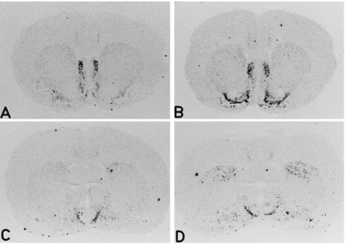
Fig. 2. Effects of chronic cocaine administration on NT mRNA expression. Rats were treated chronically with cocaine (15 mg/kg i.p. for 10 days) or saline and NT gene expression detected by in situ hybridization histochemistry. Typical film autoradiograms from saline- (A,C) and cocaine-injected rats (B,D) are shown at rostral (bregma 1.2 mm; A,B) and caudal levels (bregma -0.4 mm; C,D). Cocaine treatment increased NT gene expression in the shell of the nucleus accumbens (B) and in the dorsomedial striatum at the caudal level (D). No detectable differences were observed in the dorsal striatum at the rostral level (B).
Fig. 3. Quantification of the effects of cocaine on NT mRNA expression in different regions of the rat brain. Animals were injected daily with cocaine (COC) (15 mg/kg i.p.) or saline (SAL) for 10 days and sacrificed 1 h after the last injection. The data shown correspond to measurements performed at the following levels: shell and core regions of the nucleus accumbens (bregma 1.2 mm); dorsomedial striatum, rostral (1.2 mm) and caudal (-0.4 mm); fundus striati (1.2 mm); lateral septum (1.2 mm) and olfactory tubercle (1.7 mm). Hybridization signal is expressed in nCi/mg protein; bars, mean±S.E.M./group (n=6 rats). Cocaine treatment increased NT gene expression in the shell of the nucleus accumbens but not in the core region (A). In the striatum (B), NT mRNA signal was enhanced after cocaine treatment at the caudal, but not the rostral, level of the dorsomedial striatum, and in the ventrolateral region of the striatum, the fundus striati. No detectable differences were observed at the level of the lateral septum or olfactory tubercle (C). **P<0.01, Student's t-test.
Fig. 4. Photomicrographs from emulsion-coated sections show the effect of chronic cocaine on NT mRNA expression in the shell of the nucleus accumbens at the level of the island of Calleja major (1.2 mm to bregma) where the different subdivisions of the nucleus accumbens can be identified. Panel A is a bright-field photomicrograph of a brain section from a control animal counterstained with Toluidine blue; B and C show the same region in dark-field illumination in control (B) and cocaine-treated (C) rats. Cocaine induced an increase of the hybridization signal specific to the shell subdivision. The labeling was mainly concentrated in the ventromedial part of the shell, with little NT mRNA expression observed over the cone part. Note that the enhanced NT mRNA expression after chronic cocaine appears to be mainly due to an increase in the number of labeled cells. ICjM, island of Calleja major; LS, lateral septum; LV, lateral ventricle; ShC, shell subdivision of the nucleus accumbens, cone part; ShV, shell subdivision of the nucleus accumbens, ventromedial part. Scale BAR=100 μm.