HAL Id: hal-01302501
https://hal.sorbonne-universite.fr/hal-01302501
Submitted on 14 Apr 2016HAL is a multi-disciplinary open access archive for the deposit and dissemination of sci-entific research documents, whether they are pub-lished or not. The documents may come from teaching and research institutions in France or abroad, or from public or private research centers.
L’archive ouverte pluridisciplinaire HAL, est destinée au dépôt et à la diffusion de documents scientifiques de niveau recherche, publiés ou non, émanant des établissements d’enseignement et de recherche français ou étrangers, des laboratoires publics ou privés.
membrane oxygenation
Charles-Edouard Luyt, Nicolas Bréchot, Pierre Demondion, Tamara
Jovanovic, Guillaume Hékimian, Guillaume Lebreton, Ania Nieszkowska,
Matthieu Schmidt, Jean-Louis Trouillet, Pascal Leprince, et al.
To cite this version:
Charles-Edouard Luyt, Nicolas Bréchot, Pierre Demondion, Tamara Jovanovic, Guillaume Hékimian, et al.. Brain injury during venovenous extracorporeal membrane oxygenation. Intensive Care Medicine, Springer Verlag, 2016, 42 (5), pp.897-907. �10.1007/s00134-016-4318-3�. �hal-01302501�
Oxygenation
Charles-Edouard Luyt1,2, Nicolas Bréchot1,2, Pierre Demondion3, Tamara Jovanovic1, Guillaume Hékimian1,2, Guillaume Lebreton3, Ania Nieszkowska1,2, Matthieu Schmidt1,2, Jean-Louis Trouillet1,2, Pascal Leprince3, Jean Chastre1,2, and Alain Combes1,2
1
Service de Réanimation, Institut de Cardiologie, Groupe Hospitalier Pitié–Salpêtrière,
Assistance Publique–Hôpitaux de Paris, Paris, France; 2Sorbonne Universités, UPMC Université Paris 06, INSERM, UMRS_1166-ICAN Institute of Cardiometabolism and
Nutrition, Paris, France; 3Service de Chirurgie Thoracique et Cardiovasculaire, Institut de Cardiologie, Groupe Hospitalier Pitié–Salpêtrière, Assistance Publique–Hôpitaux de Paris,
Paris, France.
Correspondence and requests for reprints should be sent to Charles-Edouard Luyt, M.D.,
Ph.D., Service de Réanimation, ICAN, Groupe Hospitalier Pitié–Salpêtrière, 47–83,
boulevard de l’Hôpital, 75651 Paris Cedex 13, France. Tel: +33 (0)1 42 16 38 24; Fax: +33 (0)1 42 16 38 17; Email: [email protected]
Author Contributions: C.-E.L., J.C. and A.C. initiated and designed the study. All authors
contributed to the acquisition of the data, data analysis and the writing of the manuscript, and
approved the final version.
Funding/Support: None.
Short Running Head: Brain injury during venovenous-ECMO
Word count: 3242 words
Abstract
Purpose: The frequency of neurological events and their impact on patients receiving
venovenous-extracorporeal membrane oxygenation (VV-ECMO) are unknown. We therefore
study the epidemiology, risk factors and impact of cerebral complications occurring in
VV-ECMO patients.
Methods: Observational study conducted in a tertiary referral center (2006–2012) on patients
developing a neurological complication (ischemic stroke or intracranial bleeding) while on
VV-ECMO versus those who did not, and systematic review on this topic.
Results: Among 135 consecutive patients having received VV-ECMO, 18 (15 assessable)
developed cerebral complications on ECMO: cerebral bleeding in 10 (7.5%), ischemic stroke
in 3 (2%) or diffuse microbleeds in 2 (2%), occurring after respective medians (IQR) of 3 (1–
11), 21 (10–26) and 36 (8–63) days post-ECMO onset. Intracranial bleeding was
independently associated with renal failure at intensive care unit admission and rapid PaCO2 decrease at ECMO initiation, but not with age, comorbidities or hemostasis disorders. Seven
(70%) patients with intracranial bleeding and one (33%) with ischemic stroke died versus
40% of patients without neurological event. Systematic review found comparable intracranial
bleeding rates (5%).
Conclusions: Neurological events occurred frequently in patients on VV-ECMO. Intracranial
bleeding, the most frequent, occurred early and was associated with higher mortality. Because
it was independently associated with rapid hypercapnia decrease, the latter should be avoided
at ECMO onset, but its exact role remains to be determined. These findings may have major
implications for the care of patients requiring VV-ECMO.
The use of venovenous-extracorporeal membrane oxygenation (VV-ECMO) to treat patients
with refractory acute respiratory distress syndrome (ARDS) has increased in recent years [1,
2]. Among complications occurring during ECMO support, neurological injury is important
because it may be associated with increased mortality and long-term functional sequelae. In
the landmark observational study on VV-ECMO for refractory ARDS complicating
H1N1v2009 influenza, most deaths were attributed to cerebral hemorrhage [3]. To date, only
two studies have specifically investigated neurological complications of ECMO support and
both showed high numbers of brain injuries [4, 5]. However, those observational studies
concerned patients receiving venoarterial-ECMO (VA-ECMO) and most subjects were
included after cardiac surgery or cardiac arrest. VV-ECMO neurological complications have
mostly been reported as case reports [6, 7].
Because no epidemiological data on neurological complications occurring during
VV-ECMO are available, we undertook this retrospective study on information prospectively
collected from a large series of VV-ECMO patients to describe the frequency, morbidity,
mortality and risk factors of cerebral injury. We then conducted a systematic review of all
reported neurological complications in VV-ECMO series.
METHODS
All patients admitted to our intensive care unit (ICU) from 2006 to 2012 who received
VV-ECMO support were included. Because circulatory assistance of any type can be complicated
by thromboembolic phenomena, particularly cerebral infarction due to embolism, we
excluded patients having received a VA-ECMO (peripheral or central), a left ventricle assist
device (VAD), a BiVAD or a total artificial heart before, during or after VV-ECMO run.
Thus, patients switched from VA-ECMO to VV-ECMO or from VV- to VA-ECMO were
admission and during the stay was collected prospectively. In particular, any events occurring
during ECMO support were prospectively recorded in the ICU’s database.
We defined clinical neurological complication as any clinical event occurring during
ECMO course. It included any clinical sign suggestive of stroke (hemiplegia, mydriasis,
anisocoria, asymmetry on clinical examination), but also confusion, delirium, seizures, coma
despite sedation withdrawal. Patients were categorized according to the presence or not of
brain injury on brain imaging (i.e. no brain damage, ischemic stroke, intracranial bleeding and
microbleeds) and groups were compared.
ECMO Circuit
The extracorporeal system consisted of polyvinyl chloride tubing, a membrane oxygenator
(QuadroxBioline, Jostra-Maquet, Orléans, France, or Eos ECMO, Sorin, Milan, Italy), a
centrifugal pump (Rotaflow, Jostra-Maquet, or Revolution, Sorin), and drainage and
reinfusion cannulae (Biomedicus Carmeda, Medtronic, Boulogne-Billancourt, France, or
Edwards Lifesciences, Irvine, CA). An oxygen–air blender (Sechrist Industries, Anaheim,
CA) was used to ventilate the membrane oxygenator. All patients had two-site cannulation
and all cannulae were inserted percutaneously. The drainage cannula was inserted into the
femoral vein (extending into the inferior vena cava) and the reinfusion cannula was inserted
into the internal jugular vein (extending into the right atrium).
Patient Management under ECMO
All patients had a blood gas analysis in the 2 hours preceding ECMO start (blood gas analysis
pre-ECMO) and in the 2 hours following ECMO start (blood gas analysis post-ECMO).
All patients had the same protocol for anticoagulation. A heparin bolus (5000 IU) was
heparin. The heparin dose was adapted at least once daily according to activated partial
thrombin time (aPTT) value (targeting 1 to 1.5-fold the control value) and clinical tolerance;
heparin was stopped when bleeding occurred and restarted once it was controlled. Bleeding
leading to heparin withdrawal was defined as any clinical bleeding judged significant by the
physician in charge of the patient (at ECMO site, central or arterial lines, tracheal secretions,
ear nose and throat) with or without hemodynamic impact, with or without decrease in
haemoglobin level. Anticoagulant overdose was defined biologically as an aPTT
(patient/control) ratio >2.5, corresponding to an absolute value of ≥80 s.
Membrane oxygenator and circuitry were checked daily by experienced perfusionists and
were changed when fibrin deposition or thrombi had deleterious effects on blood
oxygenation, platelet count (<20,000/ml) or blood fibrinogen level (<1.5 g/L) decreased
significantly, or significant intravascular hemolysis (free plasma haemoglobin> 200 mg/L at 2
time points and no other cause of mechanical hemolysis) appeared. No systematic circuit
change was scheduled.
Daily routine neurologic examinations were performed in our patients by the
physicians and the nurses in charge of the patient. Physicians performed at least once daily
neurologic examination after sedation withdrawal, including Glasgow coma scale calculation,
response to verbal orders or pain, tendon reflexes, brainstem reflexes and plantar reflex, eye opening and pupils’ examination. Size of pupils and their light reactivity were assessed by the nurse in charge of the patient every 4 hours. Moreover, when an unexpected event occurred (such as seizures, delirium confusion, no awakening after sedation withdrawal…), this was registered in the medical chart. Once a neurologic symptom was observed (including but not
restricted to change in neurological exam, mydriasis, anisocoria, seizures, delirium, confusion, coma despite sedation withdrawal…) within 6 hours a cerebral CT scan was performed.
Literature Review
We conducted a systematic MEDLINE-Database literature review through the PubMed search
engine with a global search strategy applying prespecified selection and outcome (occurrence
of neurological complication and mortality) criteria using the terms ECMO,
venovenous-ECMO and extracorporeal oxygenation. Details on methodology and the results of this review
are presented in an Online Supplemental Data.
Statistical Analyses
Data are expressed as medians [25th–75th percentile interquartile range (IQR)] or means ( standard deviation (SD)), as appropriate. Between-group comparisons were analyzed using Student’s t-test or the Mann-Whitney U-test for continuous variables, chi-square test for categorical variables. For dichotomous variables, the median value was chosen for the cut-off
to separate the variable into two categories. To examine the univariable impact of patients’
clinical characteristics and ICU events on the development of cerebral bleeding, a
logistic-regression model tested each characteristic. Thereafter, multivariable analysis with a Cox
regression model compared the factors that were significant in the univariable analysis. The
analysis was run using backward-stepwise variable elimination (with the variable-exit
threshold set at P > 0.05). Risk factors achieving P 0.10 in our univariable analysis and parameters previously reported to be strongly associated with intracranial bleeding were
entered into the model. Interactions were tested in the model; variables strongly associated
with other(s) were not included in the multivariable model. The final model included renal
failure on admission, PaO2 change and PaCO2 change. In a sensitivity analysis, lowest
model for intracranial bleeding. Two other endpoints were investigated using Cox regression
model: ICU mortality without intracranial bleeding, and a composite endpoint; intracranial
bleeding or ICU mortality. Analyses were computed with StatView v5.0 (SAS Institute Inc,
Cary, NC) and SPSS v11.5 (SPSS Inc, Chicago, IL) software. P < 0.05 defined significance.
Ethics
In accordance with the ethical standards of our hospitals institutional review boards
(Committee for the Protection of Human Subjects), informed consent for demographic,
physiological and hospital-outcome data analyses was not obtained because this observational
study did not modify existing diagnostic or therapeutic strategies.
RESULTS
During the study period, 135 patients required VV-ECMO support. Table 1 reports their
baseline characteristics. For 23 of those 135 patients, a cerebral event could not be ruled out
because they died before neurological examination could be performed or had preexisting
neurological disease. A clinical neurological symptom appeared in 25 (19%) during
VV-ECMO support (Figure 1). Three of those 25 were clinically brain dead, confirmed by
electroencephalogram, and did not undergo cerebral imaging; therefore, hemorrhagic stroke
could not be excluded.
Brain imaging was obtained for the remaining 22 patients (computed-tomography (CT)
scan for 21 and magnetic resonance imaging (MRI, performed after ECMO removal) for one),
and five other patients without any clinical signs underwent cerebral CT scans (for one of
them with traumatic brain injury, the CT scan was obtained for follow-up) (Figure 1).
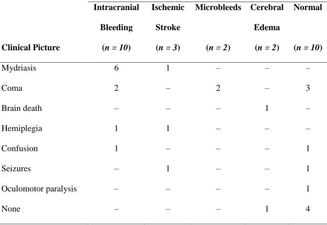
Cerebral-imaging findings as a function of clinical neurological feature(s) are given in Table
bleeding, either intraparenchymal hematoma (n = 9) or subarachnoid hemorrhage (n = 1);
three patients had ischemic strokes; two patients had diffuse microbleeds and two patients had
brain edema (for the latter, the neurological injury preceded ECMO: one suffered traumatic
brain injury 7 days before ECMO with cerebral edema before ECMO; the other had
prolonged cardiac arrest 2 days pre-ECMO and cerebral edema was attributed to cerebral
anoxia).
Thus, 18 (13%, 95% CI 7.3–18.7%) of the 135 patients had a cerebral complication that
occurred while on ECMO (three brain deaths without imaging, 10 intracranial bleeds, three
ischemic strokes and two diffuse microbleeds). Table 1 gives patients’ characteristics as a
function of the type of neurological event occurring on ECMO or not. Patients with such
complications were older and more frequently had renal failure at ICU admission (especially
patients with intracranial bleeding) than those without these events. Other parameters did not
differ between groups, although patients with intracranial bleeding tended to have higher
mortality and shorter time from ECMO onset to neurological complications.
Patients with Intracranial Bleeding
Because the predominant neurological event was intracranial bleeding, we specifically
analyzed those 10 patients to attempt to identify risk factors associated with this complication.
Figure 2 reports the pH, PaCO2 and PaO2 changes between just before and after starting ECMO in patients with and without intracranial bleeding. Comparing patients without to
those with cerebral bleeding, respectively, the latter had higher PaO2 increases just after ECMO onset (medians [IQR]: 20 [4.7–52] vs. 58 [19–214] mmHg; P = 0.04), a not
significant PaCO2 decrease just after ECMO initiation (–24 [–15 to –38] vs. –33 [–28 to –38] mmHg; P = 0.06) but no pH change difference just after starting ECMO (0.17 [0.09–0.26] vs.
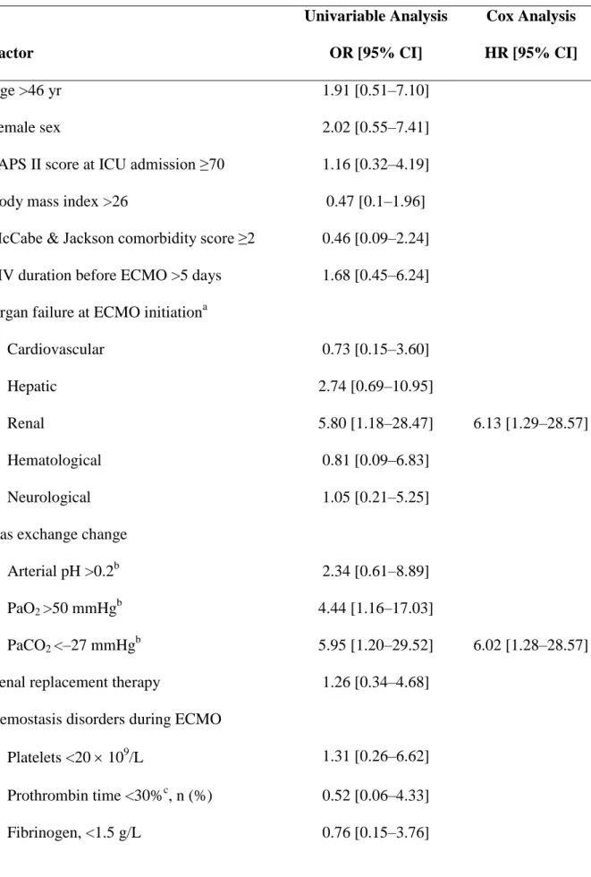
Table 3 reports the factors associated with intracranial bleeding. Renal failure at ICU
admission, PaO2 increase just after ECMO initiation >50 mmHg and PaCO2 decrease just after ECMO onset <–27 mmHg were the only factors significantly associated with brain
hemorrhage in the univariable analysis, whereas the Cox analysis retained renal failure at ICU
admission and PaCO2 decrease as the only factors independently associated with intracranial bleeding. Absolute baseline values of PaO2 and PaCO2 were not associated with intracranial
bleeding. Age, female sex and mechanical ventilation duration pre-ECMO showed a trend
towards association with intracranial bleeding. Hemostasis disorders were not associated with
intracranial bleeding in uni-or-multivariable analyses (sensitivity analysis not shown).
Table 4 lists the hemostasis-parameter values 3 days before brain injury. These patients’
heparin doses were low, as were their aPPT values and anti-Xa activity, and none had
profound thrombopenia or low fibrinogen levels.
Same risk factors were associated with an increased risk of both death and the composite
endpoint (Online Supplement, Tables E1 and E2). Among them, PaCO2 change and lowest prothrombin time during EMCO, but not renal failure, were associated with ICU mortality
(Online Supplement, Tables E1 and E2).
DISCUSSION
We report here the largest study to date of neurological complications during VV-ECMO. We
found that 25 of our 135 patients (19%) experienced a clinical neurological event, with 18
(13%, 95% CI 7.3–18.7%) of those neurological injuries occurring on VV-ECMO. Cerebral
bleeding was the predominant complication, manifesting as coma or mydriasis. Intriguingly, it
was not associated with hemostasis disorders or anticoagulant use: platelet counts,
with and without cerebral hemorrhage, and heparin doses during the 3 days preceding cerebral
bleeding were low, without overdose. The only factors independently associated with brain
hemorrhage were renal failure at ICU admission and acute PaCO2 changes at ECMO initiation.
Our results are in accordance with the literature on neurological complications occurring
in ECMO patients (Table 5). Kasirajan et al. reported 18.9% intracranial bleeding for their
series of 74 VA-ECMO patients. In that study, cerebral bleeding was independently
associated with female sex and thrombocytopenia. Mateen et al. retrospectively studied 87
VA-ECMO patients and found a high rate of neurological injury, including stroke, intracranial
bleeding and brain death [5]; the 10 brains examined at autopsy revealed more neurological
sequelae than would have been predicted by clinical findings alone. However, that study
included VA-ECMO patients after cardiac surgery or cardiac arrest. The authors of the largest
study that included 23,951 patients given ECMO support reported 10.9% neurological
complications [13]. Ischemic stroke, cerebral bleeding and seizure frequencies were 4.1%,
3.6% and 4.1%, respectively, but, as underlined by the authors themselves, they mixed adults,
children and newborns, and were unable to distinguish patients receiving VV- and/or
VA-ECMO [13]. The cerebral bleeding frequency for VV-VA-ECMO patients observed in the
systematic review (see online supplement) was 5%, similar to ours. However, this systematic
review has several limitations: first, among the 16 studies reporting neurological
complications of ECMO, most of them mixed VV- and VA-ECMO. Thus, the exact cerebral
complication frequency of VV-ECMO is difficult to calculate precisely. Another limitation is
that most previous studies reported only cerebral bleeding and no other neurological
complications, e.g. ischemic stroke. Lastly, unlike our investigation, those studies had not
been designed to evaluate factors associated with neurological complications.
differs. In VA-ECMO patients, brain damage could reflect the pre-ECMO clinical context
(low blood pressure and cerebral blood flow, hypoxia, acidosis, electrolyte disturbances,
and/or hemostasis disorders related to hepatic failure frequently observed in cardiogenic
shock), reperfusion injury at ECMO implantation, the ECMO process itself (embolic stroke
from the arterial cannula) and/or hemostasis disorders induced by the ECMO circuit. To date,
the exact mechanism of brain damage is not fully understood and probably includes a
combination of all those factors.
In VV-ECMO patients, a combination of factors also probably leads to neurological
complications, even though some of these factors are not strictly the same. Whereas disorders
created by the ECMO circuit and the oxygenator (hemolysis, thrombocytopenia, fibrinolysis,
acquired von Willebrand syndrome [14]) are the same in VA- and VV-ECMO, pre-ECMO
factors and ECMO-induced metabolic changes could differ. The pre-ECMO factors include
hypoxia, hypercapnia, respiratory acidosis and parameters associated with acute respiratory
failure before starting support. VV-ECMO specificities include abrupt O2 and CO2 changes at ECMO onset [15, 16]. Because CO2 is involved in the regulation of cerebral blood flow [17, 18], sudden CO2-level changes (from hypercapnia to normocapnia or hypocapnia) at ECMO initiation could have induced sudden cerebral blood-flow changes that could have precipitated
brain damage. Recently, Muellenbach et al. reported that patients receiving vvECMO
treatment were at risk for a decrease in cerebral regional tissue oxygen saturation at ECMO
initiation, and that this decreased is linked to PaCO2 change. This could be involved in the
pathogenesis of brain injury of ECMO patients [19, 20]. In our study, the acute PaCO2 change was independently associated with cerebral bleeding and one can hypothesize that these
abrupt changes may have facilitated brain hemorrhages. Although we have no definite proof
of this association, we think it should be taken into account when initiating VV-ECMO and in
correction. It could be achieved by starting with a low sweep gas flow and progressively
increasing it over time. Even though PaO2 change was not independently associated with cerebral bleeding, it can be recommended not to correct it too quickly by starting with a low
fraction of inspired oxygenand increasing it slowly over time. However, because rapid
decrease in PaCO2 leads to vasoconstriction, the relationship between PaCO2 change and
cerebral bleeding is difficult to understand. We cannot exclude that ECMO-induced alkalosis
have caused microbleeds or excessive local flow with altered brain blood barrier and
bleeding.
Surprisingly, hemostasis disorders were not associated with intracranial bleeding herein.
However, because lowest prothrombin time while on ECMO was strongly associated with an
increased risk of mortality, the lack of association between lowest prothrombin time and
intracranial bleeding could be an indirect effect of the increased mortality among patients
presenting this risk factor. Moreover, because our patients’ platelet function was not explored,
we cannot exclude that those with intracranial haemorrhage might have had platelet
dysfunction. Our patients received relatively low dose of heparin, but this is explained by our
anticoagulation policy; we target an aPTT ratio of 1 to 1.5, and stop heparin when clinical
bleeding occurs. Although hemostasis disorders could have played a role, the mechanism of
cerebral bleeding in those patients is probably multifactorial, including vascular injury due to
underlying disease severity, disorders created by the ECMO circuit (hemolysis,
fibrinolysis…), rapid PaO2 and PaCO2 changes and other, as yet unknown, factors. Future studies should investigate the precise mechanisms that could precipitate neurological
complications, particularly controllable factors like PaO2 and PaCO2 changes.
Our study has several limitations. First, this was a single-center study and, although we
included many VV-ECMO patients, not many developed neurological events. Thus, our
will have to be confirmed by larger studies. Moreover, because the delivery of ECMO is so
variable from centre to centre (devices used, techniques employed; management of
anticoagulation and transfusion, thresholds for addressing hypoxemia, and so on), these
results would be difficult to extrapolate to other centres. Second, although it is our policy to
carefully check hemostasis parameters in patients on VV-ECMO, we did not examine
hemostasis, specifically thrombosis and fibrinolysis, with thromboelastogram, D-dimer or
other tests [21]. Because it is well-known that ECMO patients have primary hemostasis
disorders (e.g., acquired von Willebrand disease [14]), we cannot exclude the possibility of
unrecognized hemostasis disorders that might explain the high intracranial bleeding
frequency. Third, because it was difficult to assess neurological status at ICU admission, it is
possible that, at least in some patients, neurological complications may have preceded ECMO
and that ECMO aggravated them. However, albeit impossible to demonstrate, it is not likely,
since our policy is to carefully check daily for clinical signs of brain involvement, in
particular at ICU admission. Fourth, because 23 patients could not be evaluated before dying
and because we also cannot exclude that some patients might have had an undetected
neurological injury, we may have underestimated the exact frequency of brain injury, as in
most studies published to date and reporting neurological outcomes. Fifth, because
neuroimaging was not performed in all patients, we could have underestimated the frequency
of neurological injury (missing subclinical events). Sixth, we found no direct association
between hemostasis disorders and intracranial bleeding, but lowest prothrombin time while on
ECMO was strongly associated with ICU mortality. This lack of association between lowest
prothrombin time and intracranial bleeding could be an indirect effect of the increased
mortality among patients presenting this risk factor. Lastly, we identified 2 conditions
associated with cerebral bleeding during ECMO, but their relationships with brain
we explored only the blood gas change at ECMO start, not during the whole ECMO course.
We therefore cannot be sure that rapid change of PaO2/PaCO2 could have occurred after
several hours or days and could have play a role in brain injury. Future studies should explore
these possibilities.
In conclusion, neurological complications are frequent in patients on VV-ECMO.
Intracranial bleeding, the most frequent event, occurred early during ECMO and was
associated with high mortality. The precise roles of sudden O2- and CO2-level changes need to be more precisely evaluated, not only for the sake of knowledge, but mainly because they
might have major implications in the care of patients requiring VV-ECMO.
Acknowledgment: The authors thank Janet Jacobson for editorial assistance in revising the
manuscript and Marguerite Guiguet for statistical assistance.
Conflicts of Interest: Alain Combes is the primary investigator of the EOLIA,
NCT07470703, a randomized trial of VV-ECMO supported in part by MAQUET. Alain
Combes has received honoraria for lectures by MAQUET, BAXTER, and ALUNG. Other
References
1. Luyt CE, Combes A, Becquemin MH, Beigelman-Aubry C, Hatem S, Brun AL, Zraik N,
Carrat F, Grenier PA, Richard JC, Mercat A, Brochard L, Brun-Buisson C,Chastre J
(2012) Long-term outcomes of pandemic 2009 influenza A (H1N1)-associated severe
acute respiratory distress syndrome. Chest 142:583-592.
2. Combes A, Brodie D, Bartlett R, Brochard L, Brower R, Conrad S, De Backer D, Fan E,
Ferguson N, Fortenberry J, Fraser J, Gattinoni L, Lynch W, MacLaren G, Mercat A,
Mueller T, Ogino M, Peek G, Pellegrino V, Pesenti A, Ranieri M, Slutsky A,Vuylsteke A
(2014) Position paper for the organization of extracorporeal membrane oxygenation
programs for acute respiratory failure in adult patients. Am J Respir Crit Care Med 190:
488-96.
3. Davies A, Jones D, Bailey M, Beca J, Bellomo R, Blackwell N, Forrest P, Gattas D,
Granger E, Herkes R, Jackson A, McGuinness S, Nair P, Pellegrino V, Pettila V, Plunkett
B, Pye R, Torzillo P, Webb S, Wilson M,Ziegenfuss M (2009) Extracorporeal Membrane
Oxygenation for 2009 Influenza A(H1N1) Acute Respiratory Distress Syndrome. JAMA
302: 1888-95.
4. Kasirajan V, Smedira NG, McCarthy JF, Casselman F, Boparai N,McCarthy PM (1999)
Risk factors for intracranial hemorrhage in adults on extracorporeal membrane
oxygenation. Eur J Cardiothorac Surg 15: 508-14.
5. Mateen FJ, Muralidharan R, Shinohara RT, Parisi JE, Schears GJ,Wijdicks EF (2011)
Neurological injury in adults treated with extracorporeal membrane oxygenation. Arch
Neurol 68: 1543-9.
6. Chow FC, Edlow BL, Frosch MP, Copen WA,Greer DM (2011) Outcome in patients with
H1N1 influenza and cerebrovascular injury treated with extracorporeal membrane
7. Le Guennec L, Bertrand A, Laurent C, Roze H, Chastre J, Combes A,Luyt CE (2015)
Diffuse cerebral microbleeds after extracorporeal membrane oxygenation support. Am J
Respir Crit Care Med 191: 594-6.
8. McHorney CA, Ware JE, Jr., Lu JF,Sherbourne CD (1994) The MOS 36-item Short-Form
Health Survey (SF-36): III. Tests of data quality, scaling assumptions, and reliability
across diverse patient groups. Med Care 32: 40-66.
9. Zigmond AS,Snaith RP (1983) The hospital anxiety and depression scale. Acta Psychiatr
Scand 67: 361-70.
10. Stoll C, Kapfhammer HP, Rothenhausler HB, Haller M, Briegel J, Schmidt M,
Krauseneck T, Durst K,Schelling G (1999) Sensitivity and specificity of a screening test
to document traumatic experiences and to diagnose post-traumatic stress disorder in
ARDS patients after intensive care treatment. Intensive Care Med 25: 697-704.
11. Trouillet JL, Luyt CE, Guiguet M, Ouattara A, Vaissier E, Makri R, Nieszkowska A,
Leprince P, Pavie A, Chastre J,Combes A (2011) Early percutaneous tracheotomy versus
prolonged intubation of mechanically ventilated patients after cardiac surgery: a
randomized trial. Ann Intern Med 154: 373-83.
12. Schmidt M, Zogheib E, Roze H, Repesse X, Lebreton G, Luyt CE, Trouillet JL, Brechot
N, Nieszkowska A, Dupont H, Ouattara A, Leprince P, Chastre J,Combes A (2013) The
PRESERVE mortality risk score and analysis of long-term outcomes after extracorporeal
membrane oxygenation for severe acute respiratory distress syndrome. Intensive Care
Med 39: 1704-13.
13. Nasr DM,Rabinstein AA (2015) Neurologic Complications of Extracorporeal Membrane
14. Heilmann C, Geisen U, Beyersdorf F, Nakamura L, Benk C, Trummer G, Berchtold-Herz
M, Schlensak C,Zieger B (2012) Acquired von Willebrand syndrome in patients with
extracorporeal life support (ECLS). Intensive Care Med 38: 62-8.
15. Schmidt M, Tachon G, Devilliers C, Muller G, Hekimian G, Brechot N, Merceron S, Luyt
CE, Trouillet JL, Chastre J, Leprince P,Combes A (2013) Blood oxygenation and
decarboxylation determinants during venovenous ECMO for respiratory failure in adults.
Intensive Care Med 39: 838-46.
16. Schmidt M, Pellegrino V, Combes A, Scheinkestel C, Cooper DJ,Hodgson C (2014)
Mechanical ventilation during extracorporeal membrane oxygenation. Crit Care 18: 203.
17. Lassen NA,Christensen MS (1976) Physiology of cerebral blood flow. Br J Anaesth 48:
719-34.
18. Meng L,Gelb AW (2014) Regulation of Cerebral Autoregulation by Carbon Dioxide.
Anesthesiology 122:196-205.
19. Muellenbach RM, Kilgenstein C, Kranke P, Kustermann J, Kredel M, Roewer N, Ernestus
RI,Westermaier T (2014) Effects of venovenous extracorporeal membrane oxygenation on
cerebral oxygenation in hypercapnic ARDS. Perfusion 29: 139-41.
20. Kredel M, Lubnow M, Westermaier T, Muller T, Philipp A, Lotz C, Kilgenstein C,
Kustermann J, Roewer N,Muellenbach RM (2014) Cerebral tissue oxygenation during the
initiation of venovenous ECMO. ASAIO J 60: 694-700.
21. Repesse X, Au SM, Brechot N, Trouillet JL, Leprince P, Chastre J, Combes A,Luyt CE
(2013) Recombinant factor VIIa for uncontrollable bleeding in patients with
extracorporeal membrane oxygenation: report on 15 cases and literature review. Crit Care
17: R55.
22. Kolla S, Awad SS, Rich PB, Schreiner RJ, Hirschl RB,Bartlett RH (1997) Extracorporeal
23. Linden V, Palmer K, Reinhard J, Westman R, Ehren H, Granholm T,Frenckner B (2000)
High survival in adult patients with acute respiratory distress syndrome treated by
extracorporeal membrane oxygenation, minimal sedation, and pressure supported
ventilation. Intensive Care Med 26: 1630-7.
24. Mols G, Loop T, Geiger K, Farthmann E,Benzing A (2000) Extracorporeal membrane
oxygenation: a ten-year experience. Am J Surg 180: 144-54.
25. Hemmila MR, Rowe SA, Boules TN, Miskulin J, McGillicuddy JW, Schuerer DJ, Haft
JW, Swaniker F, Arbabi S, Hirschl RB,Bartlett RH (2004) Extracorporeal life support for
severe acute respiratory distress syndrome in adults. Ann Surg 240: 595-605.
26. Brogan TV, Thiagarajan RR, Rycus PT, Bartlett RH,Bratton SL (2009) Extracorporeal
membrane oxygenation in adults with severe respiratory failure: a multi-center database.
Intensive Care Med 35: 2105-14.
27. Peek GJ, Mugford M, Tiruvoipati R, Wilson A, Allen E, Thalanany MM, Hibbert CL,
Truesdale A, Clemens F, Cooper N, Firmin RK,Elbourne D (2009) Efficacy and economic
assessment of conventional ventilatory support versus extracorporeal membrane
oxygenation for severe adult respiratory failure (CESAR): a multicentre randomised
controlled trial. Lancet 374: 1351-63.
28. Noah MA, Peek GJ, Finney SJ, Griffiths MJ, Harrison DA, Grieve R, Sadique MZ,
Sekhon JS, McAuley DF, Firmin RK, Harvey C, Cordingley JJ, Price S, Vuylsteke A,
Jenkins DP, Noble DW, Bloomfield R, Walsh TS, Perkins GD, Menon D, Taylor
BL,Rowan KM (2011) Referral to an extracorporeal membrane oxygenation center and
mortality among patients with severe 2009 influenza A(H1N1). JAMA 306: 1659-68.
29. Patroniti N, Zangrillo A, Pappalardo F, Peris A, Cianchi G, Braschi A, Iotti GA,
Arcadipane A, Panarello G, Ranieri VM, Terragni P, Antonelli M, Gattinoni L, Oleari
A(H1N1) pandemic: preparation for severe respiratory emergency outbreaks. Intensive
Care Med 37: 1447-57.
30. Pham T, Combes A, Roze H, Chevret S, Mercat A, Roch A, Mourvillier B,
Ara-Somohano C, Bastien O, Zogheib E, Clavel M, Constan A, Marie Richard JC,
Brun-Buisson C,Brochard L (2103) Extracorporeal membrane oxygenation for pandemic
influenza A(H1N1)-induced acute respiratory distress syndrome: a cohort study and
propensity-matched analysis. Am J Respir Crit Care Med 187: 276-85.
31. Lindskov C, Jensen RH, Sprogoe P, Klaaborg KE, Kirkegaard H, Severinsen IK,
Lorentsen AG, Folkersen L, Ilkjaer S,Pedersen CM (2013) Extracorporeal membrane
oxygenation in adult patients with severe acute respiratory failure. Acta Anaesthesiol
Scand 57: 303-11.
32. Michaels AJ, Hill JG, Bliss D, Sperley BP, Young BP, Quint P, Shanks TR, Dalthorp J,
Long WB,Morgan LJ (2013) Pandemic flu and the sudden demand for ECMO resources:
a mature trauma program can provide surge capacity in acute critical care crises. J Trauma
Acute Care Surg 74: 1493-7.
33. Roch A, Hraiech S, Masson E, Grisoli D, Forel JM, Boucekine M, Morera P, Guervilly C,
Adda M, Dizier S, Toesca R, Collart F,Papazian L (2014) Outcome of acute respiratory
distress syndrome patients treated with extracorporeal membrane oxygenation and
brought to a referral center. Intensive Care Med 40: 74-83.
34. Ng GW, Leung AK, Sin KC, Au SY, Chan SC, Chan OP,Wu HH (2014) Three-year
experience of using venovenous extracorporeal membrane oxygenation for patients with
severe respiratory failure. Hong Kong Med J 20: 407-12.
35. Kon ZN, Dahi S, Evans CF, Byrnes KA, Bittle GJ, Wehman B, Rector RP, McCormick
Extracorporeal Membrane Oxygenation Support for Acute Respiratory Distress
Syndrome. Ann Thorac Surg 100:2059-63.
36. Gray BW, Haft JW, Hirsch JC, Annich GM, Hirschl RB,Bartlett RH (2015)
Figure Legends
Figure 1. Flow chart of the study. VV-ECMO, venovenous-extracorporeal membrane
oxygenation.
Figure 2. Gas exchange parameter changes at the start of venovenous-extracorporeal
membrane oxygenation (VV-ECMO) in the 10 patients with intracranial bleeding (hatched
boxes) and the remaining 125 (white boxes). (A) Absolute pH change defined as
post-ECMO-onset pH – pre-ECMO pH. (B) Absolute PaCO2 change defined as post-ECMO-onset PaCO2 – pre-ECMO PaCO2. (C) Absolute PaO2 change defined as post-ECMO-onset PaO2 – pre-ECMO PaO2. The horizontal lines inside the box plots are the medians, the lower and upper limits of the box correspond to the 25th and 75th percentile interquartile ranges, the T-bars represent the 10th and 90th percentile interquartile ranges, and the circles are outliers.
Table 1. Admission Characteristics, Hemostasis Disorders during VV-ECMO and Patient
Outcomes According to Brain Damage
No Brain Patients with Brain Damage
Characteristic Damage (n = 117) Intracranial Bleeding (n = 10) Microbleeds (n = 2) Ischemic Stroke (n = 3) Age, yra 44 ± 14 49 ± 12 54 ± 4 59 ± 6 Male sex, n (%) 79 (68) 5 (50) 2 (100) 1 (33)
Body mass index, kg/m2 26 [24–31] 23 [21–27] 22 [21–22] 26 [25–47] McCabe & Jackson
comorbidity score ≥2, n (%)
41 (35) 2 (20) 0 1 (33)
ICU-admission SAPS II score 69 ± 14 71 ± 12 61 ± 1 71 ± 9
H1N1v2009 influenza-related
ARDS, n (%)
18 (15) 1 (10) 0 0
Duration of MV before ECMO,
days
5 [1–10] 8 [4–11] 5 [3–7] 11 [9–17]
Organ failure at ECMO startb
Cardiovascular 30 (25) 2 (20) 1 (50) 0
Hepatic 25 (23) 4 (40) 0 0
Renala 48 (41) 8 (80) 0 2 (67)
Hematological 15 (13) 1 (10) 0 0
Neurological 94 (80) 8 (80) 1 (50) 3 (100)
Gas exchange values
pre-ECMO
[7.16–
7.34]
7.29] 7.17] 7.40]
PaCO2 59 [49–73] 74 [57–94] 57 [53–60] 58 [53–63] PaO2 55 [47–68] 68 [66–75] 86 [42–129] 61 [55–67] Gas exchange value
post-ECMO Arterial pH 7.44 [7.37– 7.50] 7.41–7.36– 7.45] 7.43 [7.38– 7.48] 7.52 [7.35– 7.57] PaCO2 34 [28.9– 38.7] 32 [30–47] 31 [20–41] 36 [26–37] PaO2 78 [66– 118] 154 [101– 257] 140 [42– 237] 84 [63–125]
Hemostasis disorders during
ECMOc
Platelet count, 109/L 46 [30–85] 59 [26–77] 100 [76– 125]
109 [50–177]
Patients with platelets <20
109/L, n (%)
20 (17) 2 (20) 0 0
Patients with platelets
<10 109/L, n (%)
7 (6) 0 0 0
Prothrombin time, % of the
standard value
53 [37–64] 46 [39–70] 62 [43–80] 50 [29–60]
Patients with PT <30%, n
(%)
Patients with PT <20%, n (%) 13 (11) 0 0 0 Fibrinogen, g/L 2.5 [1.6– 3.9] 2.3 [1.6–4.6] 3.2 [0.5–5.5] 2.2 [0.7–3.9]
Patients with fibrinogen
<1.5 g/L, n (%)
26 (22) 2 (20) 1 (50) 1 (33)
Patients with fibrinogen <1
g/L, n (%)
16 (14) 1 (10) 1 (50) 1 (33)
Anticoagulant overdose, n (%) 15 (13) 0 0 0
Days of ECMO before
neurological eventd
– 3 [1–11] 36 [8–63] 21 [11–26]
Total duration of ECMO
support, days
10 [6–25] 3.5 [2–6] 29 [3–54] 15 [7–29]
ICU length of stay, days 18 [8–30] 3.5 [2–22] 36 [8–63] 16 [5–40]
Mortality, n (%) 45 (38) 7 (70) 0 1 (33)
Results are expressed as mean ± SD, number (%) or median [27th–75th percentile interquartile range].
Abbreviations: SAPS, Simplified Acute Physiology Score; VV-ECMO,
venovenous-extracorporeal membrane oxygenation; MV, mechanical ventilation; ARDS, acute respiratory
distress syndrome; PT, prothrombin time; ICU, intensive care unit. a
P < 0.05 for between-group comparisons.
b
Organ failure was deemed present when the corresponding Sepsis-related Organ Failure
Assessment score was >2. c
Worst value before or during ECMO. d
Table 2. Cerebral Imaging Findings and Their Corresponding Clinical Features in the 22
Patients
Cerebral Imaging Findings
Clinical Picture Intracranial Bleeding (n = 10) Ischemic Stroke (n = 3) Microbleeds (n = 2) Cerebral Edema (n = 2) Normal (n = 10) Mydriasis 6 1 – – – Coma 2 – 2 – 3 Brain death – – – 1 – Hemiplegia 1 1 – – – Confusion 1 – – – 1 Seizures – 1 – – 1 Oculomotor paralysis – – – – 1 None – – – 1 4
Table 3. Univariable and Multivariable Analyses of Factors Associated With Intracranial
Bleeding (Excluding Microbleeds) on VV-ECMO
Factor Univariable Analysis OR [95% CI] Cox Analysis HR [95% CI] Age >46 yr 1.91 [0.51–7.10] Female sex 2.02 [0.55–7.41]
SAPS II score at ICU admission ≥70 1.16 [0.32–4.19]
Body mass index >26 0.47 [0.1–1.96] McCabe & Jackson comorbidity score ≥2 0.46 [0.09–2.24] MV duration before ECMO >5 days 1.68 [0.45–6.24]
Organ failure at ECMO initiationa
Cardiovascular 0.73 [0.15–3.60]
Hepatic 2.74 [0.69–10.95]
Renal 5.80 [1.18–28.47] 6.13 [1.29–28.57]
Hematological 0.81 [0.09–6.83]
Neurological 1.05 [0.21–5.25]
Gas exchange change
Arterial pH >0.2b 2.34 [0.61–8.89] PaO2 >50 mmHgb 4.44 [1.16–17.03]
PaCO2 <–27 mmHgb 5.95 [1.20–29.52] 6.02 [1.28–28.57] Renal replacement therapy 1.26 [0.34–4.68]
Hemostasis disorders during ECMO
Platelets <20 109/L 1.31 [0.26–6.62] Prothrombin time <30%c, n (%) 0.52 [0.06–4.33] Fibrinogen, <1.5 g/L 0.76 [0.15–3.76]
Anticoagulant overdose –
Abbreviations: SAPS, Simplified Acute Physiology Score; ICU, intensive care unit; MV,
mechanical ventilation; VV-ECMO, venovenous-extracorporeal membrane oxygenation. a
Organ failure was deemed present when the corresponding Sepsis-related Organ Failure
Assessment score was >2. b
Defined as the post-ECMO pH, PaCO2 or PaO2value – the pre-ECMO pH, PaCO2 or PaO2 value.
c
Table 4. Hemostasis Parameters of Patients on Venovenous-Extracorporeal Membrane
Oxygenation with Intracranial Bleeding During the 3 Days Before the Brain Haemorrhage
Parameter
Patients with Intracranial
Bleeding (n = 10)a
aPTT, patient-to-normal value ratiob 1.14 [1.1–1.7] Anti-Xa activity, IU/Lb 0.1 [0.1– 0.1] Platelet count, 109/Lc 84 [29–135] Fibrinogen, g/Lc 2.65 [2–5.6]] Heparin dose, IU/24 hb 2,750 [0–10,000] Abbreviation: aPTT, activated partial thrombin time.
a
Values are expressed as median [25th–75th interquartile range]. b
Highest value. c
Study No. of Clinical Outcome Hemostasis Anticoagulant ECMO–Brain
Reference Design Patients Neurological Complications Deaths Disorders Use Injury Interval
Kolla [22] Cohort 100: 65 VV-ECMO; 11 VA-ECMO; 34 with both 10 (10%)a: 2 ischemic strokes 2 cerebral hemorrhages 6 brain deaths 2/2 2/2 6/6 NR NR NR
Linden [23] Cohort 17: 8 VV-ECMO;
7 VA-ECMO; 2 with both 3 (18%) a: 1 cerebral edema 1 cerebral bleeding 1 ischemic stroke 1/1 1/1 1/1 NR NR NR
Mols [24] Cohort 62: all VV-ECMO 2 (3%):
1 cerebral bleeding 1 brain death 1/1 1/1 Hemmila [25] Cohort 255: 168 VV-ECMO; 47 VA-37 (15%) a: 14 ischemic strokes 11/14 NR NR NR
ECMO; 40 with both 9 brain deaths 7 cerebral bleeds 7 seizures 9/9 6/7 4/7 Brogan [26] Registry 1473: 703 VV-ECMO; 297 VA-ECMO; 50 with both; 423 unknown 136 (9%) a:
64 cerebral strokes or bleeds
72 brain deaths 51/64 72/72 NR NR NR Peek [27] RCT 90 VV-ECMO arm: 65 received it NR 4/90 died of neurological disorders NR NR NR
Davies [3] Cohort 68 with H1N1 flu:
63 VV-ECMO; 5
VA-ECMO
6 (9%) cerebral bleeds 6/6 NR NR NR
Noah [28] Cohort 69 VV-ECMO for
H1N1 flu
Patroniti [29] Cohort 60: 59 VV-ECMO; 1 VA-ECMO 2 (3%) a: 1 (2%) cerebral bleeding 1 NR 1/1 NR NR 2 days NA Pham [30] Cohort 123:107 VV-ECMO; 16 VA-ECMO 5 cerebral bleeds (4%) a 5/5 NR NR NR Lindskov [31] Cohort 124: 110 VV-ECMO; 14 VA-ECMO NR 9 fatal cerebral infarcts with hemorrhage NR NR NR Michaels [32] Cohort 12: 7 VV-ECMO; 5 VA-ECMO 3 ischemic strokes
1 ischemic brain injury
3/3
1/1
NR NR NR
Roch [33] Cohort 85: 77 VV-ECMO;
8 VA-ECMO
2 (2%) cerebral bleeds a 2/2 NR NR NR
Ng [34] Cohort 31 VV-ECMO 1 (3%) cerebral bleeding 1/1 NR NR NR
Kon [35] Cohort 55 VV-ECMO 4 cerebral bleeds NR NR NR NR
ECMO; 91
VA-ECMO
Abbreviations: VA, venoarterial; VV, venovenous; ECMO, extracorporeal membrane oxygenation; NR, not reported; RCT, randomized–
controlled trial. a
Because the authors of these studies did not differentiate between VV- and VA-ECMO patients, the exact frequency of neurological
complications occurring on VV-ECMO cannot be calculated. b
353 adults received ECMO support for acute respiratory distress syndrome among a series 2000 adults and pediatric patients who received VA-
25 clinical brain events
(19%)
23 patients in whom a neurological event cannot be ruled out
(death or pre-existing brain injury) Normal CT scan N = 4 Brain death, no cerebral imaging N = 3 Brain injury on cerebral imaging N = 16 5 CT scan perform without
clinical signs No brain injury on cerebral imaging N = 6 Brain edema N = 1 Microbleeds N = 2 Ischemic stroke N = 3 Intracranial bleeding N = 10 Brain edema N = 1
-0.2 -0.1 0 0.1 0.2 0.3 0.4 0.5 p H c h a n g e -100 -50 0 50 100 150 200 250 P a O2 c h a n g e , m mHg