HAL Id: hal-01788611
https://hal.sorbonne-universite.fr/hal-01788611
Submitted on 9 May 2018
HAL is a multi-disciplinary open access
archive for the deposit and dissemination of
sci-entific research documents, whether they are
pub-lished or not. The documents may come from
teaching and research institutions in France or
abroad, or from public or private research centers.
L’archive ouverte pluridisciplinaire HAL, est
destinée au dépôt et à la diffusion de documents
scientifiques de niveau recherche, publiés ou non,
émanant des établissements d’enseignement et de
recherche français ou étrangers, des laboratoires
publics ou privés.
Distributed under a Creative Commons Attribution| 4.0 International License
the biogenesis of nuclear pore subunits
Jérôme Rouvière, Manuel Bulfoni, Alex Tuck, Bertrand Cosson, Frédéric
Devaux, Benoit Palancade
To cite this version:
Jérôme Rouvière, Manuel Bulfoni, Alex Tuck, Bertrand Cosson, Frédéric Devaux, et al.. A
SUMO-dependent feedback loop senses and controls the biogenesis of nuclear pore subunits. Nature
Commu-nications, Nature Publishing Group, 2018, 9, pp.1665. �10.1038/s41467-018-03673-3�. �hal-01788611�
A SUMO-dependent feedback loop senses and
controls the biogenesis of nuclear pore subunits
Jérôme O. Rouvière
1,5
, Manuel Bulfoni
2
, Alex Tuck
3,6
, Bertrand Cosson
2
, Frédéric Devaux
4
&
Benoit Palancade
1
While the activity of multiprotein complexes is crucial for cellular metabolism, little is known
about the mechanisms that collectively control the expression of their components. Here, we
investigate the regulations targeting the biogenesis of the nuclear pore complex (NPC), the
macromolecular assembly mediating nucleocytoplasmic exchanges. Systematic analysis of
RNA-binding proteins interactomes, together with in vivo and in vitro assays, reveal that a
subset of
NPC mRNAs are specifically bound by Hek2, a yeast hnRNP K-like protein.
Hek2-dependent translational repression and protein turnover are further shown to
finely tune the
levels of NPC subunits. Strikingly, mutations or physiological perturbations altering pore
integrity decrease the levels of the NPC-associated SUMO protease Ulp1, and trigger the
accumulation of sumoylated versions of Hek2 unable to bind
NPC mRNAs. Our results
support the existence of a quality control mechanism involving Ulp1 as a sensor of NPC
integrity and Hek2 as a repressor of NPC biogenesis.
DOI: 10.1038/s41467-018-03673-3
OPEN
1Institut Jacques Monod, CNRS, UMR 7592, Univ Paris Diderot, Sorbonne Paris Cité, 15 rue Hélène Brion, 75013 Paris, France.2Université Paris Diderot,
Sorbonne Paris Cité, Epigenetics and Cell Fate, UMR7216, CNRS, 35 rue Hélène Brion, 75013 Paris, France.3Wellcome Trust Centre for Cell Biology, University of Edinburgh, Max Born Crescent, Edinburgh EH9 3BF, UK.4Sorbonne Université, CNRS, Institut de biologie Paris-Seine (IBPS), UMR 7238,
Laboratoire de biologie computationnelle et quantitative, LCQB, 4 place Jussieu, 75005 Paris, France.5Present address: Department of Molecular Biology and
Genetics, Aarhus University, C.F. Møllers Allé 3, DK-8000 Aarhus C, Denmark.6Present address: Friedrich Miescher Institute for Biomedical Research,
Maulbeerstrasse 66, 4058 Basel, Switzerland. Correspondence and requests for materials should be addressed to B.P. (email:[email protected])
123456789
V
irtually all cellular processes rely on the function of
multiprotein assemblies. While their stoichiometry has to
be tightly controlled to prevent an imbalance of subunits
that could interfere with their assembly or titrate their targets,
their global abundance has also to be adjusted in response to the
cellular demand
1. Multiple layers of mechanisms have been
reported to partake in the accurate biogenesis of multisubunit
complexes. First, all the steps in the gene expression pathway,
including messenger RNA (mRNA) synthesis, processing,
trans-port, stability and translation, can be regulated in a coordinate
manner, either to lead to the proportional synthesis of the
dif-ferent subunits of multiprotein assemblies, a prominent strategy
in prokaryotes
2, or to respond to environmental or physiological
cues, as exemplified by the ribosome biosynthesis pathway
3. In
this frame, a pivotal role has emerged for transcriptional
regulators and RNA-binding proteins, the latter being in
parti-cular capable to tune the translation rate of their target messenger
ribonucleoparticles (mRNPs). Second, molecular chaperones and
assembly factors can further assist the assembly of multiprotein
complexes, as also described for ribosomes
3, in some cases in a
cotranslational manner
4. Finally, excess complexes or
unas-sembled, orphan polypeptides can be targeted for degradation by
the proteasome or the lysosome
5, with these quality control
processes being critical to adjust stoichiometry and to cope with
altered protein dosage
6,7. However, despite our improved
knowledge in proteome dynamics, the specific mechanisms at
play for most multiprotein complexes remain largely unknown.
The nuclear pore complex (NPC) provides a paradigmatic
example of an essential multisubunit complex whose homeostasis
is crucial yet poorly understood. NPCs are megadalton-sized
0 10 20 30 40 50 P ercentage of IP (%) NSP1 NUP116 NUP133 Φ RNA probe
WB:HA Input 1X Eluate 5X
RNA probe NSP1 NUP116 NUP133 Φ - Hek2 (-HA-His) 55 kDa - Hek2 (-HA-His) 55 - Hek2 (-HA-His) 55 - Hek2 (-HA-His) 55 (1) (CNN)6 (2) CAUCAUCA NSP1 NUP116 FG FG 21 80 162 221 20 r p n 20 r p n 1 2 3 4 5 6 7 8 9 10 11 12 13 14 15 CRAC CLIP MEME 0 5 10 15 20 25 30 NUP59 NSP1 P ercentage of IP (%) NUP1 NUP100 NUP116
NUP170 NUP188 NUP57 NUP2
NUP133 ASH1 rRNA
Controls Central FG Nups Outer ring Nups Nuclear FG Nups & basket Inner
ring & linker Nups No tag
HEK2-pA
RNA-binding proteins NPC
mRNAs
Nuclear FG Nups & bask
e t Other NPC-associated proteins T ransmembr ane ri ng Nups Outer r ing Nups Inner r ing & link er Nups Centr al FG Nups Cytoplasmic FG Nups Hek2 Sto1 Nab2 Me x67 Xr n1 Ski2 Yra1Npl3Nab4 Mtr4 NUP120 NUP133 NUP145C NUP84 NUP85 SEC13 SEH1 NUP145N NUP116 NUP100 NSP1 NUP57 NUP49 NUP159 NUP42 NUP1 NUP2 NUP60 MLP1 MLP2 DBP5 DYN2 GLE1 GLE2 MAD1 MAD2 PML39 ULP1 NUP157 NUP170 NUP192 NUP188 NIC96 NUP82 NUP53 NUP59 NDC1 POM152 POM34 POM33 1 2 3 4 Cytoplasm Nucleus NPC Nuclear pore complex (NPC)
Binding (RIP/CLIP hits) Binding (CRAC hits) No data Outer nuclear membrane Inner nuclear membrane
f
e
d
c
b
a
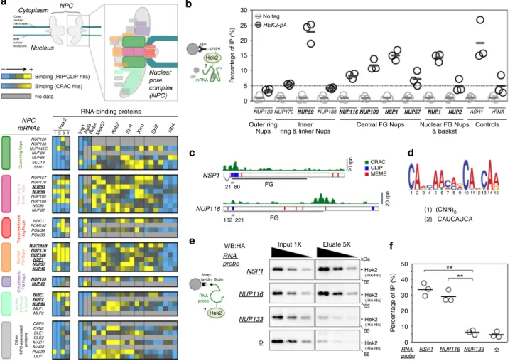
+ – IgG prot-A Hek2 mRNA ? Strep-tavidin Biotin Hek2 ** ** RNA probe ?Fig. 1 The hnRNP K-like protein Hek2 specifically associates with a subset of NPC mRNAs. a Top, Representation of the yeast nuclear pore complex (NPC), showing subcomplexes as colored boxes. Bottom, mRNAs encoding NPC components are sorted by subcomplexes and the strength of their association to the different indicated RNA-binding proteins (RBP) is represented by a color code, as scored in distinct RIP, CLIP or CRAC datasets. Bright yellow indicates the preferred association of a given mRNA to the RBP of interest. For Sto1, Mtr4, Nab2, Mex67, Xrn1 and Ski2, multiple repetitions are displayed27. For Hek2, the results from independent studies are represented: (1)24, (2)26, (3)25, (4)27. FG-Nups appear in bold, underlined. TheNUP145 mRNA gives rise to
both Nup145-N and Nup145-C nucleoporins and is displayed for each of the according subcomplexes.b Hek2-pA-associated mRNAs were immunopurified and quantified by RT-qPCR using specific primer pairs. Percentages of IP are the ratios between purified and input RNAs, normalized to the amount of purified bait and set to 1 for the “no tag”. Means and individual points (n = 3) are displayed. A schematic representation of the assay is shown. c Overview of Hek2-binding sites onNSP1 and NUP116 mRNAs. The number of CRAC hits (rpn (reads per nucleotide))27, the position of CLIP fragments26and the
occurrences of the binding site found by the MEME analysis are indicated. The positions of the FG-coding region and of minimal Hek2-binding sites used for in vitro pull down (in gray) are represented. The broken line indicates theNSP1 intron. d MEME result from NUP59, NUP116, NUP1, NSP1 and NUP100 sequences. (1, 2): previously identified Hek2-binding sequences24,26.e Left, Schematic representation of the assay. Recombinant HA-tagged Hek2 was
incubated with streptavidin beads either naive (Φ) or coated with biotinylated RNA probes encompassing Hek2-binding sites from NSP1 (21–80) or NUP116 (162–221) or a sequence from NUP133 (1429–1488). Right, Decreasing amounts of input and eluate fractions were loaded for quantification. f Percentages of IP are the ratios between Hek2 amounts in the eluate and input fractions, calculated from (e). Means and individual points (n = 3) are displayed. **P < 0.01 (Welch’s t-test)
proteinaceous assemblies embedded at the fusion points of the
nuclear envelope and formed of modular repeats of ~30 distinct
protein subunits—the nucleoporins (Nups)—which assemble
within subcomplexes and organize with a 8-fold rotational
sym-metry
8. The major task of NPCs is the selective
nucleocyto-plasmic transport of macromolecules, i.e., proteins and
RNA-containing particles, a process involving dynamic interactions
between
the
cargo-transport
factor
complexes
and
the
phenylalanine-glycine (FG) repeats-harboring nucleoporins that
lie within the central channel and the peripheral extensions of the
NPC
9. The stepwise assembly of nucleoporins to build complete
NPCs proceeds through defined pathways, either following
mitosis in conjunction with nuclear envelope reformation or
during interphase, the unique assembly mode compatible with the
closed mitosis of fungi. Nucleoporins themselves are essential
players in NPC assembly, either through scaffolding or by
med-iating interactions with chromatin and/or membranes. In
addi-tion, non-NPC factors, such as membrane bending proteins, also
contribute to NPC biogenesis
10. While multiple studies have
depicted the choreography of NPC assembly, together with their
structural organization, little is known about the mechanisms that
sustain the timely production of stoichiometric amounts of Nups
or that could possibly sense and adjust NPC biogenesis depending
on cell physiology.
The high connectivity observed between NPCs and several
bio-logical processes could place them in a strategical position to
communicate their status to the cell. Indeed, NPCs have been
described to contribute to multiple aspects of transcriptional
reg-ulation, genome stability and cell cycle progression
9. In some
situations, these connections are mediated by physical interactions
between NPCs and enzymes of the small ubiquitin-related modifier
(SUMO) pathway
11. Sumoylation is a post-translational
modifica-tion that can modulate the binding properties or the conformamodifica-tion
of its targets, ultimately impacting their stability, their localization
or their biological activity
12. Among the distinct enzymes of the
sumoylation/desumoylation machinery shown to associate with
NPCs, the conserved SUMO protease Ulp1 has essential functions
in SUMO processing and deconjugation in budding yeast. The
docking of this enzyme to the nucleoplasmic side of NPCs is
essential for viability
13,14and is believed to involve its nuclear
import through karyopherins, followed by its association with
several nucleoporins
15–19. Proper NPC localization of Ulp1 has
been shown to be critical for the spatio-temporal control of the
sumoylation of certain targets, some of them being important for
genetic integrity or gene regulation
13,16,20,21.
Here, we report an original mechanism by which the synthesis
of NPC subunits is regulated in response to changes in NPC
integrity in budding yeast. We show that a subset of
Nup-encoding mRNAs is defined by the specific binding of the
translational regulator Hek2. Hek2-regulated NPC mRNA
translation and protein turnover are further shown to
finely tune
the levels of the corresponding nucleoporins. Strikingly, Hek2
binding to NPC mRNAs is prevented by sumoylation, a process
reversed by the SUMO protease Ulp1. Mutant or physiological
situations in which NPC functionality is compromised are
asso-ciated with the loss of Ulp1 activity and the subsequent
accu-mulation of sumoylated Hek2 versions that are inactive for NPC
mRNA translational repression. We propose that Ulp1 and Hek2
are respectively the sensor and the effector of a feedback loop
maintaining nucleoporin homeostasis.
Results
A unique mRNP composition for a subset of
NPC mRNAs. In
order to unravel novel mechanisms regulating NPC biogenesis,
we systematically analyzed the association of Nup-encoding
(NPC) mRNAs with different RNA-binding proteins (RBPs) in
budding yeast. For this purpose, we took advantage of previously
published large-scale datasets obtained through RNA
immuno-precipitation
(RIP)
22–25,
crosslinking
immunoprecipitation
(CLIP)
26or crosslinking and analysis of complementary DNA
(CRAC)
27. We collected the association data for 39 NPC mRNAs
(encoding Nups and NPC-associated proteins, Fig.
1
a and
Sup-plementary Fig.
1
a) with a panel of 10 mRNA-associated factors
involved in different stages of mRNA metabolism, including
assembly into mRNP (Sto1), processing (Npl3, Nab4/Hrp1),
nuclear export (Yra1, Nab2, Mex67), degradation (Xrn1, Ski2,
Mtr4) or mRNA localization/translation (Hek2) (Fig.
1
a). This
analysis revealed that NPC mRNAs have generally the same
typical features of expressed, protein-coding RNAs, e.g., they
readily associate with mRNA export factors (Mex67, Nab2), but
not with the non-coding RNA degradation machinery (Mtr4)
(Fig.
1
a, bottom right panel). Strikingly however, a small subset of
NPC mRNAs (namely NUP170, NUP59, NUP188, NUP116,
NUP100, NSP1 and NUP1) appeared to specifically bind the
conserved Heterogeneous nuclear ribonucleoprotein K-like factor
Hek2 (a.k.a. Khd1
28,29), a feature detected in four independent
datasets (Fig.
1
a, bottom left panel). The enrichment of certain
NPC mRNAs among Hek2-bound targets appeared significant in
a Gene Set Enrichment Analysis (P
= 0.02) and was neither a
mere consequence of the different expression levels of these
particular transcripts (Supplementary Fig.
1
b) nor a general
fea-ture of any multiprotein complexes, since it was not observed
when similar analyses were performed for mRNAs encoding
proteasome or exosome subunits (Supplementary Fig.
1
c).
To further validate this
finding in vivo, we immunoprecipitated
a protein A-tagged version of Hek2 from yeast cells and analyzed
its interaction with NPC mRNAs by reverse
transcription-quantitative polymerase chain reaction (RT-qPCR). In agreement
with our previous
findings, Hek2 preferentially associated with
NUP59, NUP116, NUP100, NSP1 and NUP1 mRNAs (Fig.
1
b), to
a similar extent as its prototypal target ASH1
28,29, but not with
NUP133, NUP57 or NUP2 mRNAs (Fig.
1
b), for which Hek2
binding was in the same range as its reported, unclear association
to rRNA
27. Preferential binding to NUP170 and NUP188 mRNAs
was not confirmed, with the previous finding from genome-wide
studies possibly reflecting their different expression levels in other
genetic backgrounds. In contrast, immunoprecipitation of Hpr1,
a subunit of the mRNP packaging THO complex, did not reveal
any similar preferred association to a subset of NPC mRNAs
(Supplementary Fig.
1
d).
We then asked whether Hek2 was directly associating to this
subset of NPC mRNAs (i.e., NUP59, NUP116, NUP100, NSP1 and
NUP1), as expected from CLIP/CRAC studies
26,27. To this aim,
we
first delineated Hek2-binding sites on these mRNAs by
mining CLIP/CRAC data (Fig.
1
c) and by searching their
sequences for common motifs using the MEME software (Fig.
1
c,
d). This in silico approach revealed that these mRNAs share a
common CA-rich motif (Fig.
1
d), similar to the two previously
reported Hek2-binding sites, i.e. (CNN)
624and CAUCAUCA
26.
As anticipated from a previous study
26, this motif was
over-lapping some but not all in vivo Hek2-binding peaks as defined
by CLIP or CRAC, allowing us to define putative minimal bound
domains in NSP1 and NUP116 mRNAs (Fig.
1
c, gray bars). In an
in vitro binding assay, synthetic biotinylated RNA probes
encompassing these Hek2-binding sequences were further found
to specifically pull down recombinant, purified Hek2 (Fig.
1
e, f),
but not a control protein (Supplementary Fig.
1
e).
Altogether, our data establish that a direct association with the
hnRNP Hek2 specifically defines a subset of NPC mRNPs.
Notably, the
five Hek2-bound NPC mRNAs are coding for
FG-Nups, which are critical for nucleocytoplasmic transport
30.
wt hek2Δ A254 40S Polysomes 60S 80S 40S Polysomes 60S 80S A254 1 2 3 4 5 6 1 2 3 4 5 6 Nup133 Nup1 Nup116 0 50 100 150 200 0 50 100 150 200 0 50 100 150 200 0 50 100 150 200 0 0.4 0.6 0.8 1 1.2 0.2 0 0.4 0.6 0.8 1 1.2 0.2 0 0.4 0.6 0.8 1 1.2 0.2 0 0.4 0.6 0.8 1 1.2 0.2 Nup59 kDa - 130 - 130 kDa - 100 - 100 kDa - 115 - 115 kDa - 80 - 80 wt hek2Δ
Time of CHX treatment (min) Nup59
0 15 45 90 180
wt
hek2Δ
CHX min
Protein amounts (relative to
t=0) wt
hek2Δ
Time of CHX treatment (min) Nup116
0 15
Protein amounts (relative to
t=0) 45 90 180 wt hek2Δ CHX min wt hek2Δ
Time of CHX treatment (min) Nup1
0 15
Protein amounts (relative to
t=0) 45 90 180 wt hek2Δ CHX min wt hek2Δ
Time of CHX treatment (min)
Protein amounts (relative to
t=0) Nup133 0 15 45 90 180 wt hek2Δ CHX min
NSP1 mRNA NSP1 mRNA NUP100 mRNA NUP133 mRNA
NUP100 mRNA NUP133 mRNA DAPI DAPI wt hek2 Δ 4 6 8 10 12 14 16 18 0.5 –0.5 –1 –1.5 –2 2 1.5 1 0 Log ratio ( hek2 Δ /wt ) All mRNAs NPC mRNAs Hek2-bound NPC mRNAs
Log averaged fluorescence intensity
0 0.5 1 1.5 2 2.5 0 0.5 1 1.5 2 3 2.5 0 0.5 1 1.5 2 3 3.5 2.5 0 0.5 1 1.5 2 3 3.5 2.5 Control mRNAs 1 2 3 4 5 6 7 8 9 10 11 12 1314 1 2 3 4 5 6 7 8 9 10 11 12 13 14 1 2 3 4 5 6 7 8 9 10 11 12 1314 1 2 3 4 5 6 7 8 9 10 11 12 13 14 1 2 3 4 5 6 7 8 9 10 11 12 1314 1 2 3 4 5 6 7 8 9 10 11 12 13 14 1 2 3 4 5 6 7 8 9 10 11 12 1314 1 2 3 4 5 6 7 8 9 10 11 12 1314 NSP1 NUP100 NUP116 NUP59 NUP1 ASH1 NUP133 ACT1
mRNA levels (A.U.)
mRNA levels (A.U.)
mRNA levels (A.U.)
mRNA levels (A.U.)
wt hek2Δ wt hek2Δ Hek2-bound mRNAs
a
c
d
b
e
f
1 2 3 4 5 6 7 8 9 10 11 12 13 14 1 2 3 4 5 6 7 8 9 10 11 12 13 14A role for Hek2 in the metabolism of
NPC mRNAs. We further
investigated how Hek2 binding impacts the fate of these
parti-cular NPC mRNAs. While previous studies have revealed that
Hek2 associates with an important fraction of the transcriptome,
the consequences of this recruitment for mRNA metabolism have
only been documented in a few situations where Hek2 binding
can cause increased mRNA stability
24, asymmetrical
localiza-tion
28or translational repression
26,29.
To determine whether Hek2 binding influences the
steady-state levels of NPC mRNAs, we
first profiled the transcriptome
of hek2Δ mutant yeast cells (Fig.
2
a). Genome-wide,
Hek2-bound mRNAs showed a tendency to be less abundant upon
Hek2 inactivation (Supplementary Fig.
2
a), a trend not
observed for Nab2-associated transcripts (Supplementary
Fig.
2
b), highlighting the sensitivity and the specificity of our
analysis. However, NPC mRNAs levels were not significantly
affected by the absence of Hek2, whether or not they associate
with this factor (Fig.
2
a). We then compared the localization of
NPC mRNAs in wt and hek2Δ cells using single-molecule
fluorescence in situ hybridization (smFISH; Fig.
2
b). Detection
of NSP1, NUP100 and NUP133 mRNAs using specific sets of
probes revealed a punctuate, cytoplasmic localization for these
Nup-encoding transcripts in wt cells (Fig.
2
b, top panels). Upon
HEK2 deletion, this random distribution, as well as the total
number of detected RNA dots, were unchanged for both
Hek2-bound (NSP1, NUP100) and Hek2-unHek2-bound (NUP133) mRNAs
(Fig.
2
b, bottom panels). This set of data therefore establishes
that Hek2 binding modulates neither the levels nor the
localization of NPC mRNAs.
We then monitored the possible influence of Hek2 on NPC
mRNA translation using polysome fractionation on sucrose
gradients, which resolve free mRNPs and ribosomal subunits
from translation-engaged mRNAs (Fig.
2
c, Supplementary
Fig.
2
c). RT-qPCR analysis of the fractions of the wt polysome
gradient revealed a bimodal distribution for Hek2-bound (Fig.
2
d,
Supplementary Fig.
2
d, black lines) and Hek2–unbound (Fig.
2
e,
Supplementary Fig.
2
e, black lines) NPC mRNAs. The largest
fraction of NPC mRNAs migrated in the lightest fractions (#1–6),
corresponding to free, untranslated mRNPs and resembling the
pattern observed for the repressed ASH1 mRNA (Fig.
2
d). A less
abundant fraction of NPC mRNAs peaked with
polysome-containing fractions (#9–13), similar to the peak of the
well-translated ACT1 mRNA (Fig.
2
e). Further analysis of the
polysome profile from hek2Δ cells did not reveal any differences
in the distribution of ribosomal species as compared to wt cells
(Fig.
2
c, Supplementary Fig.
2
c). Strikingly, HEK2 inactivation
decreased the amounts of translationally repressed Hek2-bound
NPC mRNAs (Fig.
2
d, gray arrows) and triggered their
redistribution in the translated population, with a peak in heavy
polysomes fractions (≥4 ribosomes/mRNA; Fig.
2
d, red arrows).
This behavior was similar to the one reported for the
Hek2-repressed ASH1 mRNA
29(see also Fig.
2
d) and was not observed
for mRNAs which are not bound by Hek2 (e.g., NUP133 and
ACT1, Fig.
2
e).
Having established that Hek2 binding onto its NPC target
mRNAs contributes to their maintenance in a translationally
repressed state, we wondered whether it would affect the raw
levels of their cognate nucleoporins. Notably, HEK2 inactivation,
while increasing the fraction of translated NUP59, NUP116, or
NUP1 mRNAs (Fig.
2
d), did not trigger any drastic changes in the
steady-state levels of the corresponding nucleoporins (see t
= 0 in
Fig.
2
f). Since excess synthesis of subunits of multiprotein
complexes can be buffered by increased protein turnover
6, we
monitored the half-lives of these nucleoporins in wt and hek2Δ
cells. Strikingly, the degradation rates of the three nucleoporins,
as estimated from cycloheximide chase experiments, were higher
in the absence of Hek2 (Fig.
2
f), revealing that the enhanced
synthesis of nucleoporins is attenuated by their increased
turnover in these mutant cells. Consistently, the kinetics of
degradation of Nup133, whose translation is independent from
Hek2 activity, was unaffected in hek2Δ cells (Fig.
2
f). The raw
levels of this subset of nucleoporins are thereby tightly controlled
by both Hek2-mediated translational control and protein
degradation.
The latter results suggested that Hek2 function might become
crucial in conditions of disturbed proteostasis. To test this
hypothesis, we combined HEK2 inactivation with
MG132-mediated inhibition of proteasomal degradation in
drug-sensitive yeast strains, and further analyzed the cellular
localiza-tion of Nup1, whose overexpression was previously reported to
give rise to lethality
31. Strikingly, simultaneous inhibition of Hek2
and proteasome functions enhanced the formation of abnormal
cytoplasmic foci of this nucleoporin in a small fraction of cells
(Supplementary Fig.
2
f). The
fine-tuning of nucleoporin amounts
mediated by Hek2 translational repression and
proteasome-dependent turnover can thereby be critical to prevent the
accumulation of mislocalized NPC subunits.
Hek2 can be modified by SUMO. Having established that Hek2
can prevent excess Nup production, we then wondered whether
regulatory mechanisms could reverse this repressing activity in
response to an increased cellular demand for nucleoporins.
Yck1-mediated phosphorylation of Hek2 was previously reported to
disrupt its association with the ASH1 mRNA at the bud cortex
where this asymmetrically localized mRNA is targeted
29.
How-ever, this plasma membrane-anchored kinase is unlikely to
Fig. 2 Hek2-dependent translational repression and protein turnover define nucleoporin levels. a Transcriptome analysis of the hek2Δ mutant. The y-axis is the averaged log2 of thehek2Δ/wt ratios calculated from two independent microarray hybridizations. The x-axis is the log2 of the averaged fluorescence intensities. mRNAs encoding NPCs components are colored depending on their association to Hek2 (from Fig.1).b Single-molecule FISH was performed on wt and hek2Δ cells using set of probes specific for the indicated mRNAs. NSP1 and NUP100 probes were coupled to the Quasar570 fluorophore (red), and NUP133 probes to Quasar670 (far red). The z-projections are displayed, together with merged images with a nuclear staining (DAPI). Scale bar, 5 µm. c Polysome fractionation fromwt and hek2Δ cells (W303 background). The absorbance at 254 nm (A254) recorded during the collection of the fractions of
the gradient is displayed. The positions of 40S, 60S, 80S ribosomal species are indicated, as well as the number of ribosomes per mRNA in polysomes fractions.d Relative distribution of the indicated mRNAs in polysome gradients fromwt (black lines) and hek2Δ (red lines) cells. mRNAs amounts in each fraction were quantified by RT-qPCR, normalized to the sum of the fractions and to the distribution of a control spike RNA. Gray arrows indicate a decrease in the amounts of mRNAs found in the light fractions inhek2Δ cells, while red arrows point to an increase in the quantity of mRNAs found in the polysomes fractions. These results are representative of four independent experiments (two performed in the W303 background, two in the BY4742 background; see Supplementary Fig.2).e Same as (d) forNUP133 and ACT1 control mRNAs. f Protein levels of the indicated nucleoporins (Nup116, Nup1, Nup133) and of a GFP-tagged version of Nup59 were scored inwt and hek2Δ cells treated with cycloheximide (CHX) for the indicated time (min). Top, Whole-cell extracts were analyzed by western blotting using anti-GFP, anti-GLFG, anti-FSFG or anti-Nup133 antibodies. Bottom, The relative amounts of the indicated proteins (mean and individual points;n = 3) were quantified over the time following CHX treatment and expressed relative to t = 0
similarly target cytoplasm-localized NPC mRNPs (Fig.
2
b). In
view of the functional relationships between sumoylation and
NPCs
11and of the multiple examples of nucleic acid-binding
proteins whose activity is controlled by SUMO
32, we rather
wondered whether Hek2 could be regulated by this modification.
To answer this question, cellular SUMO conjugates were
purified by denaturing Ni
2+chromatography from strains
expressing a poly-histidine-tagged version of SUMO and the
hemagglutinin (HA)-tagged version of Hek2 (Fig.
3
a). This assay
specifically detected slower-migrating species of Hek2 in the
b
c
d
h
j
a
e
f
g
i
Hek2 -SUMO Hek2 n.d. n.d. Φ NSP1 NUP116 NUP133 Percentage of IP (%) 0 5 10 15 20 25 * * RNA probe kDa 70 55 100 -kDa 70 55 100-Input 1X Eluate 5X Input 1X Eluate 5X
Input 1X Eluate 5X Input 1X Eluate 5X
WB: HA RNA probe RNA probe NUP133 Φ NSP1 NUP116 - Hek2 (-HA-His) - Hek2-SUMO (-HA-His) - n.s. - Hek2 (-HA-His) [short exposure] - Hek2 (-HA-His) [short exposure] - Hek2 (-HA-His) - Hek2-SUMO (-HA-His) kDa - 70 - 55 - 170 0 0.2 0.4 0.6 0.8 1 1.2 wt ulp1 Hek2/Mlp2 (relative to wt) WB: Hek2 (-HA) Pab1 Mlp2 (-pA) Input Eluate HEK2-HA No tag MLP2-pA No tag MLP2-pA wt wt ulp1 wt wt ulp1 WB: kDa - 70 - 55 - 55 wt ulp1 0 0.2 0.4 0.6 0.8 1 1.2 Hek2/Cbc2 (relative to wt) Hek2 (-HA) Pab1 Cbc2 (-pA) Input Eluate HEK2-HA No tag CBC2-pA No tag CBC2-pA wt wt ulp1 wt wt ulp1 His6-SUMO wt wt K15R K29-30R wt ulp1 HEK2-HA ulp1 HEK2-HA HEK2-HA + – + + – + + + - Hek2 (-HA) - Hek2 (-HA) Hek2-SUMO (-HA) - Hek2 (-HA) - Hek2 (-HA) Hek2-SUMO (-HA) - Hek2 (-HA) - Hek2 (-HA) Hek2-SUMO (-HA) Eluates WB: HA Inputs WB: HA kDa 100 70 55 -kDa 100 70 55 -His6 SUMO Hek2 IgG prot-A Cbc2 Hek2 MIp2 IgG prot-A ? ? Nuclear mRNP NPC-bound mRNP RNA probe cap ? Ni2+ Strep-tavidin biotin SUMO Hek2 Hek2
SUMO-conjugate fraction of cells co-expressing Hek2-HA and
His-SUMO (Fig.
3
b). Importantly, these modified Hek2 forms
were not detected upon inactivation of the unique
SUMO-conjugating enzyme Ubc9 (Supplementary Fig.
3
a). Conversely,
these species accumulated in cells carrying a thermosensitive
allele of the NPC-associated SUMO-protease Ulp1 (ulp1-333
33,
reported to disturb both Ulp1 activity and NPC localization, and
thereafter referred as ulp1; Fig.
3
c). This pattern was not observed
upon inactivation of Ulp2, the alternative yeast
SUMO-deconjugating enzyme localized in the nucleoplasm
18,34(Supple-mentary Fig.
3
b). Furthermore, modified species accumulating in
the ulp1 mutant migrated slightly slower when they were purified
from cells expressing doubly tagged His-Flag-SUMO
(Supple-mentary Fig.
3
c). Taken together, these data demonstrate the
existence of SUMO-modified versions of Hek2 that are
deconjugated by Ulp1 in a specific manner.
The apparent molecular weights of these Hek2 forms are
compatible with mono-sumoylations occurring on distinct lysine
residues. To identify their positions, we generated several
plasmid-based hek2 mutants where multiple lysines were mutated
to arginines to prevent SUMO conjugation without disturbing the
charge of the protein (Supplementary Fig.
3
d), and expressed
them in hek2Δ cells. While mutations of all Hek2 lysines
(K1-30R) completely abolished sumoylation, mutations of residues 19
to 30 (K19-30R), 25 to 30 (K25-30R) or 29/30 (K29-30R) were
found to prevent the formation of most of the lower sumoylated
version of Hek2 (Supplementary Fig.
3
e, lanes 5, 15, 32, 35), and
mutations of lysines 8 to 18 (K8-18R), 13 to 18 (K13-18R) or 15
alone (K15R) strongly decreased its major upper sumoylation
band (Supplementary Fig.
3
e, lanes 4, 13, 22, 24). Consistently,
the
K15R
K29-30R
combined
mutant
strongly
reduced
Hek2 sumoylation (Fig.
3
d). Importantly, the turnover of Hek2
was unaffected in conditions where its sumoylation was enhanced
(ulp1 cells) or decreased (hek2-K15 K29-30R cells), demonstrating
that this modification does not regulate its stability
(Supplemen-tary Fig.
3
f).
Hek2 binding to
NPC mRNAs requires desumoylation by
Ulp1. In order to determine whether Hek2 sumoylation could
rather regulate its interaction with its target mRNAs, we
com-bined the following approaches. First, we purified two different
subsets of mRNPs from wt and ulp1 cells and analyzed their
association with Hek2 (Fig.
3
e). mRNPs were isolated using as
baits either Cbc2, a subunit of the nuclear cap-binding complex
(Cbc2-pA, Fig.
3
f), or Mlp2, which anchors mRNPs to NPCs
prior to nuclear export (Mlp2-pA, Fig.
3
g)
35. Strikingly, ULP1
loss of function triggered a clear decrease in the amounts of Hek2
recovered in both mRNP populations (Fig.
3
f, g), while it did not
affect the recruitment of canonical mRNP components such as
the poly-A-binding protein Pab1, in agreement with our previous
study
35. Second, we specifically looked at the association of Hek2
with NPC mRNAs in wt and ulp1 cells through Hek2-pA
immunoprecipitation followed by RT-qPCR. This assay further
confirmed that ULP1 inactivation leads to a decrease in the
association of Hek2 with its target mRNAs (Supplementary
Fig.
3
g).
These two experiments demonstrate that the SUMO protease
Ulp1 is required for both Hek2 desumoylation and binding to
NPC mRNAs, suggesting that this association could be directly
repressed by SUMO. To further challenge this hypothesis, we
went on to compare the binding of unmodified and sumoylated
Hek2 to NPC mRNAs in a reconstituted in vitro assay (Fig.
3
h).
For this purpose, we
first achieved the in vitro sumoylation of
recombinant Hek2 in the presence of purified versions of the
SUMO-activating enzyme (Aos1-Uba2), the SUMO-conjugating
enzyme (Ubc9) and SUMO, partly reproducing the observed
in vivo sumoylation pattern (Supplementary Fig.
3
h,
first lane).
When further used in the in vitro RNA-binding assay, the
sumoylated version of Hek2 was unambiguously less prone to
bind RNA that its unmodified counterpart (Fig.
3
i, j). Altogether,
our data thereby establish that Hek2 sumoylation negatively
regulates its association to NPC mRNAs and that Ulp1
desumoylating activity is required for optimal binding.
Compromised NPC integrity alters Ulp1 and Hek2 activities.
The fact that the SUMO protease that controls the binding of
Hek2 to NPC mRNAs is itself associated to nuclear pores
prompted us to test whether it could be part of a feedback
mechanism sensing NPC integrity and further modulating Nups
biogenesis. We therefore asked whether mutant or physiological
situations associated with defects in nuclear pore functions would
result in changes in the activity of Ulp1 towards Hek2.
Mutants of distinct NPC subcomplexes, e.g., the outer ring
Nup84 complex and the nuclear basket Nup60-Mlp1/2 complex,
were previously shown to exhibit decreased levels of Ulp1 at the
nuclear envelope
15,16. To complement these
findings, we
system-atically analyzed the localization of Ulp1 in
ΔFG mutants in
which the genetic removal of FG domains from specific
nucleoporins leads to defects in nucleocytoplasmic transport,
including karyopherin-dependent import
30. In wt cells, the green
fluorescent protein (GFP)-tagged version of Ulp1 exhibited a
discontinuous rim-like staining of the nuclear periphery typical of
its NPC-associated localization (Fig.
4
a). In most
ΔFG mutants
however,
the
Ulp1-GFP
nuclear
envelope
staining
was
Fig. 3 Hek2 sumoylation prevents its association to mRNAs. a Principle of the purification of sumoylated Hek2. Extracts from cells expressing a His-tagged version of SUMO were used for denaturing nickel chromatography.b–d Extracts from wt and HEK2-HA cells (b), HEK2-HA and HEK2-HA ulp1 cells (c) or HEK2-HA ulp1 and HEK2 K15R K29-30R-HA ulp1 cells (d) expressing or not His6-SUMO (+/−) were used for nickel chromatography. Total lysates
(“Inputs”) and purified His-SUMO conjugates (“Eluates”) were analyzed by western blotting using anti-HA antibodies. The positions of the sumoylated and unmodified versions of Hek2-HA, as well as molecular weights, are indicated. Note the non-specific binding of a fraction of non-sumoylated Hek2-HA (also observed in the absence of His-SUMO, second lanes in (b, c)), a classical issue in SUMO-conjugates purification. e Principle of the mRNP purification procedure. Cbc2 or Mlp2 are purified through a protein-A tag, and the protein content of the associated mRNPs is analyzed by western blot. Note that RNAse A treatment experiments confirmed the RNA dependence of the interactions scored in such assays35.f, g Top, Soluble extracts (“Input”, left panels) and Cbc2-pA-associated mRNPs (f) or Mlp2-pA-associated mRNPs (g) (“Eluate”, right panels) isolated from wt and ulp1 cells were analyzed by immunoblotting using the indicated antibodies. Bottom, The relative amounts of Hek2 associated to Cbc2- and Mlp2-bound mRNPs are represented (mean and individual points;n = 3 for Cbc2-pA, n = 2 for Mlp2-pA). h Principle of the in vitro RNA-binding assay. i An in vitro sumoylation mixture containing both unmodified and sumoylated Hek2 was incubated with streptavidin beads either naive (Φ) or previously coated with biotinylated RNA probes encompassing Hek2-binding sites fromNSP1 or NUP116 or a sequence from NUP133. Decreasing amounts of input and eluate fractions were loaded to allow quantification. j Percentages of IP are the ratios between unmodified (or sumoylated) Hek2 amounts in the eluate and in the input fractions and were calculated from (i). Means and individual points (n = 3) are displayed. Note that sumoylated Hek2 was not detectable (n.d.) and thereby not quantified on control pull downs. *P < 0.05 (Welch’s t-test)
significantly reduced (Fig.
4
a, b). This phenotype was unlikely to
be caused by a reduction in the number of NPCs, according to a
previous characterization of these mutants
30, but rather reflected
a decrease in the karyopherin-dependent import step that
precedes Ulp1 anchoring at NPCs. Consistently, we did not
observe this reduced Ulp1 staining in the nup1ΔFG mutant
(Fig.
4
a, b) which is unexpected to impair karyopherin function
30.
To further characterize this phenotype, we pursued the analysis
of the nsp1ΔFGΔFxFG mutant in which removal of the FG
domains from a single nucleoporin is sufficient to decrease Ulp1
levels at the nuclear envelope (Fig.
4
a, b). In agreement with the
previously reported interdependence between Ulp1 NPC
localiza-tion
and
stability
15,16,
western
blot
analysis
of
this
nsp1ΔFGΔFxFG mutant further revealed a reduction in the total
His6-SUMO kDa 100 70 -55 - - Hek2-HA Hek2-HA (SUMO) + + HEK2-HA Untreated Ethanol kDa 100 70 -55 - - Hek2-HA
kDa + + + His6 -SUMO
Hek2-HA (SUMO) 100 70 -55 - - Hek2-HA wt nsp1 ∆FG ∆FxFG ulp1 HEK2-HA Eluates WB: HA Inputs WB: HA kDa 100 70 55 -- Hek2--HA kDa 100 70 55 35 -WB: SUMO SUMO conugates wt wt nsp1 ΔFG ΔFxFG ulp1 -100 kDa wt nsp1 ΔFGΔ FxFG wt 0 0.2 0.4 0.6 0.8 1 1.2 wt nsp1 ΔFG ΔFxFG Ulp1 levels (relative to wt) WB: Ulp1(GFP) Ponceau
Ulp1 signal at the NE (A.U.)
0 50 100 150 200 250 300 350
***
***
***
(1) (2) (3) (4) (5) nup145ΔGLFG nup100ΔGLFG nup57ΔGLFG nup145ΔGLFG nup100ΔGLFG nsp1ΔFGΔFxFG nsp1ΔFGΔFxFG wt nup1ΔFxFG (1) (2) (3) (4) (5) ULP1-GFPa
d
e
f
c
b
Fig. 4 Defects in nuclear pore integrity impact Ulp1 activity and Hek2 sumoylation. a Fluorescence microscopy analysis of Ulp1-GFP inwt, nup145ΔGLFG nup100ΔGLFG nup57ΔGLFG, nup145ΔGLFG nup100ΔGLFG nsp1ΔFGΔFxFG, nsp1ΔFG·FxFG and nup1ΔFxFG cells grown at 30 °C. Scale bar, 5 µm. b Quantification of the Ulp1 nuclear envelopefluorescence intensity in the different strains. The numbers refer to the genotypes as depicted in (a). For each strain, at least 150 cells were analyzed. Boxplots were generated using KaleidaGraph (Synergy Software): each box encloses 50% of the measured values, the median is displayed as a line, and the bars extending from the top and bottom of each box mark the minimum and maximum values within the dataset falling within an acceptable range. Values falling outside of this range are displayed as individual points. ***P < 0.001 (Mann–Whitney–Wilcoxon test). c Ulp1-GFP amounts were measured inwt and nsp1ΔFGΔFxFG cells by western blotting using anti-GFP antibodies (top panel). Ponceau staining was used as a loading control (lower panel). A serial dilution of thewt sample was used for quantification. Ulp1-GFP amounts normalized to ponceau are represented (mean and individual points,n = 2). d Whole cell extracts of the indicated strains were analyzed by western blotting using anti-SUMO antibodies. The bands that are modified in the nsp1ΔFGΔFxFG mutant are also typically altered in ulp1 cells (arrows). e Hek2 sumoylation was detected in wt and nsp1ΔFGΔFxFG cells as in
Fig.3. Total lysates (“Inputs”) and purified His-SUMO conjugates (“Eluates”) were analyzed by western blotting using anti-HA antibodies. The pattern of
Hek2 sumoylation inulp1 cells was analyzed as a control. The positions of the sumoylated and unmodified versions of Hek2-HA, as well as molecular weights, are indicated.f Hek2 sumoylation was similarly detected inwt cells, either untreated, or treated with 10% ethanol for 1 h
amounts of cellular Ulp1 as compared to wt cells (Fig.
4
c).
Consistently, analysis of the global pattern of cellular SUMO
conjugation in this same mutant highlighted a number of discrete
changes, in particular the accumulation of high-molecular-weight
SUMO conjugates, resembling those caused by ULP1 inactivation
(Fig.
4
d, arrows). We then wondered whether the changes in Ulp1
levels and activity detected in this mutant were sufficient to
modulate Hek2 sumoylation. Remarkably, nsp1ΔFGΔFxFG cells
exhibited a clear increase in the levels of sumoylated Hek2
(Fig.
4
e). Loss of NPC integrity upon genetic alteration of several
distinct NPC components can therefore impact the levels of active
Ulp1, which is sufficient to trigger the accumulation of
sumoylated, inactive versions of Hek2.
We
finally asked whether physiological changes in NPC
integrity could also lead to the accumulation of inactive Hek2
in wt cells. Environmental stresses can trigger changes in NPC
integrity, as exemplified by the specific delocalization of certain
NPC components, including Ulp1, upon exposition to elevated
alcohol levels
36–38. We then analyzed the sumoylation levels of
Hek2 in wt cells exposed to ethanol stress (Fig.
4
f). Strikingly,
increased levels of sumoylated Hek2 were detected in this
situation (Fig.
4
f). Changes in NPC integrity, triggered by either
genetic alterations or physiological changes, can thereby translate
into the accumulation of inactive versions of Hek2.
Discussion
By combining the analysis of genomic data with in vivo and
in vitro interaction assays, we have established that a subset of the
mRNAs that encode the subunits of nuclear pores display a
unique mRNP composition characterized by the binding of the
hnRNP Hek2/Khd1 (Fig.
1
). This conserved RNA-binding
pro-tein was previously reported to have various effects on the
metabolism of its target mRNAs
24,26,28,29, possibly reflecting
coregulations involving other RBPs
39, including the Hek2 paralog
Pbp2/Hek1, or transcript specificities, as in the case of the
bud-localized mRNA ASH1. Here, we show that Hek2 binding to
Nup-encoding mRNAs affects neither their steady-state levels nor
their subcellular localization (Fig.
2
a, b), in contrast with other
target mRNAs (Supplementary Fig.
2
a-b)
28. However, Hek2
binding appears to regulate the translation of NPC mRNAs.
Indeed, upon HEK2 inactivation, the percentage of translated
Hek2 target mRNAs increases and peaks with the heavy
poly-somes containing the most actively translating ribopoly-somes, a
phenotype that is not observed for control transcripts (Fig.
2
d, e).
In this frame, the regulation of NPC mRNAs is reminiscent of the
one scored for ASH1 and FLO11, two mRNAs for which Hek2
binding represses translation initiation (Fig.
2
d)
26,29. In the case
of the ASH1 transcript, it was demonstrated that Hek2 directly
binds to the translation factor eIF4G1, likely constraining its
initiation-promoting activity
29, a mechanism of repression
pos-sibly also at play on NPC mRNAs. Notably, our study uncovers
that in wt cells, these mRNAs distribute in two populations, one
being actively translated and the other translationally repressed.
Such a bimodal distribution is rather uncommon in yeast, in
which whole-genome polysomal profiles previously revealed that
most mRNAs are associated with translating ribosomes during
exponential growth
40, and likely indicates undergoing
transla-tional controls. However, it has to be noted that Hek2 binding is
unlikely to be the only determinant of this particular translational
regulation. Indeed, a large fraction of each Hek2-bound mRNAs
(e.g., NSP1 and NUP1, Fig.
2
d) remains untranslated in the
absence of Hek2. In addition, the NPC mRNAs that are not
among Hek2 preferred targets (e.g., NUP133, Fig.
2
e) also exist for
the most part in a translation-inactive fraction. Whether alternate
RBPs, specific for distinct subsets of NPC mRNAs, or other layers
of regulations also partake in the
fine-tuning of the translation of
these transcripts remains to be investigated.
While Hek2 represses NPC mRNA translation, protein
turn-over also contributes to the definition of the cellular levels of
nucleoporins. Indeed, excess Nups likely synthesized in the
absence of Hek2-dependent translational repression appear to be
buffered by an increase in their degradation rates (Fig.
2
f). This
mechanism is reminiscent of the post-translational attenuation
described to occur for multiprotein complex subunits when they
are naturally produced in super-stoichiometric amounts
7, or
overexpressed due to genomic amplification
6. Excess subunits of
NPCs, which do not assemble into stable complexes and could be
possibly unfolded, are thereby expected to undergo increased
ubiquitin-dependent, proteasome-mediated degradation. Several
conserved ubiquitin ligases are susceptible to partake in this
process, including (i) Hul5 and San1, which recognize misfolded
proteins in the cytoplasm and the nucleus, respectively
41,42; (ii)
Tom1, which couples ubiquitin to unassembled ribosomal
pro-teins
43; or (iii) any yet-to-be characterized quality control factor
specialized in the degradation of orphan polypeptides, as recently
identified in mammals
44. The fact that the cellular concentration
of Hek2-regulated nucleoporins such as Nup59, Nup1 and
Nup116 is tightly restricted by both translational repression and
protein degradation suggests that their accumulation could be
detrimental, with these hydrophobic proteins being potentially
prone to form toxic aggregates. Consistently, we found that Nup1
can form cytoplasmic foci when Hek2 and proteasome functions
are inhibited (Supplementary Fig.
2
f), and overexpressed Nup59
was similarly reported to accumulate within cytoplasmic
struc-tures
45. Interestingly, overproduction of Nup170, a direct partner
of Nup59, was described to trigger the formation of cytoplasmic
foci containing distinct unassembled NPC subunits
46, suggesting
that these excess, mislocalized nucleoporins might also interfere
with the NPC assembly process.
In agreement with the physiological importance of such
Hek2-mediated regulations, it is not surprising that the activity of this
protein is itself under control. We found that sumoylation of Hek2
occurs on two different domains, thus generating two distinct
monosumoylated versions of the protein (Fig.
3
b–d, Supplementary
Fig.
3
d, e). Both modified regions are located at the vicinity of the
third K-homology (KH) domain (Supplementary Fig.
3
d), the
major RNA-interacting motif of the protein
24, providing a possible
molecular rationale for the SUMO-mediated decrease in RNA
binding scored in vivo (Fig.
3
f, g, Supplementary Fig.
3
g) and
in vitro (Fig.
3
i, j). In this respect, inhibition of RNA recognition
could be caused by steric hindrance, as already reported for several
sumoylated DNA- or RNA-binding proteins
32, or, alternatively,
occurs through changes in the oligomerization status of the protein,
as proposed in the case of human hnRNP C1
47. Furthermore, the
spatio-temporal control of Hek2 function is likely to depend on a
combination of post-translational modifications including, besides
its sumoylation, its reported phosphorylation by Yck1
29and its
ubiquitination detected in proteome-wide analyses
48. Notably,
Hek2 sumoylation appears to have significant effects at low
stoi-chiometry, a paradox commonly observed for SUMO targets
12.
However, the real stoichiometry of Hek2 sumoylation may be
under-estimated in view of the intrinsic difficulty to preserve this
labile modification
49,50. Alternatively, transient sumoylation may
promote permanent changes in Hek2 association with RNA or
yet-to-be identified protein partners that would be maintained after
removal of the modification, as already shown for other factors
51.
Finally, the stoichiometry of sumoylation may be much greater for
the small pool of Hek2 actually involved in RNA binding. In
sup-port of this last hypothesis, Hek2 recruitment onto mRNAs
pri-marily occurs prior to nuclear export, as shown by its association
with nuclear, partly unprocessed mRNPs (Fig.
3
f, g)
27; this Hek2
population, a minor fraction of this predominantly cytoplasmic
protein (Supplementary Fig.
4
a), would be the only one targeted by
the nuclear sumoylation machinery
18. Desumoylation by Ulp1
could then favor its binding onto mRNAs at the nucleoplasmic side
of NPCs (Supplementary Fig.
4
b). The cytoplasmic fate of certain
mRNPs would then be determined prior to export, as in the case of
ASH1 whose asymmetrical localization and translation depends on
Hek2 binding. This molecular mechanism could also explain why
ASH1 asymmetry requires Nup60
52, since this nucleoporin is one of
the major determinants of Ulp1 stability at NPCs
15,16.
The control of Hek2 function through Ulp1-mediated
desu-moylation is also likely to adjust its RNA-binding activity in
response to the status of nuclear pores in the cell. Since several
distinct nucleoporin subcomplexes are indeed required to
posi-tion and stabilize Ulp1 at the pore (Fig.
4
a, b)
15,16, the level of
activity of this SUMO protease provides a readout for the number
and the functionality of NPCs. Consistently, changes in NPC
composition in mutant or perturbed physiological situations
impact Ulp1 activity and trigger the accumulation of sumoylated,
inactive versions of Hek2 (Fig.
4
e, f). In view of the function of
Hek2 in controlling NPC mRNA translation (Fig.
2
), this could in
turn result in the increased synthesis of nucleoporins in a
feed-back process (Supplementary Fig.
4
c). Their recruitment into
NPCs would then compete their proteasomal degradation and
contribute to restore NPC integrity. Strikingly, some of the
nucleoporins that are targeted by this mechanism appear to be the
most limiting ones for completing fully assembled NPCs
(Sup-plementary Fig.
4
d). Among them, Nsp1 is also critical to define
NPC number during the asymmetric division of budding
yeast
53,54. While the pathway described here could indeed
con-nect the cellular availability of specific nucleoporins to the status
of NPCs, other quality control mechanisms are known to control
NPC homeostasis. In yeast, aberrant NPC assembly intermediates
are cleared from the nuclear envelope by the activity of
ESCRT-III/Vps4 complexes
55, while in mammals, defects in the assembly
of nuclear pore baskets triggers a cell cycle delay
56.
Localization of SUMO proteases at NPCs has been conserved
in all eukaryotes
11and also involves several distinct
NPC-associated determinants in mammalian cells
57,58. Sumoylation of
KH domain containing Hek2 orthologs such as hnRNP K,
hnRNP E1 and hnRNP E2 has also been reported
59–61. Strikingly,
hnRNP K desumoylation involves SENP2, the NPC-localized
ortholog of Ulp1 in mammals
62. In view of the association
between hnRNP K and a subset of NPC mRNAs in a
genome-wide survey of human RBPs
63, the conservation of the pathway
described here will certainly deserve further investigation.
Methods
Yeast strains and plasmids. Unless otherwise indicated, all the strains used in this study (listed in Supplementary Table1) are isogenic to BY4742/BY4741 and were grown in standard culture conditions. Experiments using the ulp1 allele were performed at semi-permissive temperature (30 °C) as previously described35. Experiments with the ubc9 thermosensitive mutant were performed following 2 h of shift at 37 °C. When indicated, cycloheximide (0.1 mg per ml, Sigma), MG132 (100 µM, Sigma) or ethanol (10% v/v) were added to the medium for the indicated time. Drug-sensitive erg6Δ strains were used for MG132 treatment16. Construction
of plasmids (listed in Supplementary Table2) was performed using standard PCR-based molecular cloning techniques and was checked by sequencing.
Bioinformatic analysis of RNA immunoprecipitation datasets. RIP, CLIP or CRAC data were collected for the following RNA-binding proteins: Yra1 (RIP followed by microarray analysis, one replicate22), Nab2 (CRAC, three replicates27), Npl3 (RIP followed by microarray analysis, one replicate23), Nab4/Hrp1 (RIP followed by microarray analysis, one replicate23), Mex67 (CRAC, three
repli-cates27), Sto1 (CRAC, three replicates27), Xrn1 (CRAC, two replicates27), Ski2
(CRAC, four replicates27), Mtr4 (CRAC, three replicates27) and Hek2 (CRAC, one replicate27; CLIP, one replicate26; RIP followed by microarray analysis, one
repli-cate in two distinct studies24,25). For each dataset, all protein-coding RNAs were
ranked and given a color according to their relative binding to the corresponding
RBP. Scores available from microarray or sequencing analyses22–25were used to split the RNAs in four equally sized groups corresponding respectively to“high” (light yellow),“medium” (dark yellow), “low” (dark blue) and “very low/no” (light blue) binding. CLIP data were used to define bound (light yellow) and unbound (light blue) mRNAs according to the published peak calling analysis26. CRAC hits werefirst normalized by hits per million within each RBP CRAC dataset, then for each mRNA (∑i2= 1) to account for differences in mRNA abundances, and scaled to occupy the 0–1 range. Colors ranging from light blue (0) to light yellow (1) were used to depict the binding of a given mRNA to a RBP. Binding categories were further displayed for NPC mRNAs (Fig.1a) or proteasome/exosome RNAs (Sup-plementary Fig.1c). Gene set enrichment analyses were performed as previously described64. The MEME software (v4.11.3)65was applied to the sequences of NUP59, NUP116, NUP1, NSP1 and NUP100 mRNAs. Out of 6 retrieved motifs, 5 corresponded to FG-coding sequences, while one, found with an e-value of 4.8e−7, matched the known Hek2-binding site (Fig.1d).
mRNP and RNA immunoprecipitation. Cbc2-pA- and Mlp2-pA-associated mRNPs complexes were purified as previously described35: cells were lysed by bead
beating using a Fastprep (Qbiogene) in the following extraction buffer: 20 mM Hepes pH 7.5, 110 mM KOAc, 2 mM MgCl2, 0.1% Tween-20, 0.5% Triton X-100,
1 mM dithiothreitol (DTT), 1× protease inhibitors cocktail, complete EDTA-free, Roche, and antifoam B, Sigma, 1:5000. After 10,000 × g centrifugation at 4 °C for 5 min, the soluble extract was incubated with IgG-conjugated magnetic beads for 10 min at 4 °C. Beads were washed 3 times with extraction buffer and eluted with sodium dodecyl sulfate (SDS) sample buffer.
Hek2-pA-associated mRNA purifications were performed according to the same procedure in the presence of RNAsin (Promega, 40 U per ml of buffer). Hpr1 RNA immunoprecipitation was performed as previously described35: cells were crosslinked with 1% formaldehyde for 10 min at 25 °C. Cells were further lysed by bead beating in the following lysis buffer: 50 mM Hepes pH 7.5, 140 mM NaCl, 1 mM EDTA, 1% Triton X-100, 0.1% deoxycholate, 1× protease inhibitors cocktail, complete EDTA-free, Roche. Soluble extracts were recovered following centrifugation at 10,000 × g for 5 min at 4 °C and immunoprecipitated overnight at 4 °C in the presence of anti-Hpr1 antibodies35. Immuno-complexes were captured on protein-G sepharose beads (GE Healthcare) and washed as follows: twice with lysis buffer, twice with lysis buffer containing 360 mM NaCl; twice with 10 mM Tris pH 8, 250 mM LiCl, 0.5% Nonidet-P40, 0.5% deoxycholate, 1 mM EDTA and once with 10 mM Tris-HCl pH 8, 1 mM EDTA. Elution was achieved through 20 min of incubation at 65 °C in the presence of 50 mM Tris pH 8, 10 mM EDTA, 1% SDS. The eluate was deproteinized with proteinase K (Sigma, 0.2 mg per ml) and uncrosslinked for 30 min at 65 °C. Total and immunoprecipitated RNAs were purified with the Nucleospin RNAII kit (Macherey Nagel) and reverse transcribed with Superscript II reverse transcriptase (Life Technologies). cDNAs were further quantified by real-time PCR with a LightCycler 480 system (Roche) according to the manufacturer’s instructions. The sequences of the primers used for qPCR in this study are listed in Supplementary Table3. Controls without reverse transcriptase allowed estimating the lack of contaminating DNA.
Polysome profiling analysis. The protocol was adapted from a published pro-cedure40. A total of 100 ml cultures were grown in YPD media to midlog phase (OD600= 0.4–0.6). Prior to harvest, cycloheximide (CHX) (Sigma) was added to
final a concentration of 0.1 mg per ml. All subsequent procedures were carried out on ice with pre-chilled tubes and buffers. Cultures were cooled on ice and pelleted by centrifugation at 2600 × g for 5 min at 4 °C. Pellets were washed twice in 2.5 ml of ice-cold lysis buffer (20 mM Tris-HCl pH 8, 140 mM KCl, 1.5 mM MgCl2, 1%
(v/v) Triton X-100, 0.5 mM DTT, 0.1 mg per ml CHX and 1 mg per ml heparin), resuspended in 0.7 ml of ice-cold lysis buffer and lysed by bead beating using a Fastprep (Qbiogene, 3 × 30 s). Cell debris and glass beads were removed by cen-trifugation at 2600 × g for 5 min at 4 °C. The supernatant was transferred to a 1.5 ml tube and clarified by centrifugation at 10,000 × g for 10 min at 4 °C. 10 A254
units of extract were layered onto an 11 ml 20–50% (wt/vol) sucrose gradient prepared in the lysis buffer without Triton X-100. The samples were ultra-centrifuged at 39,000 × g for 2.5 h at 4 °C in a SW41 rotor. The gradients were fractionated in 14 fractions of 0.9 ml using an ISCO fractionation system with concomitant measurement of A254. Total lysates and fractions were supplemented
with 50 µl of 3 M NH4Ac, 5 ng of Luciferase RNA (Promega), 1 µl of Glycoblue
(Ambion) and 1.2 ml of ethanol. Samples were vortexed and precipitated overnight at−20 °C. The pellets were collected by centrifugation at 10,000 × g for 10 min at 4 °C, washed once in 75% ethanol and resuspended in 100 µl DEPC-treated H2O.
RNAs were further purified using the Nucleospin RNAII kit (Macherey Nagel) following the RNA clean-up procedure. Equal volumes of all samples were reverse transcribed with Superscript II reverse transcriptase (Life Technologies) and cDNAs were further quantified by real-time PCR as described above.
Recombinant protein production. His and GST fusion proteins were expressed in Rosetta (DE3) Escherichia coli cells transformed with the corresponding plasmids and grown in LB medium supplemented with the required antibiotics. Expression of the recombinant proteins was achieved by submitting bacterial cultures to cold and chemical shocks (4 °C, 2% ethanol), and inducing them with 0.2 mM