HAL Id: hal-02565710
https://hal.archives-ouvertes.fr/hal-02565710
Submitted on 6 May 2020
HAL is a multi-disciplinary open access
archive for the deposit and dissemination of
sci-entific research documents, whether they are
pub-lished or not. The documents may come from
teaching and research institutions in France or
abroad, or from public or private research centers.
L’archive ouverte pluridisciplinaire HAL, est
destinée au dépôt et à la diffusion de documents
scientifiques de niveau recherche, publiés ou non,
émanant des établissements d’enseignement et de
recherche français ou étrangers, des laboratoires
publics ou privés.
Distributed under a Creative Commons Attribution| 4.0 International License
Altered muscle membrane potential and redox status
differentiates two subgroups of patients with chronic
fatigue syndrome
Yves Jammes, Nabil Adjriou, Nathalie Kipson, Christine Criado, Caroline
Charpin, Stanislas Rebaudet, Chloé Stavris, Régis Guieu, Emmanuel
Fenouillet, Frédérique Retornaz
To cite this version:
Yves Jammes, Nabil Adjriou, Nathalie Kipson, Christine Criado, Caroline Charpin, et al.. Altered
muscle membrane potential and redox status differentiates two subgroups of patients with chronic
fatigue syndrome. Journal of Translational Medicine, BioMed Central, 2020, 18 (1),
�10.1186/s12967-020-02341-9�. �hal-02565710�
RESEARCH
Altered muscle membrane potential
and redox status differentiates two subgroups
of patients with chronic fatigue syndrome
Yves Jammes
1,2, Nabil Adjriou
1, Nathalie Kipson
1, Christine Criado
1, Caroline Charpin
2, Stanislas Rebaudet
2,
Chloé Stavris
2, Régis Guieu
1, Emmanuel Fenouillet
1,3and Frédérique Retornaz
2*Abstract
Background: In myalgic encephalomyelitis/chronic fatigue syndrome (ME/CFS), altered membrane excitability often
occurs in exercising muscles demonstrating muscle dysfunction regardless of any psychiatric disorder. Increased oxi-dative stress is also present in many ME/CFS patients and could affect the membrane excitability of resting muscles.
Methods: Seventy-two patients were examined at rest, during an incremental cycling exercise and during a 10-min
post-exercise recovery period. All patients had at least four criteria leading to a diagnosis of ME/CFS. To explore mus-cle membrane excitability, M-waves were recorded during exercise (rectus femoris (RF) musmus-cle) and at rest (flexor digi-torum longus (FDL) muscle). Two plasma markers of oxidative stress (thiobarbituric acid reactive substance (TBARS) and oxidation–reduction potential (ORP)) were measured. Plasma potassium (K+) concentration was also measured at
rest and at the end of exercise to explore K+ outflow.
Results: Thirty-nine patients had marked M-wave alterations in both the RF and FDL muscles during and after
exercise while the resting values of plasma TBARS and ORP were increased and exercise-induced K+ outflow was
decreased. In contrast, 33 other patients with a diagnosis of ME/CFS had no M-wave alterations and had lower base-line levels of TBARS and ORP. M-wave changes were inversely proportional to TBARS and ORP levels.
Conclusions: Resting muscles of ME/CFS patients have altered muscle membrane excitability. However, our data
reveal heterogeneity in some major biomarkers in ME/CFS patients. Measurement of ORP may help to improve the diagnosis of ME/CFS.
Trial registration Ethics Committee “Ouest II” of Angers (May 17, 2019) RCB ID: number 2019-A00611-56
Keywords: Muscle excitability, Oxidative stress, Potassium outflow, Myalgic encephalomyelitis/chronic fatigue
syndrome
© The Author(s) 2020. This article is licensed under a Creative Commons Attribution 4.0 International License, which permits use, sharing, adaptation, distribution and reproduction in any medium or format, as long as you give appropriate credit to the original author(s) and the source, provide a link to the Creative Commons licence, and indicate if changes were made. The images or other third party material in this article are included in the article’s Creative Commons licence, unless indicated otherwise in a credit line to the material. If material is not included in the article’s Creative Commons licence and your intended use is not permitted by statutory regulation or exceeds the permitted use, you will need to obtain permission directly from the copyright holder. To view a copy of this licence, visit http://crea-tivecommons.org/licenses/by/4.0/. The Creative Commons Public Domain Dedication waiver ( http://creativecommons.org/publicdo-main/zero/1.0/) applies to the data made available in this article, unless otherwise stated in a credit line to the data.
Background
Adult patients with chronic fatigue may be suffering from chronic fatigue syndrome, also known as myalgic encephalomyelitis/chronic fatigue syndrome (ME/CFS). ME/CFS often occurs in the absence of any disease that could be responsible for body fatigue and is characterized
by intense fatigue which is worsened by physical/men-tal activity and is often associated with post-exertional malaise (PEM) [1, 2]. Current definitions of ME/CFS do not use biological markers.
Several body systems including the muscular and nerv-ous systems are affected in ME/CFS [3–5]. Potential causes of muscle dysfunction in ME/CFS patients may include oxidative stress with reduced heat-shock pro-tein production. Experimental evidence indicates that exercising skeletal muscles are often affected in ME/CFS
Open Access
*Correspondence: F.retornaz@hopital-europeen.fr
2 Department of Internal Medicine, European Hospital, Marseille, France
Page 2 of 8 Jammes et al. J Transl Med (2020) 18:173
because marked alterations in myopotential occur in response to direct muscle stimulation (M-wave) [5–11]. M-wave alterations show impaired muscle membrane excitability and begin early in the exercising muscles before culminating during the 10-min recovery period [6–8]. In contrast, M-wave alterations are absent in healthy subjects in whom the myopotential amplitude often increases with the incremental pedalling force [12,
13]. No data are available on altered excitability in the resting muscles of ME/CFS patients.
Fulle et al. [7] observed a dysregulation of the Na+/K+
ATPase pump in ME/CFS patients and suggested that this dysregulation could result from increased fluidity of the sarcoplasmic reticulum membrane in relation to the excessive production of reactive oxygen species (ROS). Indeed, an increase in both resting levels and exercise-induced production of ROS is often found in ME/CFS patients [8–11, 14–16]. In these studies, thiobarbituric acid reactive substance (TBARS), an element related to overall redox equilibrium and a marker of lipid peroxida-tion, was used on several occasions to measure oxidative stress. Conversely, oxidation-reduction potential (ORP) per se was not addressed in this context. ORP reflects the capacity of an aqueous medium to either release or accept electrons produced by chemical reactions. ORP therefore measures the overall balance between oxidants and reductants in a system. When the oxidant activity exceeds reductant activity, a high ORP value is measured [17–19].
Because oxidative stress conditions are detected in the blood of ME/CFS patients, we hypothesized that M-wave alterations may not only affect the exercising muscles but also affect resting ones. We therefore investigated the presence of altered muscle excitability in exercising and resting muscles of patients with a clinical diagnosis of ME/CFS. We simultaneously recorded M-waves in the rectus femoris (RF) leg muscle and flexor digitorum longus (FDL) forearm muscle, during and after an incre-mental cycling exercise. The redox status of the patients was also assessed by measuring resting plasma levels of TBARS and ORP.
Methods Study population
Subjects aged ≥ 18-years referred to our hospital for the diagnosis of chronic fatigue were recruited between April 2018 and June 2019. Inclusion criteria included: at least 6 months of chronic fatigue and at least four symp-toms of inability as required for the diagnosis of ME/ CFS [1, 2]. Exclusion criteria were: a history of any other pathology confounding the ME/CFS diagnosis, includ-ing multiple sclerosis, auto-immune disease (lupus and sicca syndrome), diabetes, congestive heart disease,
non-treated hypothyroidism, inflammatory muscle dis-ease, or a significant psychiatric illness (e.g. major depres-sion or psychosis) or pregnancy. Patients with drug abuse during the previous 12 months were also excluded.
The study protocol was approved by the French insti-tutional review boards for human studies (ANSM and CPP Ouest II) and written consent was obtained from all patients to measure their maximal capacity at work.
Electromyography (EMG) recording and analysis
Bipolar Ag–AgCl surface electrodes (Medtronic, 13 L 20 Skovlunde, Denmark) were used to measure EMG volt-age from the RF and FDL muscles on the dominant side of the body, as in previous studies [8–13]. The EMG sig-nal was amplified (Nihon Kohden, Tokyo, Japan; com-mon mode rejection ratio, 90 dB; input impedance, 100 mOhm; gain, 1000–5000) with a frequency band rang-ing from 10‒10,000 Hz. Compound muscle mass action potentials (M-waves) were evoked by direct muscle stim-ulation, using a constant-current neurostimulator (Grass, Quincy, MA, USA) delivering supramaximal shocks with 0.1 ms rectangular pulses through an isolation unit. Stim-ulating skin electrodes were fixed perpendicularly to the muscle axis from either side of the recording electrodes. An oscilloscope (model DSO 400; Gould, Ballainvilliers, France) was used to measure the average M-waves from eight successive potentials and to calculate the peak M-wave amplitude and its duration, and the conduction time, that is the time between the stimulus artefact and the peak.
Maximal handgrip strength
Maximal handgrip strength was also measured to deter-mine the degree of force failure. Maximal handgrip strength was measured with the wrist in the neutral posi-tion with a pronated forearm to hold the handgrip device (model 5401; Takei Scientific Instruments Co Ltd, Nii-gata-City, Japan), as recommended by de Ponte et al. [20]. Study participants were instructed to perform three max-imal handgrips sustained for 3 s. The highest handgrip strength of three contractions, expressed in Newtons (N), was considered the maximum. Each forearm was tested. The reference values were those reported by Steiber [21].
Blood measurements
Five ml of heparinized blood was taken from each subject to measure the biochemical variables. Potassium con-centration was measured at rest and at the end of exer-cise to evaluate K+ outflow in relation to exercise. Two
blood markers of oxidative stress (TBARS and ORP) were used. Plasma TBARS concentration was measured according to the method of Uchiyama and Mihara [22]. Plasma ORP was measured using a combined platinum
electrode with an Ag/AgCL reference and a potentiostat (ArrowDox; Lazar Research Laboratories, Los Angeles CA, USA). Electrode calibration was performed using 10 different molarities of HCl. Two to three measurements of each plasma sample, separated by a 1-h interval, were performed. Between each measurement, the ORP of pure water was recorded and used to determine the shift with time of the rest value provided by the electrode, which was eventually used to correct the ORP sample values. The results are the average of 2–3 replications with a standard deviation of < 5%.
Exercise protocol
All patients underwent an exercise session on a cycle ergometer up to their maximal work rate supported for a 1-min period, often limited by muscle fatigue. As in our previous studies [8–11, 13], the protocol consisted of: (i) a 2-min rest period, during which all variables were meas-ured and venous blood samples collected; (ii) a 1-min 20-W work load period used to reach the 1 Hz cycling frequency; (iii) a work period; and (iv) a 10-min recovery period. During the work period, the load was increased by 20 W every 60 s until fatigue obliged the subject to stop the exercise. Peak oxygen uptake (VO2max) was
measured at the maximal work rate (Wmax) as was the maximal increase in heart rate. M-waves were recorded at 20, 60, 100 W, and at the maximal work rate reached. The ergometer was then unloaded and the subject con-tinued to cycle for a 2-min recovery period to facilitate venous blood return from the legs. During recovery, M-waves were recorded at 2, 5 and 10 min. During the exercise session and the post-exercise recovery period the right forearm remained totally relaxed, the hand being placed on the ergocycle handlebar without grasping.
Statistical analyses
Power analysis for determination of sample size was founded on an assumption of 95% confidence intervals [95% CI] and 80% power. The Holm-Sidak test was used for both pairwise comparisons and comparisons versus a control situation (rest) to determine the significance of changes in M-wave amplitude and duration. The Stu-dent’s t-test was used to assess intergroup differences in M-wave changes between resting values of TBARS and ORP, and maximal changes in M-wave amplitude and duration. Linear regressions between maximal M-wave variations and resting levels of TBARS and ORP were investigated.
Results
Seventy-two patients were included in the study. After completion of the whole exercise trial, two groups of patients were distinguished: (i) 39 ME/CFS patients
without comorbidities with M-wave alterations (group 1); and (ii) 33 ME/CFS patients who did not present M-wave changes (group 2). In group 2, 15 patients suffered from other diseases (obstructive sleep apnoea (n = 4), ankylos-ing spondylitis (n = 2), sequelae of poliomyelitis (n = 1), Lyme disease (n = 4), Ehlers Danlos syndrome (n = 2) or a psychiatric disorder (burn out) (n = 2)).
There was no difference in age, sex ratio, exercise power characteristics and maximal handgrip strength between the two groups (Table 1), the later observation suggesting that the patients reached the same level of muscle fatigue. There was also no difference in the onset of fatigue. However, the maximal increase in plasma K+
concentration, measured at the end of exercise (Delta K+
max), was lower and resting TBARS and ORP levels were higher in group 1 vs. group 2.
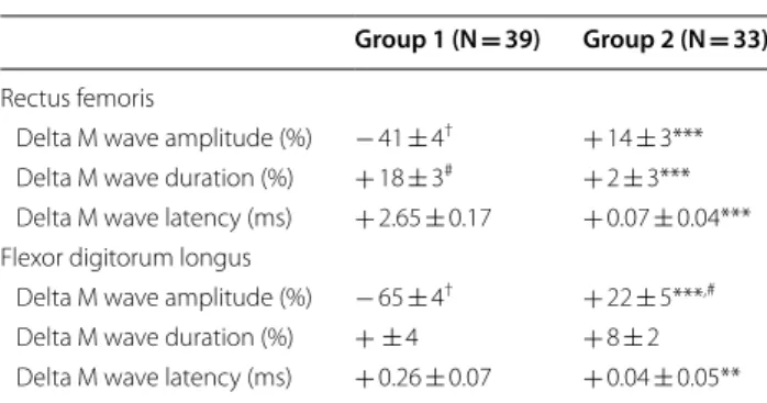
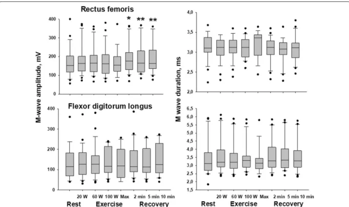
Maximal M-wave variations measured in the RF and FDL muscles are shown in Table 2. In group 1, M-wave alterations were present in both the exercis-ing (RF) and restexercis-ing (FDL) muscles. More specifically, the M-wave amplitude and length and the conduction time were affected in RF whereas only the M-wave amplitude varied in FDL. The kinetics of the M-wave changes in group 1 are shown in Fig. 1: compared to resting values, significant changes in M-wave amplitude (F value > 12) occurred close to the end of exercise and often progressed until the end of the recovery period. A significant increase in M-wave duration (F = 4.42)
Table 1 Characteristics of the study population
Group 1 had marked changes in M-wave amplitude and duration whereas no M-wave changes were measured in group 2. All values shown are mean ± standard deviation
TBARS thiobarbituric acid reactive substance, ORP oxidation reduction potential, VO2 oxygen uptake, HR heart rate
Maximal increase in plasma potassium (K+) concentration was measured at the end of exercise. Asterisks denote significant intergroup difference (**p < 0.01; ***p < 0.001)
Group 1 (N = 39) Group 2 (N = 33)
Age (years) 43 ± 3 47 ± 2
Weight (kg) 64 ± 2 68 ± 2
Sex ratio (F/M) 28/11 17/16 Onset of fatigue (months) 78 ± 19 69 ± 14 TBARS rest (nmol/ml) 1.36 ± 0.03 1.01 ± 0.05***
ORP (mV) 165 ± 5 127 ± 4***
VO2max (mlO2/min/kg) 19 ± 1 19 ± 1
VO2max (% predicted) 67 ± 3 63 ± 3 Maximal work rate (W) 124 ± 5 118 ± 7 Maximal work rate (% predicted) 90 ± 3 83 ± 3 HR max (bpm) 151 ± 3 148 ± 3 Delta K+ max (mmol/l) 0.42 ± 0.07 0.65 ± 0.07**
Handgrip strength (N 278 ± 22 315 ± 33 Handgrip strength (% predicted) 62 ± 7 71 ± 7
Page 4 of 8 Jammes et al. J Transl Med (2020) 18:173
only occurred in RF. In group 2, the M-wave amplitude tended to increase in RF (F = 10.24) with no change in duration or conduction time. No M-wave change occurred in FDL (Table 2 and Fig. 2).
Regarding plasma markers, a significant positive correlation was found between TBARS and ORP val-ues (Fig. 3). When plotting all values measured in both groups of patients (i.e. maximal variations in M-wave amplitude vs. resting levels of TBARS or ORP), negative linear regressions were found (Fig. 4). No significant cor-relations were found between M-wave changes in the two muscle groups and K+ outflow magnitude.
Discussion
This study distinguished two groups of patients with ME/ CFS. In group 1, M-wave alterations were found during and after exercise in both the exercising leg muscle and the resting forearm muscle. In the exercising leg muscle, a decreased in amplitude and longer duration of M-waves were measured whereas in the resting forearm muscle only an alteration in M-wave amplitude was observed. By referring to the redox data presented here, our study shows that high ORP and TBARS values were associ-ated with disorders in both exercising and resting mus-cles. Finally, exercise-induced K+ outflow was reduced
and M-wave alterations were proportional to the mag-nitude of oxidative stress. In group 2, with no M-wave
Table 2 M-wave characteristics
Maximal variations in M-wave amplitude and duration expressed as a percentage of resting values. Latencies measured at rest and at the end of the post-exercise recovery period. Symbols indicate that the changes in each group differ significantly from control values (i.e. rest data) (#p < 0.01; †p < 0.001). Asterisks denote significant intergroup differences (***p < 0.001). Post-exercise variations in M-wave latencies are not significant
Group 1 (N = 39) Group 2 (N = 33) Rectus femoris
Delta M wave amplitude (%) − 41 ± 4† + 14 ± 3*** Delta M wave duration (%) + 18 ± 3# + 2 ± 3*** Delta M wave latency (ms) + 2.65 ± 0.17 + 0.07 ± 0.04*** Flexor digitorum longus
Delta M wave amplitude (%) − 65 ± 4† + 22 ± 5***,#
Delta M wave duration (%) + ± 4 + 8 ± 2 Delta M wave latency (ms) + 0.26 ± 0.07 + 0.04 ± 0.05**
Fig. 1 Time course of changes in M-wave amplitude and duration during exercise (rectus femoris) and at rest (flexor digitorum longus) in group 1 patients. Median values are given at rest, at the four steps of exercise, and at the 2nd, 5th, and 10th min of post-exercise recovery. Asterisks (*p < 0.05; **p < 0.01; ***p < 0.001) indicate significant decreases in M-wave amplitude and increased duration
alterations, low baseline levels of TBARS and ORP were measured. It should be noted however that half the sub-jects in this group suffered from comorbidities which could contribute to body fatigue.
There are several important strengths of this study. First, we used a population with homogeneous criteria for ME/CFS. Second, all patients completed a blinded exercise protocol.
The main limitation to this study is its monocentric nature. Thus, we could not exclude potential referral bias.
The present observations confirm the existence of redox disorders in many ME/CFS patients [3–11, 14–16]. These data also show that abnormal redox status is gen-erally present in ME/CFS patients with abnormal mus-cle membrane excitability, in contrast to patients with no muscle abnormalities. We therefore conclude that high ORP values can be used to distinguish these two groups of patients. A recent study has already reported that plasma ORP changes give a rapid, accurate and user-friendly measurement of increased oxidative stress [23]. We confirm this idea and conclude that ORP meas-urement is cheaper, quicker to perform and provides information on the overall imbalance between oxidants and anti-oxidants whereas TBARS measurements need Fig. 2 Time course of changes in M-wave amplitude and duration during exercise (rectus femoris) and at rest (flexor digitorum longus) in group 2 patients. Median values are given at rest, at the four steps of exercise, and at the 2nd, 5th, and 10th min of post-exercise recovery. Asterisks (*p < 0.05; **p < 0.01) indicate significant increases in M-wave amplitude
Fig. 3 Regression with 95% CIs obtained between resting TBARS and ORP values. The regression equation and significance against zero of the R coefficient value is indicated
Page 6 of 8 Jammes et al. J Transl Med (2020) 18:173
complex biochemical processing and only explore a sin-gle element of the overall redox equilibrium.
The present study also shows that the decreased M-wave amplitude in both exercising and resting mus-cles was proportional to the magnitude of oxidant/ anti-oxidant imbalance in plasma, which supports the primary hypothesis of Fulle et al. [14]. This also supports the hypothesis that M-wave alterations in non-exercising muscles result from a systemic disorder of muscle mem-brane excitability due to dysregulation of the oxidant/ anti-oxidant balance in blood. Increased ROS production following exercise inhibits Na+–K+ pump activity [7],
reducing the K+ outflow and muscle membrane
excit-ability [24]. Previous studies have shown that increased plasma oxidant level is associated with reduced K+
out-flow from exercising muscles in healthy subjects [25] and ME/CFS patients [10]. In the present study, it should be noted that K+ outflow measured at the end of exercise
was higher in group 2 vs. group 1. This indicates that the dysregulation of oxidant/anti-oxidant status in plasma
alters the ionic flux through the muscle membrane, which may partly promote altered muscle membrane excitability.
The existence of a disorder of muscle excitability after exercise could partly explain the occurrence of PEM reported by many ME/CFS patients. However in our series of patients PEM was present in both groups. Pie-trangelo et al. [26] hypothesized that patients with ME/ CFS are subjected to problems of muscle aging (sarco-penia) and that intergroup differences in biological data (M-wave, redox status, K+ outflow) may have resulted
from differences in gene expression. Gene expression subtypes have been reported in ME/CFS patients [27] and the authors isolated 40 abnormal gene pathways. However, the intergroup differences between oxidative abnormalities could not result from age and/or sex dif-ferences as reported in healthy subjects by Fano et al. [28] because in our study these parameters were similar in the two groups. Furthermore, the comorbidities pre-sent in group 2 patients cannot explain the absence of Fig. 4 Maximal variations in M-wave amplitude expressed as a percentage of resting values plotted against resting levels of TBARS and ORP. Linear regressions with 95% CIs are shown as well as the regression equations and the significance against zero of the R coefficient value
marked oxidative damage. Indeed, high levels of oxida-tive stress are often measured in patients with ankylosing spondylitis [29] or Lyme disease [30], these comorbidi-ties being present in six of our patients. Additionally, fatigue that leads to physical deterioration is another important symptom that is observed in 90% of patients with post-polio syndrome, probably resulting from spine frontal horn motor neuron damage during acute polio-virus infection [31]. However this comorbidity was only reported in one of our patients. We therefore have no satisfactory explanation for the intergroup differences observed here.
Conclusion
Future studies in different patient populations may help to further refine the biological criteria of ME/CFS and in particular address the reason(s) for the presence or absence of peripheral muscle fatigue in patients. Our data also highlight the heterogeneity of some biomarker meas-urements in patients with a diagnosis of ME/CFS. Finally, our data support the use of ORP to address the redox stress in a ME/CFS context with diagnostic relevance.
Abbreviations
CFS: Chronic fatigue syndrome; EMG: Electromyography; FDL: Flexor digitorum longus; K+: Potassium; ME: Myalgic encephalomyelitis; M-wave: Evoked muscle
action potential; ORP: Oxidation–reduction potential; PEM: Post-exertional malaise; RF: Rectus femoris; ROS: Reactive oxygen species; TBARS: Thiobarbitu-ric acid reactive substance; VO2max: Maximal oxygen uptake; Wmax: Maximal
work rate.
Acknowledgements
None.
Authors’ contributions
YJ, FR, EF and RG designed the research study, analyzed the data and wrote the paper. YJ, EF, FR, CCharpin, SR, CS, NA, NK and CCriado performed the acquisition of data. YJ, FR, and EF analyzed and interpreted the data. All authors read and approved the final manuscript.
Funding
This study did not receive any funding body for its design, analysis and inter-pretation of data, or for writing the manuscript.
Availability of data and materials
The datasets used and/or analyzed during the current study are available from the corresponding author on reasonable request.
Ethics approval and consent to participate
The protocol was approved by the Ethics Committee “Ouest II” of Angers (May 17, 2019) RCB ID: number 2019-A00611-56 and the study was carried out according to the Code of Ethics of the World Medical Association (Declaration of Helsinki). Procedures were carried out with the adequate understanding and written consent of the subjects.
Consent for publication
Our manuscript does not contain any identifiable individual’s data in any form (including individual details, images or videos).
Competing interests
The authors declare that they have no competing interests.
Author details
1 UMR 1263 C2VN INRA INSERM, Faculty of Medicine, Aix Marseille University,
Marseille, France. 2 Department of Internal Medicine, European Hospital,
Mar-seille, France. 3 Institut National des Sciences Biologiques, CNRS, Paris, France.
Received: 27 January 2020 Accepted: 9 April 2020
References
1. Carruthers BM. Definitions and aetiology of myalgic encephalomyelitis: how the Canadian consensus clinical definition of myalgic encephalomy-elitis works. J Clin Pathol. 2007;60:117–9.
2. Institute of Medicine of the National Academies. Beyond myalgic encephalomyelitis/chronic fatigue syndrome: redefining an illness. Com-mittee on the Diagnostic Criteria for Myalgic Encephalomyelitis/Chronic Fatigue Syndrome; Board on the Health of Select Populations. Washing-ton: National Academies Press; 2015. https ://doi.org/10.17226 /19012 . 3. Gerwyn M, Maes M. Mechanisms explaining muscle fatigue and
muscle pain in patients with myalgic encephalomyelitis chronic fatigue syndrome (ME/CFS): a review of recent findings. Curr Rheumatol Rep. 2017;19:1.
4. Rutherford G, Panning P, Newton JL. Understanding muscle dysfunc-tion in chronic fatigue syndrome. J Aging Res. 2016. https ://doi. org/10.1155/2016/24973 48.
5. Jammes Y, Retornaz F. Understanding neuromuscular disorders in chronic fatigue syndrome. F1000 Research. 2019. https ://doi.org/10.12688 /f1000 resea rch.18660 .1.
6. Samii A, Wassermann EM, Ikoma K, Mercuri B, George MS, O’Fallon A, et al. Decreased post-exercise facilitation of motor evoked potentials in patients with chronic fatigue syndrome or depression. Neurology. 1996;47:1410–4.
7. Fulle S, Belia S, Vecchiet J, Morabito C, Vecchiet L, Fano G. Modification of the functional capacity of sarcoplasmic reticulum membranes in patients suffering from chronic fatigue syndrome. Neuromusc Disorders. 2003;13:479–84.
8. Jammes Y, Steinberg JG, Mambrini O, Brégeon F, Delliaux S. Chronic fatigue syndrome: assessment of increased oxidative stress and altered muscle excitability in response to incremental exercise. J Intern Med. 2005;257:299–310.
9. Jammes Y, Steinberg JG, Delliaux S. Chronic fatigue syndrome: acute infection and history of physical activity affect resting levels and response to exercise of plasma oxidant/antioxidant status and heat shock proteins. J Intern Med. 2012;272:74–84.
10. Jammes Y, Steinberg JG, Guieu R, Delliaux S. Chronic fatigue syndrome with history of severe infection combined altered blood oxidant status, and reduced potassium efflux and muscle excitability at exercise. Open J Intern Med. 2013;3:98–105.
11. Fenouillet E, Vigouroux A, Steinberg JG, Chagvardieff A, Retornaz F, Guieu R, et al. Association of biomarkers with health-related quality of life and history of stressors in myalgic encephalomyelitis/chronic fatigue syndrome patients. J Transl Med. 2016;31(14):251.
12. Arnaud S, Zattara Hartmann MC, Tomei C, Jammes Y. Correlation between muscle metabolism and changes in M-wave and surface electromyo-gram: dynamic constant load leg exercise in untrained subjects. Muscle Nerve. 1997;20:1197–9.
13. Jammes Y, Zattara Hartmann MC, Caquelard F, Arnaud S, Tomei C. Electro-myographic changes in vastus lateralis during dynamic exercise. Muscle Nerve. 1997;20:247–9.
14. Fulle S, Mecocci P, Fanó G, Vecchiet I, Vecchini A, Racciotti D, et al. Specific oxidative alterations in vastus lateralis muscle of patients with the diag-nosis of chronic fatigue syndrome. Free Radic Biol Med. 2000;29:1252–9. 15. Richards RS, Roberts TK, McGregor NR, Dunstan RH, Butt HL. Blood
parameters indicative of oxidative stress are associated with symptom expression in chronic fatigue syndrome. Redox Rep. 2000;5:35–41. 16. Vecchiet J, Cipollone F, Falasca K, Mezzetti A, Pizzigallo E, Bucciarelli T,
et al. Relationship between musculoskeletal symptoms and blood mark-ers of oxidative stress in patients with chronic fatigue syndrome. Neurosci Lett. 2003;335:151–4.
Page 8 of 8 Jammes et al. J Transl Med (2020) 18:173
•fast, convenient online submission •
thorough peer review by experienced researchers in your field • rapid publication on acceptance
• support for research data, including large and complex data types •
gold Open Access which fosters wider collaboration and increased citations maximum visibility for your research: over 100M website views per year •
At BMC, research is always in progress. Learn more biomedcentral.com/submissions
Ready to submit your research? Choose BMC and benefit from:
17. Dickinson BC, Chang CJ. Chemistry and biology of reactive oxygen spe-cies in signaling or stress responses. Nat Chem Biol. 2011;7:504–11. 18. Bobe G, Cobb TJ, Leonard SW, Aponso S, Bahro CB, Koley D, et al.
Increased static and decreased capacity oxidation-reduction potentials in plasma are predictive of metabolic syndrome. Redox Biol. 2017;12:121–8. 19. Daniels RC, Jun H, Tiba MH, McCracken B, Herrera-Fierro P, Collinson M,
et al. Whole blood redox potential correlates with progressive accumu-lation of oxygen debt and acts as a marker of resuscitation in a swine hemorrhagic shock model. Shock. 2018;49:345–51.
20. de Ponte ÁM, Guirro EC, Pletsch AH, Dibai-Filho AV, Brandino HE, Guirro RR. The forearm positioning changes electromyographic activity of upper limb muscles and handgrip strength in the task of pushing a load cart. J Body Mov Ther. 2015;19:597–603.
21. Steiber N. Strong or weak handgrip? Normative reference values for the German population across the life course stratified by sex, age, and body height. PLoS ONE. 2016;11:e0163917.
22. Uchiyama M, Mihara M. Determination of malonedialdehyde precursor in tissues by thiobarbituric acid test. Anal Biochem. 1978;86:271–8. 23. Polson D, Villalba N, Freeman K. Optimization of a diagnostic platform
for oxidation-reduction potential (ORP) measurement in human plasma. Redox Rep. 2018;23:125–9.
24. Sjøgaard G. Exercise-induced muscle fatigue: the significance of potas-sium. Acta Physiol Scand Suppl. 1990;593:1–63.
25. Hermann A, Sitdikova GF, Weiger TM. Oxidative stress and maxi calcium-activated potassium (BK) channels. Biomolecules. 2015;5:1870–911.
26. Pietrangelo T, Fulle S, Coscia F, Gigliotti PV, Fanò Illic G. Old muscle in young body: an aphorism describing the chronic fatigue syndrome. Eur J Transl Myol. 2018;28:7688.
27. Kerr JR, Petty R, Burke B, Gough J, Fear D, Sinclair LI, et al. Gene expression subtypes in patients with chronic fatigue syndrome/myalgic encephalo-myelitis. J Infect Dis. 2008;197:1171–84.
28. Fanò G, Mecocci P, Vecchiet J, Belia S, Fulle S, Polidori MC, et al. Age and sex influence on oxidative damage and functional status in human skeletal muscle. J Muscle Res Cell Motil. 2001;22:345–51.
29. Pishgahi A, Abolhasan R, Danaii S, Amanifar B, Soltani-Zangbar MS, Zam-ani M, et al. Immunological and oxidative stress biomarkers in ankylosing spondylitis patients with or without metabolic syndrome. Cytokine. 2020;128:155002.
30. Peacock BN, Gherezghiher TB, Hilario JD, Kellermann GH. New insights into Lyme disease. Redox Biol. 2015;5:66–70.
31. Pastuszak Ż, Stępień A, Tomczykiewicz K, Piusińska-Macoch R, Galbarczyk D, Rolewska A. Post-polio syndrome. Cases report and review of literature. Neurol Neurochir Pol. 2017;51:140–5.
Publisher’s Note
Springer Nature remains neutral with regard to jurisdictional claims in pub-lished maps and institutional affiliations.

