HAL Id: inserm-00880088
https://www.hal.inserm.fr/inserm-00880088
Submitted on 5 Nov 2013
HAL is a multi-disciplinary open access
archive for the deposit and dissemination of
sci-entific research documents, whether they are
pub-lished or not. The documents may come from
teaching and research institutions in France or
abroad, or from public or private research centers.
L’archive ouverte pluridisciplinaire HAL, est
destinée au dépôt et à la diffusion de documents
scientifiques de niveau recherche, publiés ou non,
émanant des établissements d’enseignement et de
recherche français ou étrangers, des laboratoires
publics ou privés.
Thomas Karaouzène, Michèle El Atifi, Jean-Paul Issartel, Marianne Grepillat,
Charles Coutton, Delphine Martinez, Christophe Arnoult, Pierre Ray
To cite this version:
Thomas Karaouzène, Michèle El Atifi, Jean-Paul Issartel, Marianne Grepillat, Charles Coutton, et
al.. Comparative testicular transcriptome of wild type and globozoospermic Dpy19l2 knock out mice.
Basic and Clinical Andrology, 2013, 23 (1), pp.7. �inserm-00880088�
R E S E A R C H A R T I C L E
Open Access
Comparative testicular transcriptome of wild type
and globozoospermic Dpy19l2 knock out mice
Thomas Karaouzène
1,2, Michèle El Atifi
3,4,5, Jean-Paul Issartel
3,4,5, Marianne Grepillat
1,2,6, Charles Coutton
1,2,7,
Delphine Martinez
1,6, Christophe Arnoult
1,2and Pierre F Ray
1,2,6*Abstract
Background: Globozoospermia is a male infertility phenotype characterized by the presence in the ejaculate of near 100% acrosomeless round-headed spermatozoa with normal chromosomal content. Following
intracytoplasmic sperm injection (ICSI) these spermatozoa give a poor fertilization rate and embryonic development. We showed previously that most patients have a 200 kb homozygous deletion, which includes DPY19L2 whole coding sequence. Furthermore we showed that the DPY19L2 protein is located in the inner nuclear membrane of spermatids during spermiogenesis and that it is necessary to anchor the acrosome to the nucleus thus performing a function similar to that realized by Sun proteins within the LINC-complex (Linker of Nucleoskeleton and
Cytoskeleton). SUN1 was described to be necessary for gametogenesis and was shown to interact with the
telomeres. It is therefore possible that Dpy19l2 could also interact, directly or indirectly, with the DNA and modulate gene expression during spermatogenesis.
In this study, we compared the transcriptome of testes from Dpy19l2 knock out and wild type mice in order to identify a potential deregulation of transcripts that could explain the poor fertilization potential of Dpy19l2 mutated spermatozoa.
Methods: RNA was extracted from testes from DPY19L2 knock out and wild type mice. The transcriptome was carried out using GeneChip® Mouse Exon 1.0 ST Arrays. The biological processes and molecular functions of the differentially regulated genes were analyzed with the PANTHER software.
Results: A total of 76 genes were deregulated, 70 were up-regulated and 6 (including Dpy19l2) were down-regulated. These genes were found to be involved in DNA/RNA binding, structural organization, transport and catalytic activity.
Conclusions: We describe that an important number of genes are differentially expressed in Dpy19l2 mice. This work could help improving our understanding of Dpy19l2 functions and lead to a better comprehension of the molecular mechanism involved in spermatogenesis.
Keywords: Male infertility, Globozoospermia, Spermatogenesis, Dpy19l2, Transcriptome
* Correspondence:pray@chu-grenoble.fr
1Université Joseph Fourier, Grenoble F-38000, France 2
Laboratoire AGIM, CNRS FRE3405, Equipe “Génétique, Infertilité et Thérapeutiques”, La Tronche F-38700, France
Full list of author information is available at the end of the article
© 2013 Karaouzène et al.; licensee BioMed Central Ltd. This is an Open Access article distributed under the terms of the Creative Commons Attribution License (http://creativecommons.org/licenses/by/2.0), which permits unrestricted use, distribution, and reproduction in any medium, provided the original work is properly cited.
Karaouzène et al. Basic and Clinical Andrology 2013, 23:7 http://www.bacandrology.com/content/23/1/7
Résumé
Contexte: La globozoospermie est caractérisée par la présence dans l’éjaculat de près de 100% de spermatozoïdes ronds et dépourvus d’acrosome qui présentent un contenu chromosomique normal. L’injection intracytoplasmique (ICSI) de ces spermatozoïdes donne cependant un taux de fécondation et de développement embryonnaire particulièrement bas. Nous avons montré précédemment que la plupart des patients globozoospermes présentent une délétion
homozygote de 200 Kb qui inclue la totalité de la séquence codante du gène DPY19L2. De plus nous avons montré que la protéine DPY19L2 était localisée dans la membrane interne des noyaux des spermatides pendant la spermatogénèse et qu’elle est nécessaire pour fixer l’acrosome au noyau, réalisant ainsi une fonction similaire à celle des protéines Sun au sein du complexe LINC (Linker of Nucleoskeleton and Cytoskeleton). Il a par ailleurs été montré que SUN1 était
nécessaire à la spermatogénèse et que cette protéine interagit avec les télomères chromosomiques. Il est donc possible que Dpy19l2 interagisse également, directement ou indirectement avec l’ADN et module l’expression génique lors de la spermatogénèse. Dans cette étude nous avons donc comparé le transcriptome de testicules de souris invalidées (KO) pour le gène Dpy19l2 à celui de souris sauvage afin d’identifier une éventuelle dérégulation génique qui pourrait expliquer le faible potentiel reproductif des spermatozoïdes globozoocéphales.
Méthode: L’ARN a été extrait de testicules de souris KO pour Dpy19l2 et de souris sauvages. Le transcriptome a été réalisé en utilisant des puces d’expression ® Mouse Exon 1.0 ST Arrays. Les processus biologiques et les fonctions des gènes dérégulés ont été analysés en utilisant le logiciel PANTHER.
Résultats: Un total de 76 gènes a été identifié comme étant dérégulés, 70 gènes étaient surexprimés et 6 (incluant Dpy19l2) étaient sous-exprimés. Il s’agit de gènes principalement impliqués dans des interactions avec des acides nucléiques (ADN/ARN), ou ayant un rôle structural, dans le transport, ou présentant une activité catalytique.
Conclusions: Cette étude nous a permis d’identifier et de décrire un nombre important de gènes exprimés de manière différentielle chez les souris KO pour Dpy19l2. Ce travail peut permettre d’améliorer notre compréhension des fonctions de Dpy19l2 et peut contribuer à obtenir une meilleure compréhension des mécanismes moléculaires nécessaire à la spermatogénèse.
Keywords: Male infertility, Globozoospermia, Spermatogenesis, Dpy19l2, Transcriptome
Background
A recent study supported by the World Health Organi-zation indicates than in 2010, an estimated 48.5 million couples worldwide were unable to have a child after five years [1]. Male factors are believed to be responsible for 30-50% of all infertility cases, but micro deletions of the Y chromosome are the only genetic defects altering hu-man spermatogenesis, which are diagnosed routinely.
To be able to fertilize the oocyte, the spermatozoon needs to cross the zona pellucida (ZP), which is a glycopro-tein layer surrounding the oocyte. The acrosomal reaction (AR), during which the acrosome (a giant vesicle of secre-tion) releases its content, plays an important role in the fertilization process. Enzymes released from the acrosome locally digest and soften the ZP so that the spermatozoon can penetrate deeper and fertilize the oocyte. The acro-some, a highly specialized organelle found only in sperm, is tightly bound to the nucleus via the acroplaxome (a net-work of proteins including keratin 5 and β-actin) [2].
Globozoospermia is a severe teratozoospermia character-ized by the presence of 100% of round-headed spermato-zoa devoid of acrosome. Men with globozoospermia have a primary infertility due to this absence of acrosome, which prevents their sperm from fertilizing the oocytes in vivo [3]. Spermatozoa from globozoospermic patients have near
normal levels of aneuploidy but give a poor fertilization rate and embryonic development even when performing Intra Cytoplasmic Sperm Injection (ICSI) [3]. Studies by immunocytochemistry showed that most round headed sperm lacked the phospholipase zeta protein (PLCzeta), a protein normally located around a the sperm’s head [4-7] and required to induce oocyte intracellular calcium oscilla-tion and oocyte activaoscilla-tion [8,9]. It has therefore been pos-tulated that it is the absence of PLCzeta which might be responsible for the poor fertilization potential of round-headed spermatozoa [10]. In the course of this work we wanted to assess if the absence of PLCzeta in round-headed spermatozoa results from a transcriptional repres-sion of the gene and if other transcriptional deregulations could also contribute to the poor fertilization potential of these gametes.
The syndrome of globozoospermia was first described in the seventies [7,11] and cases have been described regularly since [12-20]. Familial cases rapidly pointed to a genetic cause for this syndrome. In the recent years, SPATA16 has been described to be involved in globozoospermia [21]. We demonstrated recently that DPY19L2 was in fact the main locus associated with globozoospermia as 15 out of 20 analysed patients presented a 200 Kb homozygous dele-tion removing the entire gene [22]. We then identified
DPY19L2 point mutations and heterozygous deletions and demonstrated that 84% of the 31 globozoospermic patients analysed had a molecular alteration of DPY19L2 [23]. We finally confirmed that the recurrent deletion observed in a majority of men with globozoospermia was caused by non-allelic homologous recombination (NAHR), between two highly homologous sequences, or low-copy repeats (LCR), located on each side of DPY19L2 [24].
We previously characterized Dpy19l2 Knockout mice (Dpy19l2−/−)and showed that these mice present the same
phenotype than men carrying mutations in DPY19L2, ie round-head spermatozoa without acrosome. It also permitted us to determine that i.) DPY19L2 is located in the inner nuclear membrane of wild type mouse spermatids, ii.) DPY19L2 is required for acrosome attach-ment to the nucleus and iii.) the detachattach-ment of the acro-some in Dpy19l2−/−mice prevents correct anchoring of
the manchette. Moreover we described that SUN5 and DPY19L2 partially colocalized in transfected HEK cells [25]. SUN-domain proteins are known to interact with chromosome-binding proteins and various KASH-domain partners to form SUN-domain-dependent ‘bridges’ across the inner and outer nuclear membranes. These bridges physically connect the nucleus to every major component of the cytoskeleton [26]. SUN1, one of the members of the family, was described to be necessary for gametogenesis and was shown to interact with the telomeres [27]. We can hypothesize that Dpy19l2 could interact directly or indir-ectly with the DNA and thus have an effect on the regula-tion of transcripregula-tion. It is thus possible that the absence of Dpy19l2 could cause some modification in the germ cell transcription pattern.
The goal of this study was to determinate if Dpy19l2 knock out mice present significant testis transcriptional modifications compared to wild type and in particu-lar modifications that may explain the poor success rate encountered by globozoospemic patients following ICSI- IVF.
Methods
Ethical statement
Animal housing and sacrificing was in accordance with French guidelines on the use of animals in scientific inves-tigations with the approval of the local Ethical Committee.
Animals
Dpy19l2 knock out mice were obtained from Mutant Mouse Regional Resource Center, University of California, Davis, CA. The mouse colony used in this study was initi-ated from two couples. The first one consisted of an het-erozygous female and a wild type male. The second was composed of two heterozygous mice for the Dpy19l2 dele-tion. Reproduction of these two couples achieved wild type, heterozygous and homozygous Dpy19l2 deleted mice. Mice
were sacrificed at 2 months old, which means that they were pubescent and that their reproductive organs were fully established. A total of four animals were sacrificed. RNA was extracted from two homozygous WT and two homozygous KO animals.
Genotyping PCRs
Genotyping was done on DNA isolated from tail biopsies. Tail biopsies (ca. 2 mm in length) were digested in 200 μl lysis Direct PCR LYsis Reagent (Tail) (Viagen Biotech inc, CA, USA) and 0,2 mg of proteinase K for 12–15 hours at 55°C and 1 hour at 85°C. The DNA was directly used for PCRs.
PCR was done for 35 cycles, with an annealing temp-erature of 57°C, and an elongation time of 60 seconds at 72°C. The primers used are described in Figure 1. PCRs products were separated on agarose gel electrophoresis. Genotypes were determined according to the migration pattern (Figure 1).
Tissue collection
Mice were sacrificed and testes were collected. Tissues were snap frozen in liquid nitrogen prior storage at −80°C. Two mice in each group were used for the micro-array analysis.
B
A
1 - 2 1 - 3Dpy19l2
Htz
(+/-)
KO
(-/-)
WT
(+/+)
T0
Figure 1 Strategy for Dpy19l2 KO mice genotyping. A) Scheme of the Dpy19l2 alleles and primers (red arrows) used for their detection. Primer sequence is as follow: 1:GAAGGCTACACCTCTTGCA, 2:GCTGCAGCAACGACCACTTC; 3:CCTAGGAATGCTCGTCAAGA. B) Examples of PCRs with (from left to right) Dpy19l2+/− mouse, Dpy19l2−/− mouse, Dpy19l2+/+ mouse and water (control).
Karaouzène et al. Basic and Clinical Andrology 2013, 23:7 Page 3 of 10
RNA extraction
Total RNA was extracted from tissues using mirVana isolation kit™ (Ambion, Applied Biosystems, Foster City, CA) as per the manufacturer’s instructions. RNA purity and quantity was assessed using the NanoDrop c1000 (ThermoFisher Scientific, Waltham, USA). Quality was de-termined by both evaluation of the integrity of rRNA bands using RNA Nano 6000 kit (Bio-Analyser, Agilent Tech-nologies, Palo Alto, CA) and absorbtion readings ant 260 and 280 nm. For detail see Additional file 1: Table S1.
Array hybridization
For each group, two biological replicates were used. The replicates came from four separate RNA extractions: two from homozygous WT and two from homozygous KO ani-mals. cDNA synthesis, amplification, enzymatic fragmenta-tion and biotinylafragmenta-tion were performed using the Ambion WT Expression Kit (Ambion, Austin, TX, USA). Samples were hybridized to Affymetrix GeneChip® Mouse Exon 1.0 ST Arrays as per the manufacturer’s instructions. The Affimetrix Mouse Exon 1.0 ST array, contains probe sets for 35,557 genes. Briefly, 5 μg of fragmented biotinylated ssDNA was hybridised for 16 hrs at 45°C, 60 rpm to the array chip on a GeneChip® Hybridization Oven 640. After 16 hrs, GeneChips® were washed on a GeneChip® Fluidics station 450 using the washing script Prime 450 with buffers and stains supplied with the GeneChip Hybridisation Wash and Stain Kit from Affymetrix.
Data acquisition and analysis
Data was acquired on a GeneChip® Scanner 3000 7G and .CEL file generation performed using AGCC. Expression Console with Robust Multi-chip Average (RMA) was used initially to extract probe intensity data. RMA background correction was applied including pre-background adjust-ment for GC content and quantile normalization across all chips in the experiment. Probe data was log2 transformed.
Gene level expression analysis
Two separate experiments (experiment 1 and 2) were car-ried out, each time with one testis from homozygous wild type and homozygous KO mice. Hence for each gene a total of two values were obtained in WT (Dpy +/+ (1) and (2)) and KO mice (Dpy −/− (1) and (2)).
For each gene transcripts we calculated 4 ratios corre-sponding to the 4 possible combinations
R1¼ Dpy−=− 1ð Þ Dpyþ = þ 1ð Þ R3¼ Dpy−=− 2ð Þ Dpyþ = þ 1ð Þ R2¼ Dpy−=− 1ð Þ Dpyþ = þ 2ð Þ R4¼ Dpy−=− 2ð Þ Dpyþ = þ 2ð Þ
For each gene, if at least three of these ratios ap-peared > = 1.7 fold up or down, the transcript was
considered to be significantly differentially expressed. These values and the log2 ratio for all deregulated genes are shown in Additional file 2: Table S2. The histogram of the log2 ratio of each deregulated gene is shown in Figure 2.
Gene ontology analysis
The lists of genes expressed differently in Dpy19l2−/− mice were imported into PANTHER (http://www.pantherdb. org/) to identify the biological process, molecular functions and gene networks significantly deregulated in Dpy19l2−/−
testis compared to WT controls. Results
Gene expression profile
Array hybridization was performed with the Affimetrix Mouse Exon 1.0 ST array, which contains probe sets for 35,557 genes. Of these, we identified that 76 genes had a level of testicular expression that was different between WT and Dpy19l2−/−mice (transcripts with an expression
ratio > = 1.7 fold up- or down regulated). Among them, 6 genes were underexpressed and 70 genes were over-expressed (Figure 2 and Additional file 2: Table S2). As expected Dpy19l2 was found part of the down-regulated genes, thus validating the experimental approach we used. Interestingly, we did not observe any difference in the expression level of PLCzeta in the testes from KO and WT mice.
Panther gene ontology analysis
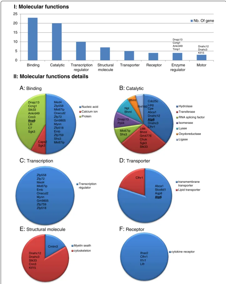
The 76 genes that were differentially regulated were up-loaded into the PANTHER software (Gene List Analysis). Among them 64 were recognized by the PANTHER soft-ware. The molecular functions and biological process pre-dictions that are generated from PANTHER are based on the direction of expression of a number of downstream genes which have been previously shown to be associated with these functions. The list of each function associated to all deregulated genes is provided in Additional file 3: Table S3. Several molecular functions were found to be enriched in the testis of Dpy19l2−/−mice (Figure 3). Genes
encoding proteins witch are able to bind nucleic acids or proteins were most frequently deregulated (23 genes), es-pecially those encoding for protein binding to the nucleic acids (12 genes), confirming that Dpy19l2 could interact with DNA. Other functions such as catalytic activity, tran-scription regulator activity, structural functions were also deregulated in the KO mice testes. Because of its lo-cation in the inner nucleus membrane, DPY19L2 could be a bridge between the nucleus and the cytoplasm. We observed that 5 genes encoding for transporters are deregulated in KO mice: among them, four are transmem-brane transporters and one is a lipid transporter. Moreover globozoospermia is characterized by structural deficiency
of spermatozoon head and we see that 6 deregulated genes encode for proteins with structural molecular function.
Numerous biological processes are also deregulated in the testis of Dpy19l2−/− mice (Figure 4). Metabolic
pro-cesses and cellular propro-cesses are most often deregulated. We see that 6 genes predicted to be involved in repro-duction biological process separated deregulated. Among those, no genes were described to be involved in the acro-some formation but two genes encode for dyneins and one for a protein predicted to be involved in sperm motility. Discussion
Spermiogenesis is the final stage of spermatogenesis. Dur-ing this step, the nucleus condenses, acquires its specific shape, and the flagellum and the acrosome are formed. The acrosome is essential for the spermatozoa to cross of the ZP and is thus necessary for in vivo fertilization. Globozoospermia is a teratozoospermia characterised by the formation of round-head spermatozoa without acro-some. This pathology has been described to be associated with the absence of the protein PLCzeta which is also known to be essential for fertilization and oocyte activation [4-7]. We previously demonstrated that this pathology is mostly due to a homozygous deletion of the testis-specific gene DPY19L2 [22,23] and that DPY19L2 is expressed in spermatids and it is located only in a restricted zone of the nuclear membrane facing the acrosome.
This study revealed that 76 genes were deregulated in the testis of Dpy19l2 KO mice. This result could be con-cordant with a very specific regulatory role of Dpy19l2 at the transcription level. On the other hand we note that the micro-array contains 35,557 probe-set for almost as many genes. It is therefore a small minority (0.2%) of genes that is deregulated in DPy19l2 KO mice. It is interesting to note that almost all of these genes appeared as up-regulated and that only 5 of them were down- regulated. If Dpy19l2 has a direct influence on gene regulation we can therefore say that it mainly act as a repressor of gene expression. We note that apart from Dpy19l2, which is obviously absent from the KO and is found (due to background fluorescence levels) to have a 4 fold decrease in expression compared to controls, the most down-regulated gene, ATP6, has a 2.6 fold decreased expression and the most up-regulated gene, Cepp, has a 2.2 fold increased expression. The observed level of transcription modifications is therefore moderate.
Dpy19l2 co-localises with SUN5 [28] and we hypothe-sized that SUN5 is a likely partner of Dpy19l2 [25]. In mouse, Sun1, another Sun protein, was also described to
-4.50 -3.50 -2.50 -1.50 -0.50 0.50 1.50 2.50 R4 R3 R2 R1 Aqp8 Gm10629 Med4 Lipa Il1r1 Cmtm3 Gm4776 Lifr Lipg Ttc27 Lrg1 Mki67ip Abca1 Cpe Cage1 Fam178a Caps2 Cylc2 Cdc25c Lias Ppil4 Chuk Akd1 Npl Tmem47 Timp1 Zfp759 Hspd1 Ifnar2 Mum1l1 Cfhr3 Sgk3 Nup210l Cnn3 Gm4942 Dnahc14 Cfhr1 Gm13034 Zfp518 Kif15 Ccdc144b Emb Cenpp Mynn Zfp72 Ccng1 Stk33 Gstk1 Dnahc12 Ankrd49 Cc2d2b Spef2 Ankrd32 C7 Nuf2 Dnajc13 Dnahc3 Ppp2r3c Shcbp1 Sfrs3 Zfp558 Setd2 Malat1 Rif1 Dennd4a Rnf160 Cep290 Bhmt Slco6d1 Gm9805 Dpy19l2 ATP6 Snord32a Onecut2 Dnajb8 Ifna9
Figure 2 Histogram of the log2 ratio of all deregulated transcripts. Genes that are upregulated in Dpy19l2 KO mice have positive values while genes that are downregulated have negative values. Values can be seen in Additional file 2: Table S2.
Karaouzène et al. Basic and Clinical Andrology 2013, 23:7 Page 5 of 10
Nucleic acid Calcium ion Protein
A:
Binding Med4 Zfp558 Mki67ip Onecut2 Zfp72 Gm9805 Mynn Zfp518 Emb Zfp759 Sfrs3 Mki67ip Caps2 Sgk3 Dnajc13 Ccng1 Stk33 Ankrd49 Cnn3 Ifna9 Lifr C7 Sgk3 Hydrolase Transferase RNA splicing factor Isomerase Lyase Oxydoreductase Ligase Mki67ip Sfrs3 Cdc25c Lipg Cpe Abca1 Dnahc12 Atp6 Dnahc3 Cfhr1 Dnajc13 Ppil4 Rnf160 Npl Bhmt Bhmt Lias Bhmt Gm4776 Chuk Sgk3 Stk33B:
Catalytic transmembrane transporter Lipid transporter Abca1 Slco6d1 Aqp8 Atp6 Cfhr1D:
Transporter Transcription régulator Zfp558 Zfp72 Med4 Mki67ip Emb Onecut2 Mynn Gm9805 Zfp759 Zfp518C:
Transcription Myelin seath cytoskeleton Dnahc12 Dnahc3 Stk33 Cnn3 Kif15 Cmtm3E:
Structural molecule cytokine receptorF:
Receptor Ifnar2 Cfhr1 Il1r1 LifrII: Molecular functions details
Dnahc12 Dnahc3 Kif15 0 5 10 15 20 25
Binding Catalytic Transcription regulator
Structural molecule
Transporter Receptor Enzyme regulator Motor Nb. Of gene
I: Molecular functions
Dnajc13 Ccng1 Ankrd49 Timp1Figure 3 I, Histogram presenting all PANTHER molecular function of genes that are deregulated in Dpy19l2−/−mice testes. II, A-F,
Gamete generation Fertilization Dnahc12 Dnahc3 Ccng1 Zfp558 Dnahc12 Gm4776 Dnahc3 Dnajb8 F: Reproduction Protein Vesicle-mediated Nuclear Ion Lipid Dnahc12 Dnajc13 Dnahc3 Kif15 Dnajc13 Dnahc12 Kif15 Dnahc3 Slco6d1 ATP6 Nup210l Ppil4 Abca1 E: Transport 0 5 10 15 20 25 30 35 Nb of gene Ifnar2 Cfhr1 Dnajb8 Gm4776 Lifr Chuk Ifna9 C7 Stk33 Gm4776 Dnahc12 Dnahc3 Lifr Mki67ip C7 Kif15 Stk33 Ifnar2 Dnajb8 Sgk3 Tmem47 Ifna9 Ankrd32 Dnahc12 Dnahc3 Kif15 Stk33 Gm4776 Dnahc13 Stk33 Cmtm3 Cnn3 Cfhr1 Cell communication Cell cycle Cellular component organization Chromosomes segregation Cell motion Cytokinesis Cell adhesion Cfhr1 Kif15 Dnahc12 Dnahc3 C7 Dnahc12 Dnahc3 Nuf2 Kif15 Ankrd32 Stk33 Dnahc12 Dnahc3 Kif15 Cdc25c Ccng1 Dnajc13 Dnahc12 Ankrd32 Ankrd49 Gm4776 Kif15 Nuf2 Mki67ip Dnahc3 Stk33 Sgk3 Ifnar2 Dnajc13 Cfhr1 Emb Gm4776 Chuk Ifna9 Lifr Cmtm3 Stk33 Il1r1 C7 Sgk3 A: Cellular process Protein nucleobase, nucleoside, nucleotide and nucleic acid Lipid
Cellular amino acid Carbohydrate Cdc25c Dnajc13 Cpe Gstk1 Cfhr1 Ppp2r3c Dnajb8 Chuk Gm4776 Mki67ip Rnf160 Sgk3 Stk33 Timp1 Hspd1 Ppil4 Dennd4a Sfrs3 Med4 Zfp558 Zfp72 Ankrd32 Onecut2 Mki67ip Mynn Gm9805 Emb Zfp518 Zfp759 Lipg Abca1 Npl Bhmt Gm4776 B: Metabolic process Signal trasduction Cell-cell signaling Ifnar2 Lifr Dnajc13 C7 Ifna9 Ifnar2 Cfhr1 Emb Chuk Gm4776 Lifr Stk33 Cmtm3 C7 Sgk3 Il1r1 Ifna9 C: Cell communication Other Mitosis Meiosis D: Cell cycle Stk33 Kif15 Dnahc12 Ccng1 Nuf2 Kif15 Dnahc3 Cdc25c Mki67ip Dnajc13 Ankrd32 Ankrd49 Gm4776 Sgk3
II: Biological process
I: Biological process
Figure 4 I, Histogram presenting all PANTHER biological process of genes deregulated in Dpy19l2−/−mice testes. II, A-F, Details of some
of PANTHER biological process. Up-regulated genes are in bold and underlined.
Karaouzène et al. Basic and Clinical Andrology 2013, 23:7 Page 7 of 10
be necessary for gametogenesis and was shown to inter-act in the nucleus with the telomeres [27]. We observed that most of the deregulated genes (70/76) were up reg-ulated in KO animal. We can hypothesize that Dpy19l2 could also interact, directly or indirectly, potentially via Sun5, with germ cell DNA and thus could have an effect on the regulation of transcription in spermatogenic cells. Heterochromatin is constituted by highly compact trans-criptionaly repressed DNA. It regroups down-regulated genes and is particularly abundant at the periphery of the nucleus where it interacts with factors located in the nuclear lamina. We can thus speculate that Dpy19l2 could intervene during spermiogenesis to include selected genes in heterochromatin repressive domains. In the ab-sence of Dpy19l2, these genes would not be repressed and appear as up regulated. This regulations could be a limited to selected loci as electron microscopic observations of round spermatids nuclei from Dpy19l2 KO animals do not show any obvious difference in the abundance of hetero-chromatin [25].
The PANTHER software allows a classification of genes according to their predicted molecular functions (Figure 3). We see that the most represented gene function that is deregulated in Dpy19L2 mice is “binding” (23 genes). This group is divided in three sub categories: nucleic acid, pro-tein and calcium ion (Additional file 2: Table S2). [Ca2+]Iis
known to play an important role in male fertility. [Ca2+]I
signaling is the primary regulator of sperm flagellum beat-ing and calcium intracellular rise is known to be essential for the acrosome reaction [29]. Indeed, solubilisation of the zona pelucida stimulates generation of IP3in mouse sperm
[30] which is known to mobilize the acrosomal Ca2+ stored to permit acrosomal reaction [31,32]. The bio-chemical nature of the Ca2 + −binding sites are globally unknown but recently a calcium-binding protein has been isolated from the acrosomal membrane of bovine sperm-atozoa [33]. We observe that in Dpy19l2 KO mice two cal-cium binding proteins are up-regulated : Caps2 and Sgk3 (Figure 2 and 3). Ten of the deregulated genes are de-scribed to encode proteins with DNA binding abilities. Al-though we did not find direct evidence that these encoded proteins have transcriptional regulation activities, they might be involved in the regulation of gene expression and play a role in the up- and -down regulation of some of the other genes we found to be deregulated in this transcrip-tome analysis.
We did not observe a down-regulation of PLCzeta that could account for its absence from round-headed sperms. This suggests that in Dpy19l2−/− mice PLCzeta is nor-mally expressed but that the absence of Dpy19l2 and the abnormalities it induces on sperm morphology likely pre-vents the correct positioning of PLCzeta, which is likely to be eliminated in the residual body. This hypothesis is consolidated by the fact that several studies show that
treatment with a calcium ionophore improves ICSI suc-cess rates? results for men with globozoospermia [5]. We note however that fertilization and pregnancies can be achieved by ICSI on DPY19L2 deleted men [34]. This can probably be explained by the fact that remains of misplaced PLCzeta often position near the manchette can be observed on a small proportion of round-headed sperm [6].
This study also reveals that several genes encoding for transporters were deregulated in Dpy19l2 KO mice. Among them four are transmembrane transporters and one is a lipid transporter. We note the deregulation of the gene Abca1, which is expressed in mouse spermatozoa within the seminiferous tubules and the epididymis, and is a key regulator of cholesterol efflux. Depletion of the chol-esterol from the cytoplasmic plasma membrane and modi-fication of its lipid composition is one of the key events in the process of spermatozoa capacitation, which ultimately leads to the acrosome reaction and egg fertilization. Trans-porters and in particular those mediating cholesterol efflux, are thus particularly important. The deregulation of Abca1 could therefore alter the physiological composition of ma-ture sperm and contribute to the poor fertilization poten-tial of Dpy19l2 mutant sperm.
The analyze of biological process regulated in Dpy19l2−/−
mice reveals 6 genes predicted to be involved in reproduct-ive functions and particularly in gamete generation and fertilization. Surprisingly half of these genes code for dy-neins, which are important constituents of the microtu-bules. The others are involved in the processes of sperm motility and cytoskeleton structure. These results can be linked to our previous observation that the absence of Dpy19l2 leads to the destabilization of both the nuclear dense lamina and the junction between the acroplaxome and the nuclear envelope. This destabilization causes a fail-ure of the linkage of the acrosome and the manchette to the acroplaxome, a cytoskeletal plate anchored to the nu-clear envelope. The manchette is a transient microtubular structure necessary during spermatid elongation. Moreover, the manchette is necessary for protein trafficking and its defects could disturb the overall distribution of proteins in spermatids [35].
Conclusions
We showed that Dpy19l2−/− induced globozoospermia
altered gene expression in mice testis but the overall modifications at the transcript level remained modest. We showed that PLCzeta was not down-regulated in KO mice indicating that the absence of the protein observed in the sperm of globozoospermic patient is not due to a transcriptional deregulation. This likely indicates that PLCzeta cannot reach its physiological localization on round-headed spermatozoa and that it is probably lost with the cytoplasmic elimination (residual body) during
spermiogenesis. We also observed that several genes en-coding proteins involved in transports, and in particular Abca1, involved in the cholesterol efflux, were deregulated. This could also contribute to the poor fertilization poten-tial of the round-headed spermatozoa. Secondary anomal-ies stemming from the morphological abnormalitanomal-ies of the sperm could also lead to a wide range of protein deregula-tion as exemplified by the absence of PLCzeta. A prote-omic analysis of these deregulations could permit to have a functional view of the extent of the molecular anomal-ies present in Dpy19l2 KO mice. Further work will per-mit a better comprehension of molecular mechanism involved in spermatogenesis and in the physiopathology of globozoospermia.
Additional files
Additional file 1: Table S1. RNA quantification.
Additional file 2: Table S2. Ratios of transcripts values measured in Dpy19l2 WT and KO mice.
Additional file 3: Table S3. PANTHER output of all deregulated genes in Dpy19l2 KO mice.
Abbreviations
AR:Acrosomal reaction; DNA: Deoxyribonucleic acid; dNTP: Deoxynucleotide triphosphates; FISH: Fluorescent in situ hybridization; ICSI: Intracytoplasmic sperm injection; IVF: In vitro fertilization; Kb: Kilobase (1000 nucleotides); KO: Knock-out; LCR: Low-copy repeats; LINC: Linker of nucleoskeleton and cytoskeleton; NAHR: Non-allelic homologous recombination; PCR: Polymerase chain reaction; PLC zeta: Phospholipase zeta; RNA: Ribonucleic acid; WT: Wild type; ZP: Zona pellucida.
Competing interests
The authors have no competing interests.
Authors’ contributions
TK realised most of molecular work. MEA, MG, CC, MD and CA provided technical help. JPI and CA provided conceptual help. PFR conceived, designed the experiments and supervised the work. TK and PR wrote the manuscript. All authors read and approved the final manuscript.
Acknowledgements
This work was supported by the program GENOPAT 2009 from the French Research Agency (ANR). The funders had no role in study design, data collection and analysis, decision to publish, or preparation of the manuscript.
Author details
1Université Joseph Fourier, Grenoble F-38000, France.2Laboratoire AGIM,
CNRS FRE3405, Equipe “Génétique, Infertilité et Thérapeutiques”, La Tronche F-38700, France.3Team7 Nanomedicine and Brain, INSERM U836, Grenoble,
France.4Institut des Neurosciences, Université Joseph Fourier, Grenoble, France.5Clinical Transcriptomics and Proteomics Platform, Centre Hospitalier
Universitaire et Grenoble Institut des Neurosciences, Grenoble, CNRS, Grenoble, France.6CHU de Grenoble, UF de Biochimie et Génétique
Moléculaire, Grenoble cedex 9 F-38043, France.7CHU de Grenoble, Département de Génétique et Procréation, Grenoble cedex 9 F-38043, France.
Received: 29 April 2013 Accepted: 22 July 2013 Published: 3 September 2013
References
1. Mascarenhas MN, Flaxman SR, Boerma T, Vanderpoel S, Stevens GA: National, regional, and global trends in infertility prevalence since 1990: a systematic analysis of 277 health surveys. PLoS Med 2012, 9:e1001356. 2. Kierszenbaum AL, Tres LL: The acrosome-acroplaxome-manchette
complex and the shaping of the spermatid head. Arch Histol Cytol 2004, 67:271–84.
3. Dam AH, Feenstra I, Westphal JR, Ramos L, van Golde RJ, et al: Globozoospermia revisited. Hum Reprod Update 2007, 13:63–75. 4. Heytens E, Parrington J, Coward K, Young C, Lambrecht S, et al: Reduced
amounts and abnormal forms of phospholipase C zeta (PLCzeta) in spermatozoa from infertile men. Hum Reprod 2009, 24:2417–28. 5. Taylor SL, Yoon SY, Morshedi MS, Lacey DR, Jellerette T, et al: Complete
globozoospermia associated with PLCzeta deficiency treated with calcium ionophore and ICSI results in pregnancy. Reprod Biomed Online 2010, 20:559–64.
6. Kashir J, Sermondade N, Sifer C, Oo SL, Jones C, et al: Motile sperm organelle morphology evaluation-selected globozoospermic human sperm with an acrosomal bud exhibits novel patterns and higher levels of phospholipase C zeta. Hum Reprod 2012, 27:3150–60.
7. Holstein AF, Schirren C, Schirren CG: Human spermatids and spermatozoa lacking acrosomes. J Reprod Fertil 1973, 35:489–91.
8. Swann K, Lai FA: PLCzeta and the initiation of Ca(2+) oscillations in fertilizing mammalian eggs. Cell Calcium 2013, 53:55–62.
9. Swann K, Larman MG, Saunders CM, Lai FA: The cytosolic sperm factor that triggers Ca2+ oscillations and egg activation in mammals is a novel phospholipase C: PLCzeta. Reproduction 2004, 127:431–9.
10. Yoon SY, Jellerette T, Salicioni AM, Lee HC, Yoo MS, et al: Human sperm devoid of PLC, zeta 1 fail to induce Ca(2+) release and are unable to initiate the first step of embryo development. J Clin Invest 2008, 118:3671–81.
11. Kullander S, Rausing A: On round-headed human spermatozoa. Int J Fertil 1975, 20:33–40.
12. Alvarez Sedo C, Rawe VY, Chemes HE: Acrosomal biogenesis in human globozoospermia: immunocytochemical, ultrastructural and proteomic studies. Hum Reprod 2012, 27:1912–21.
13. Dam AH, Ramos L, Dijkman HB, Woestenenk R, Robben H, et al: Morphology of partial globozoospermia. J Androl 2011, 32:199–206. 14. Escalier D: Failure of differentiation of the nuclear-perinuclear skeletal
complex in the round-headed human spermatozoa. Int J Dev Biol 1990, 34:287–97.
15. Florke-Gerloff S, Topfer-Petersen E, Muller-Esterl W, Mansouri A, Schatz R, et al: Biochemical and genetic investigation of round-headed spermatozoa in infertile men including two brothers and their father. Andrologia1984, 16:187–202.
16. Perrin A, Coat C, Nguyen MH, Talagas M, Morel F, et al: Molecular cytogenetic and genetic aspects of globozoospermia: a review. Andrologia2013, 45:1–9.
17. Perrin A, Louanjli N, Ziane Y, Louanjli T, Le Roy C, et al: Study of aneuploidy and DNA fragmentation in gametes of patients with severe
teratozoospermia. Reprod Biomed Online 2011, 22:148–54.
18. Perrin A, Morel F, Moy L, Colleu D, Amice V, et al: Study of aneuploidy in large-headed, multiple-tailed spermatozoa: case report and review of the literature. Fertil Steril 2008, 90:1201.e13–1201.e17.
19. Sermondade N, Hafhouf E, Dupont C, Bechoua S, Palacios C, et al: Successful childbirth after intracytoplasmic morphologically selected sperm injection without assisted oocyte activation in a patient with globozoospermia. Hum Reprod 2011, 26:2944–9.
20. Zhu F, Gong F, Lin G, Lu G: DPY19L2 gene mutations are a major cause of globozoospermia: identification of three novel point mutations. Mol Hum Reprod2013, 19(6):395–404.
21. Dam AH, Koscinski I, Kremer JA, Moutou C, Jaeger AS, et al: Homozygous mutation in SPATA16 is associated with male infertility in human globozoospermia. Am J Hum Genet 2007, 81:813–20.
22. Harbuz R, Zouari R, Pierre V, Ben Khelifa M, Kharouf M, et al: A Recurrent Deletion of DPY19L2 Causes Infertility in Man by Blocking Sperm Head Elongation and Acrosome Formation. Am J Hum Genet 2011, 88:351–61.
23. Coutton C, Zouari R, Abada F, Ben Khelifa M, Merdassi G, et al: MLPA and sequence analysis of DPY19L2 reveals point mutations causing globozoospermia. Hum Reprod 2012, 27:2549–58.
Karaouzène et al. Basic and Clinical Andrology 2013, 23:7 Page 9 of 10
24. Coutton C, Abada F, Karaouzene T, Sanlaville D, Satre V, et al: Fine Characterisation of a Recombination Hotspot at the DPY19L2 Locus and Resolution of the Paradoxical Excess of Duplications over Deletions in the General Population. PLoS Genet 2013, 9:e1003363.
25. Pierre V, Martinez G, Coutton C, Delaroche J, Yassine S, et al: Absence of Dpy19l2, a new inner nuclear membrane protein, causes
globozoospermia in mice by preventing the anchoring of the acrosome to the nucleus. Development 2012, 139:2955–65.
26. Tzur YB, Wilson KL, Gruenbaum Y: SUN-domain proteins: ‘Velcro’ that links the nucleoskeleton to the cytoskeleton. Nat Rev Mol Cell Biol 2006, 7:782–8. 27. Ding X, Xu R, Yu J, Xu T, Zhuang Y, et al: SUN1 is required for telomere
attachment to nuclear envelope and gametogenesis in mice. Dev Cell 2007, 12:863–72.
28. Frohnert C, Schweizer S, Hoyer-Fender S: SPAG4L/SPAG4L-2 are testis-specific SUN domain proteins restricted to the apical nuclear envelope of round spermatids facing the acrosome. Mol Hum Reprod 2011, 17:207–18.
29. Darszon A, Nishigaki T, Beltran C, Trevino CL: Calcium channels in the development, maturation, and function of spermatozoa. Physiol Rev 2011, 91:1305–55.
30. Strunker T, Goodwin N, Brenker C, Kashikar ND, Weyand I, et al: The CatSper channel mediates progesterone-induced Ca2+ influx in human sperm. Nature2011, 471:382–6.
31. O’Toole CM, Arnoult C, Darszon A, Steinhardt RA, Florman HM: Ca(2+) entry through store-operated channels in mouse sperm is initiated by egg ZP3 and drives the acrosome reaction. Mol Biol Cell 2000, 11:1571–84. 32. Herrick SB, Schweissinger DL, Kim SW, Bayan KR, Mann S, et al: The
acrosomal vesicle of mouse sperm is a calcium store. J Cell Physiol 2005, 202:663–71.
33. Nagdas SK, Buchanan T, McCaskill S, Mackey J, Alvarez GE, et al: Isolation of a calcium-binding protein of the acrosomal membrane of bovine spermatozoa. Int J Biochem Cell Biol 2013, 45:876–84.
34. Kuentz P, Vanden Meerschaut F, Elinati E, Nasr-Esfahani MH, Gurgan T, et al: Assisted oocyte activation overcomes fertilization failure in
globozoospermic patients regardless of the DPY19L2 status. Hum Reprod 2013, 28:1054–61.
35. Kierszenbaum AL, Rivkin E, Tres LL: Cytoskeletal track selection during cargo transport in spermatids is relevant to male fertility. Spermatogenesis 2011, 1:221–230.
doi:10.1186/2051-4190-23-7
Cite this article as: Karaouzène et al.: Comparative testicular transcriptome of wild type and globozoospermic Dpy19l2 knock out mice. Basic and Clinical Andrology 2013 23:7.
Submit your next manuscript to BioMed Central and take full advantage of:
• Convenient online submission
• Thorough peer review
• No space constraints or color figure charges
• Immediate publication on acceptance
• Inclusion in PubMed, CAS, Scopus and Google Scholar
• Research which is freely available for redistribution
Submit your manuscript at www.biomedcentral.com/submit