HAL Id: inserm-00769008
https://www.hal.inserm.fr/inserm-00769008
Submitted on 27 Dec 2012
HAL is a multi-disciplinary open access
archive for the deposit and dissemination of
sci-entific research documents, whether they are
pub-lished or not. The documents may come from
teaching and research institutions in France or
abroad, or from public or private research centers.
L’archive ouverte pluridisciplinaire HAL, est
destinée au dépôt et à la diffusion de documents
scientifiques de niveau recherche, publiés ou non,
émanant des établissements d’enseignement et de
recherche français ou étrangers, des laboratoires
publics ou privés.
Renal arterial resistive index is associated with severe
histological changes and poor renal outcome during
chronic kidney disease.
Naïke Bigé, Pierre Lévy, Patrice Callard, Jean-Manuel Faintuch, Valérie
Chigot, Virginie Jousselin, Pierre Ronco, Jean-Jacques Boffa
To cite this version:
Naïke Bigé, Pierre Lévy, Patrice Callard, Jean-Manuel Faintuch, Valérie Chigot, et al.. Renal arterial
resistive index is associated with severe histological changes and poor renal outcome during chronic
kidney disease.. BMC Nephrology, BioMed Central, 2012, 13 (1), pp.139. �10.1186/1471-2369-13-139�.
�inserm-00769008�
R E S E A R C H A R T I C L E
Open Access
Renal arterial resistive index is associated with
severe histological changes and poor renal
outcome during chronic kidney disease
Naïke Bigé
1*, Pierre Patrick Lévy
2,3,4, Patrice Callard
5, Jean-Manuel Faintuch
6, Valérie Chigot
6, Virginie Jousselin
1,
Pierre Ronco
1,7,8and Jean-Jacques Boffa
1,7,8Abstract
Background: Chronic kidney disease (CKD) is a growing public health problem and end stage renal disease (ESRD) represents a large human and economic burden. It is important to identify patients at high risk of ESRD. In order to determine whether renal Doppler resistive index (RI) may discriminate those patients, we analyzed whether RI was associated with identified prognosis factors of CKD, in particular histological findings, and with renal outcome. Methods: RI was measured in the 48 hours before renal biopsy in 58 CKD patients. Clinical and biological data were collected prospectively at inclusion. Arteriosclerosis, interstitial fibrosis and glomerulosclerosis were
quantitatively assessed on renal biopsy in a blinded fashion. MDRD eGFR at 18 months was collected for 35 (60%) patients. Renal function decline was defined as a decrease in eGFR from baseline of at least 5 mL/min/ 1.73 m2/year or need for chronic renal replacement therapy. Pearson’s correlation, Mann–Whitney and Chi-square tests were used for analysis of quantitative and qualitative variables respectively. Kaplan Meier analysis was realized to determine renal survival according to RI value using the log-rank test. Multiple logistic regression was performed including variables with p < 0.20 in univariate analysis.
Results: Most patients had glomerulonephritis (82%). Median age was 46 years [21–87], eGFR 59 mL/min/ 1.73m2[5–130], percentage of interstitial fibrosis 10% [0–90], glomerulosclerosis 13% [0–96] and RI 0.63 [0.31-1.00]. RI increased with age (r = 0.435, p = 0.0063), pulse pressure (r = 0.303, p = 0.022), renal atrophy (r = −0.275, p = 0.038) and renal dysfunction (r = −0.402, p = 0.0018). Patients with arterial intima/media ratio ≥ 1 (p = 0.032), interstitial fibrosis > 20% (p = 0.014) and renal function decline (p = 0.0023) had higher RI. Patients with baseline RI ≥ 0.65 had a poorer renal outcome than those with baseline RI < 0.65 (p = 0.0005). In multiple logistic regression, RI≥0.65 was associated with accelerated renal function decline independently of baseline eGFR and proteinuria/ creatininuria ratio (OR=13.04 [1.984-85.727], p = 0.0075). Sensitivity, specificity, predictive positive and predictive negative values of RI ≥ 0.65 for renal function decline at 18 months were respectively 77%, 86%, 71% and 82%. Conclusions: Our results suggest that RI ≥ 0.65 is associated with severe interstitial fibrosis and arteriosclerosis and renal function decline. Thus, RI may contribute to identify patients at high risk of ESRD who may benefit from nephroprotective treatments.
Keywords: Chronic kidney disease, Arteriosclerosis, Fibrosis, Renal doppler
* Correspondence:naikebige@gmail.com
1Department of Nephrology, AP-HP, Hôpital Tenon, 4 rue de la Chine, Paris
F-75020, France
Full list of author information is available at the end of the article
© 2012 Bigé et al.; licensee BioMed Central Ltd. This is an Open Access article distributed under the terms of the Creative Commons Attribution License (http://creativecommons.org/licenses/by/2.0), which permits unrestricted use, distribution, and reproduction in any medium, provided the original work is properly cited.
Background
Chronic kidney diseases (CKD) represent a growing pub-lic health problem [1]. Only few patients will experi-enced rapid renal function decline [2] and fewer will reach end stage renal disease [3]. Prediction of renal function outcome is a critical issue. Predictive factors in-clude arterial hypertension, proteinuria and baseline renal function. In addition, interstitial fibrosis closely correlates to renal function and long-term prognosis [4] but in most patients, renal histology assessment is not performed. We interested in renal arterial resistive index (RI) because it can be measured not invasively by Doppler analysis of intrarenal arterial blood flow veloci-ties and because its prognosis value has been proven in various clinical settings. These include the thera-peutic management of renal artery stenosis [5,6]. Dur-ing renal transplantation, increase in RI early after surgery is a marker of tubular necrosis [7] and, later on, is predictive of long-term graft dysfunction [8].
However, the clinical interest of RI in the course of CKD is still unclear. Twenty years ago, Platt et al. showed that RI was significantly higher in nephropathies with tubulo-interstitial and/or vascular injury than in isolated glomerulopathies. Later on, four studies ana-lyzed the correlation between RI and histological changes associated with the progression of CKD [9-12]. However, several points make these results questionable. First of all, pathological criteria were not clearly defined in two of these studies [10,11]. Moreover, although authors agreed that RI increased with tubulo-interstitial injury, three studies [10-12] did not distinguish chronic lesions, such as tubular atrophy and interstitial fibrosis, from interstitial oedema and cellular infiltration, which may result from acute injury. Three studies tested the correlation between RI and glomerulosclerosis and found conflicting results [9,10,12]. Only two groups examined simultaneously the association of RI with vari-ous lesions associated with CKD, i.e. tubulo-interstitial, glomerular and vascular lesions. Whereas the first study showed an association of RI with arteriosclerosis, but not with arteriolosclerosis [10], the most recent one found that vascular lesions were globally associated with RI without distinction between arteriolar and arterial lesions [9]. Furthermore, the most accurate threshold of RI in clinical practice is still debated [12,13]. In parallel, several studies reported the correlation of RI with renal outcome in CKD [9,11,13-17].
Despite these encouraging results, renal Doppler remains underemployed for the management of CKD in clinical practice. We conducted a prospective study in patients who underwent renal biopsy for diagnosis of CKD. Our primary goal was to assess the association be-tween pathological lesions and RI. The second objective was to determine the relation of RI with renal function
outcome. The last purpose was to establish the most relevant threshold of RI in clinical practice.
Methods Ethics statement
The study was approved by the ethic committee CPP Ile-de France 5 and written informed consent was obtained from all participants.
Patients
We carried out a prospective study from October 2006 to November 2007 in 58 consecutive patients referred to the Nephrology department of the Tenon Hospital in Paris, France, who underwent a diagnosis renal biopsy. Inclusion criteria were the following: i) existence of a chronic kidney disease according to the KDOQI defin-ition [18], i.e. estimated glomerular filtration rate (eGFR) < 60 mL/min/ 1.73 m2 and/or albuminuria ii) presence of at least 5 glomeruli on the biopsy sample, iii) renal US Doppler performed within two days before renal bi-opsy with a standardized measurement of RI. Patients were excluded from the study if they had renal artery stenosis, acute cardiac failure or hepato-renal syndrome.
Clinical and biological data
Clinical (age, sex, treatments and blood pressure) and bio-logical data (serum creatinine, eGFR according to the modified MDRD formula and proteinuria/creatininuria ratio) were collected prospectively at inclusion. Biochemical parameters were all measured in the biochemistry labora-tory of the hospital.
MDRD eGFR at 6, 12 and 18 months after renal biopsy was collected in 46 (79%), 43 (74%) and 35 (60%) patients, respectively. In cohort studies, slope of eGFR decline is less than -5 mL/min/ 1.73 m2 in the majority of CKD patients [2,19-23]. In order to be close to clinical practice, we chose to define renal function decline as a decrease in eGFR of at least 5 mL/min/ 1.73 m2/year from baseline or need for chronic renal replacement therapy (RRT).
Ultrasonographic doppler examination
US Doppler examination was performed in a standar-dized fashion by one of the two well-trained ultrasono-graphers selected for the study, in fasted patients. A SIEMENS ELEGRA SS device and a 3.5 MHz probe were used. For each patient, the maximal length of both kidneys was measured and added to obtain combined renal length. Arterial velocity signals were obtained from segmental or interlobar arteries in one kidney. Three records were performed at superior, medium and infer-ior poles. RI was calculated according to the Pourcelot’s formula: [(peak systolic velocity- end diastolic velocity)/ peak systolic velocity]. The mean of the three poles mea-sures was used as the reference value of RI for each
Bigé et al. BMC Nephrology 2012, 13:139 Page 2 of 9 http://www.biomedcentral.com/1471-2369/13/139
patient. In 15 patients, RI was measured in both kidneys with a Pearson’s correlation coefficient > 0.99.
Histological examination
For each patient, histological analysis was performed by a unique senior pathologist who was unaware of US Doppler results. Light microscopy examination was performed on samples stained by H & E, PAS, Jones and Masson’s tri-chrome. The whole cortex was analyzed on eight serial sec-tions of each biopsy under 25 to 400 × magnification.
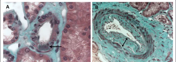
Interstitial fibrosis was assessed visually as the percentage of fibrotic interstitial cortical tissue visible on Masson’s stain by 5%-stages [24]. Glomerulosclerosis was defined as the percentage of totally sclerotic glomeruli. Arteriolo-sclerosis was defined as the presence of hyaline deposits in the wall of at least one preglomerular arteriole (Figure 1A). Absence of arteriolosclerosis was asserted when none of the eight sections display hyaline deposits. Arteriosclerosis was defined as a thickening of the intima of at least one ar-tery. Patients were classified in three groups according to the maximal intima thickness visible on the biopsy sample (Figure 1B): i) absence of arteriosclerosis: normal intima thickness, ii) moderate arteriosclerosis: thickening of intima with an intima/media ratio < 1, iii) severe arteriosclerosis: thickening of intima with an intima/media ratio ≥ 1.
Statistical analysis
Number and percentage of patients, median and minimum-maximum values are provided for qualitative and quantitative values respectively. Linear relationship of RI with other variables was tested with Pearson’s correl-ation test. Analysis of parameters associated with RI and renal function decline were compared with Chi-square and Mann–Whitney tests respectively for qualitative and
quantitative variables. Kaplan Meier analysis was realized to determine renal survival according to RI value using the log-rank test. In order to determine independent para-meters associated with RI or renal function decline, we performed a multiple linear or logistic regression, respect-ively, including variables with p < 0.20 in univariate analysis.
Statistical analysis was performed using http://marne. u707.jussieu.fr/biostatgv/ website, GraphPad Prism 5.0 and StatView 5.0 softwares. A two-tailed p value < 0.05 was considered to be significant.
Results
Characteristics of patients
Fifty-eight patients were enrolled in the study accord-ing to the inclusion criteria. Their characteristics are summarized in Table 1. They were predominantly male (68.9%). Their median age was 49 years [23–89]. Their median eGFR and proteinuria/creatininuria ratio were respectively 59 mL/min/ 1.73 m2[5–130] and 245 mg/ mmol [7–2000]. Forty seven (81%) patients presented glomerular nephropathy. On biopsy, median sclerotic glomeruli and interstitial fibrosis percentage were re-spectively 13% [0–96] and 10% [0–90]. The presence of arteriolosclerosis or arteriosclerosis could not be stud-ied in 5 and 12 patients, respectively, because of the absence of visible arteriole or artery on biopsy sample. Hyaline arterial deposits were observed on 25 of the 53 biopsies with visible arteriolar sections (47.2%). Arterio-sclerosis was observed in 21 of the 46 patients (45.6%) for whom at least one artery was present on the bi-opsy. Eight (17.3%) patients had severe arteriosclerosis with an intima/media ratio ≥ 1 (Table 1). Median RI was 0.62 [0.31 – 1.0].
Figure 1 Vascular lesions analyzed on renal biopsy (optic microscopy, Masson’s trichromic staining). A: Arteriolosclerosis was defined as the presence of hyaline deposits (arrow) in the wall of at least one arteriole. B: Arteriosclerosis was defined as a thickening of intima. Maximal intima thickness visible on the sample biopsy was measured (double arrow). Here, intima/media ratio was superior to 1, which corresponds to severe arteriosclerosis.
Clinical and biological parameters associated with renal arterial resistive index
We found a positive correlation between RI and age (r = 0.435, p = 0.0063), pulse pressure (r = 0.303, p = 0.022) which are both associated with elevated arterial stiffness.
Furthermore, RI was inversely correlated with baseline eGFR (r = −0.402, p = 0.0018) and to a lesser extent with combined renal length (r = −0.275, p = 0.038). In a mul-tiple linear regression analysis including age, baseline eGFR, pulse pressure and combined renal length, only age (p = 0.0052) and baseline eGFR (p = 0.015) were in-dependently associated with RI. We did not find any sig-nificant correlation of RI with proteinuria/creatininuria ratio (r = 0.141, p = 0.29), systolic (r = 0.080, p = 0.55) and diastolic blood pressure (r = −0.169, p = 0.21). RI was not different between patients who received an anti-hypertensive treatment and those who did not (p = 0.89). In the same way, RI did not differ between patients treated with renin angiotensin system (RAS) blockers (p = 0.65) and those who were not. RI was not influenced by the number of antihypertensive drugs pre-scribed (p = 0.87).
Histological parameters associated with renal arterial resistive index
RI was not different whether hyaline deposits were present or not (0.62 [0.31-1.00] vs 0.63 [0.50-0.71], p = 0.99) (Figure 2A). RI value was similar in patients with normal intima or moderate intima thickening (intima/media ratio < 1) (0.62 [0.52-0.83] vs 0.60 [0.31-0.69], p = 0.71). In con-trast, patients with severe arteriosclerosis (intima/media ratio ≥ 1) had a significantly higher RI than those with no or moderate arteriosclerosis (0.73 [0.56-1.00] vs 0.61 [0.31-0.83], p = 0.032) (Figure 2B). No association was found between RI value and glomerulosclerosis. RI tended to increase with the percentage of interstitial fibrosis (n = 58, r = 0.222, p = 0.10) and was significantly higher when interstitial fibrosis exceeded 20% (0.67 [0.55-1.00] vs 0.61 [0.31- 0.83], p = 0.014) (Figure 2C). Finally, patients with isolated glomerular involvement had significant lower RI than those with interstitial fibrosis > 5% and/or vascular lesions (hyaline arterial deposits and/or intima thickening) (0.60 [0.47-0.64] vs 0.64 [0.31-1.00], p = 0.05).
Association of renal arterial resistive index with renal function outcome
Renal function outcome was assessed at 18 months in 35 (60%) patients (Table 2). Among them, 13 (37%) patients experienced renal function decline. Seven patients had a decrease in eGFR of at least 5 mL/min/ 1.73 m2/year and 6 needed chronic RRT. Their RI was significantly higher than RI of patients with stable or improved renal function (0.69 [0.63-1.00] vs 0.61 [0.31-0.70], p = 0.0023) (Table 2). In univariate analysis, other factors associated with renal function decline at 18 months were: age (p = 0.0035), baseline eGFR (p = 0.052) and proteinuria/creatininuria ratio (p = 0.049) (Table 2). Baseline RI ≥ 0.65 (p = 0.0075) and age (p = 0.037) were the only independent factors associated
Table 1 Characteristics of patients at baseline
Number of patients 58 (100%) Age 49 [23–89] Male 49 (68.9%) Blood pressure (mmHg) Systolic 130 (88–181) Diastolic 78 (59–115) Pulse pressure 50 (28–80) Renal function
Serum creatinine (μmol/L) 124 (54–906) eGFR (ml/min/1,73m2) 59 (5–130) Proteinuria/creatininuria(mg/mmol) 245 (7–2000) Antihypertensive treatments No antihypertensive drug 27 (46.5%) 1 antihypertensive drug 11 (19%) 2 antihypertensive drugs 9 (15.5%) ≥3 antihypertensive drugs 11 (19%) RAS blockers 23 (39.6%) Renal biopsy % sclerotic glomeruli 13 (0–96) % interstitial fibrosis 10 (0–90) Vascular lesions 34/51 (66.7%) Arteriolar hyaline deposits 25/53 (47.2%) Intima/media ratio Normal 25 (54.4%) < 1 13 (28.3%) ≥1 8 (17.3%) Diagnosis FSGN/HIVAN 9 (15.5%) IgA nephropathy 11 (19%) Membranous nephropathy 7 (12.1%) Minimal change disease 3 (5.2%) Lupus 3 (5.2%) Vascularitis 3 (5.2%) Membranous proliferative nephritis 2 (3.4%) Diabetes 2 (3.4%) Amyloidosis 2 (3.4%) Other glomerulonephritis 5 (8.6%) Acute tubulo-interstitial nephritis 3 (1.7%) Chronic tubulo-interstitial nephritis 2 (3.4%) Vascular nephropathy 2 (3.4%) Other 4 (6.9%) Renal arterial resistive index 0.62 (0.31-1.00)
Bigé et al. BMC Nephrology 2012, 13:139 Page 4 of 9 http://www.biomedcentral.com/1471-2369/13/139
with renal function decline at 18 months identified by multiple logistic regression (Table 3).
Which RI threshold should be used in clinical practice?
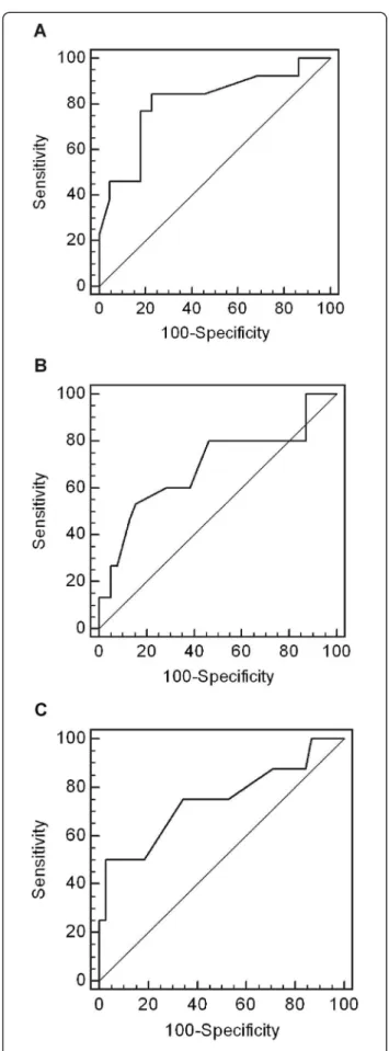
In order to define the most accurate threshold, we draw receiver operating characteristic (ROC) curves (Figure 3). According to the ROC curves analysis, 0.65 threshold
was the most discriminant for renal function decline at 18 months, interstitial fibrosis exceeding 20% and severe arteriosclerosis. Analysis of renal survival using Kaplan-Meier curves confirmed that patients with baseline RI ≥ 0.65 had a poorer renal outcome than those with base-line RI < 0.65 (p = 0.0005, Log-Rank test) (Figure 4). Sensitivity, specificity, predictive positive and predictive negative values of RI ≥ 0.65 for renal function decline at 18 months were respectively 77%, 86%, 71% and 82%.
Discussion
The present study shows that initial measurement of RI in patients with various nephropathies at time of renal biopsy is clinically relevant for several reasons. We show that RI is associated with renal function and pulse pres-sure, a surrogate marker of arterial stiffness. More im-portantly, RI is associated with severe interstitial fibrosis and arteriosclerosis and eGFR decline. Previous studies reported either the association of RI with interstitial fi-brosis, tubulo-interstitial lesions [9-12,25], or vascular lesions [9-11] or renal outcome in CKD [9,11,13,15,16]. To our knowledge, none of these studies simultaneously evaluated the association of RI with the main chronic renal histological lesions and with renal function out-come. Furthermore, the most relevant threshold of RI for clinical practice was still debated. Here, the cut-off value of 0.65 was the most discriminant for severe ar-teriosclerosis, extended fibrosis and renal function decline.
In this study, we analyzed the association of RI with pathological changes and renal function outcome in an unselected population of 58 patients with various nephropathies and renal function alterations. In order to be closer to clinical practice, our inclusion criteria differ from other studies which were done in specific renal dis-eases [12,25]. Our results emphasize the general predict-ive value of RI in CKD patients independently of the type of nephropathy. Secondly, only well-defined chronic renal lesions as interstitial fibrosis, percentage of scler-otic glomeruli, arteriosclerosis lesions were taken into account in our study. These criteria differ from previous
studies which used combined scores as
tubulo-interstitial injury or tubulo-interstitial fibrosis/tubular atrophy, which could reflect acute kidney injury [9-12]. Despite a relative small population of CKD patients, we found a significant association of RI with severe renal lesions and renal function decline, consistently with previous studies [9-16,20].
As previous authors, we found a positive correlation between RI and age [10,26-29] and in a lesser extent with pulse pressure [26,30-33]. Prior studies reported an association between RI and other markers of arterial stiffness as pulse wave velocity [31,34] and ankle-brachial blood pressure index [35]. In our study, RI was
Figure 2 Association of RI with histological parameters. Boxes show the first and third quartiles, with the median as a thick line. Whiskers extend to minimum and maximum values. A: RI according to arteriolosclerosis, defined as the presence of hyaline deposits in the wall of at least one preglomerular arteriole. B: RI according to maximal intima thickness (moderate arteriosclerosis was defined as a thickening of intima with intima/media ratio < 1 and severe arteriosclerosis as an intima/media ratio ≥ 1). * p<0.05 versus normal and intima/media<1. C: RI according to interstitial fibrosis. * p<0.05 versus interstitial fibrosis<20%.
not associated with systolic nor with diastolic blood pressure despite its relationship with pulse pressure. This suggests that the elevation of RI rather reflects the vascular consequences of hypertension than hyperten-sion itself. An alternative explanation is the lack of power of our study. Nevertheless, this result was demon-strated by previous authors who showed that RI is a marker of target organ damage in essential hypertension [30,32,34,36,37] as left ventricular hypertrophy, carotid and coronary atherosclerosis. Other studies demon-strated that high RI was also associated with systemic atherosclerosis in diabetic patients [31,38] and renal transplant recipients [35]. Moreover, Pearce et al. re-cently showed that elevated renal RI is predictive of car-diovascular events in the elderly [39].
The elevation of RI with severe arteriosclerosis may account for its association with cardiovascular risk. As previous authors [9,25], we actually found an association between RI and renal arteriosclerosis. It is interesting to note that only patients with severe intima thickening exhibited high RI. Nor hyaline arteriolar deposits nor moderate intima thickening were associated with increased RI. This could be due to the lack of power of
our study. Nevertheless, it could also suggest that the presence of moderate vascular lesions is not sufficient to induce an elevation of RI and that these alterations must be important enough to reduce artery lumen, raise arter-ial stiffness and vascular resistance, and consequently generate an increase in RI. Overall, these findings sug-gest that high RI reflects severe renal arteriosclerosis and maybe systemic arteriosclerosis.
Our study also questioned the correlation of RI with renal fibrosis. We did not find any relationship between RI and glomerulosclerosis. Moreover, RI was significantly lower in case of isolated glomerular involvement, i.e. without any vascular and/or tubulo-interstitial damage. This result corroborates previous findings of Platt et al. [25]. Only one among three previous studies found a sig-nificant association between glomerulosclerosis and RI [10]. This correlation was weak in a second study [12] and not significant in a multivariate analysis in the third one [9]. Altogether, these findings suggest that glomeru-losclerosis does not influence the value of RI.
On the other hand, as other authors, we found an asso-ciation between RI value and the extension of interstitial fibrosis [9,11,12,25] and the severity of renal impairment [13,26,40-44]. Median percentage of interstitial fibrosis was six fold higher in patients with RI exceeding 0.65. As arteriosclerosis, interstitial fibrosis appears as an import-ant determinimport-ant of RI. Three hypotheses can be drawn about the physiopathological mechanisms involved in ele-vation of RI with the progression of CKD: i) decrease in arterial compliance and increase in vascular resistance be-cause of renal arteriosclerosis, ii) elevation of pressure exerted by interstitial fibrosis on adjacent vessels, iii) vaso-constriction secondary to the hypoxia induced by the pre-vious phenomena and by the loss of capillaries associated with renal fibrosis. These mechanisms are probably com-bined and our results do not allow us to precise which one contributes the most to the elevation of RI.
Table 2 Univariate analysis of parameters associated with renal function decline at 18 months (defined as a decrease in eGFR of at least 5 mL/min/ 1.73 m2/year or need for RRT) (Mann–Whitney and Fisher’s exact tests)
No decline Decline p Number (%) of patients 22 (63%) 13 (37%)
Age (years) 38 (23–68) 55 (24–79) 0.0035 Baseline eGFR (mL/min/1.73m2) 65 (17–108) 12 (4–65) 0.052
eGFR(mL/min/1.73m2) at 18 months 69 (24–123) 8 (5–70) 0.000015 SBP (mmHg) 127 (98–160) 129 (98–163) 0.30 DBP (mmHg) 77 (62–91) 70 (59–96) 0.89 Proteinuria/creatininuria (mg/mmol) 96 (7–1216) 492 (69–1742) 0.049 % sclerotic glomeruli 16 (0–71) 4 (0–80) 0.88 % interstitial fibrosis 18 (0–80) 18 (0–90) 0.76 RI 0.61 (0.31-0.70) 0.69 (0.63-1.00) 0.0023 Number (%) of patients with RI ≥ 0.65 4 (18%) 10 (77%) 0.0011
Table 3 Multivariate analysis of parameters associated with renal function decline at 18 months (defined as a decrease in eGFR of at least 5 mL/min/ 1.73 m2/year or
need for RRT) (logistic regression, n=35 patients)
OR 95% CI P Proteinuria/creatininuria (mg/mmol) 1.001 0.998-1.003 0.56 Baseline eGFR (mL/min/1.73m2) 0.977 0.936-1.021 0.30
Age (years) 1.079 0.996-1.169 0.062 RI ≥ 0.65 7.751 1.045-57.479 0.045 OR 95% CI P Age (years) 1.078 1.004-1.158 0.037 RI ≥ 0.65 13.04 1.984-85.727 0.0075
Bigé et al. BMC Nephrology 2012, 13:139 Page 6 of 9 http://www.biomedcentral.com/1471-2369/13/139
In the second part of our study, we found that high RI was independently associated with accelerated renal function decline. Sugiura et al. [13] reported similar re-sult in a larger cohort of 311 CKD patients followed-up for two years. Such results were also found by other authors in CKD [11,14-17], essential hypertension [17] and renal transplantation [8,45]. Our results extend pre-vious findings to a population of patients with various nephropathies. The concomitant association of RI with interstitial fibrosis and arteriosclerosis which are known to be major determinants of the progression of CKD [4] may explain its prognosis value.
Finally, we attempted to define the most relevant thresh-old of RI in clinical practice. Consistently with the first findings of Sugiura et al. [12], we found that 0.65 was the most accurate threshold to detect extent interstitial fibro-sis, but also severe arteriosclerosis. Nevertheless, more re-cently, Sugiura et al. suggested that 0.70 threshold was better than 0.65 cut-off to predict renal function decline [13]. In contrast, we found by ROC curves analysis that RI ≥ 0.65 has the best sensitivity (77%) and specificity (86%) to discriminate renal function decline. This discrepancy may be explained by the different definitions of renal func-tion decline used in the two studies. Using the criteria of at least 10 mL/min/ 1.73 m2/year, Sugiura et al. may have selected more severe patients. In cohort studies, mean slope of eGFR decline is less than 5 mL/min/ 1.73 m2in most CKD patients [2,19-23]. A decrease of 5 mL/min/ 1.73 m2/year appears to be closer to clinical practice and more helpful to detect a larger of number patients at high risk of accelerated CKD progression.
Our study has several limits. We mostly included glom-erulonephritis and few vascular and tubulo-interstitial dis-eases. Because of the weak proportion of chronic vascular and tubulo-interstitial diseases, we can wonder whether our results could apply to those nephropathies. Never-theless, several previous studies found an association between high RI and poor renal outcome in essential hypertension and chronic tubulo-interstitial nephropa-thies [13,14,17,34,37]. The main limit of our study is the non exhaustive collection of renal function data. However, our results corroborate those of several pre-vious studies [9,11,13,15,16].
Conclusion
Our results show that RI ≥ 0.65 in CKD patients with various nephropathies is associated with extended inter-stitial fibrosis, severe arteriosclerosis and renal function
Figure 3 Receiver operating characteristic (ROC) curves for RI to discriminate. A: renal function decline at 18 months (AUC = 0.809, p = 0.0002). B: interstitial fibrosis > 20% (AUC = 0.690, p = 0.037). C: severe arteriosclerosis (AUC = 0.740, p = 0.039).
decline. Consistently with previous findings, these results suggest that non invasive US Doppler measurement of RI could give the opportunity to identify CKD patients at high risk of ESRD and help clinicians in their manage-ment. Our results need to be confirmed in a larger tar-geted intervention multicentric study of outpatients.
Abbreviations
CKD: Chronic kidney disease; DBP: Diastolic blood pressure; eGFR: Estimated glomerular filtration rate; ESRD: End stage renal disease; FSGN: Focal segmental glomerulonephritis; HIVAN: HIV-associated nephropathy; RI: Renal doppler resistive index; RRT: Renal replacement therapy; SBP: Systolic blood pressure.
Competing interests
None of the authors have conflicts of interest to declare. Authors’ contributions
JJB designed the study. NB and JJB had full access to all the data and take responsibility for the integrity of the data and the accuracy of the data analysis. NB and VJ collected data. PC realized histological study. VC and JMF performed renal US Doppler. NB and PL realized statistical analysis. NB and JJB wrote the manuscript. PL and PR made critical revision of the manuscript. All authors approved the final version of this manuscript.
Author details
1Department of Nephrology, AP-HP, Hôpital Tenon, 4 rue de la Chine, Paris
F-75020, France.2Public Health Department, AP-HP, Hôpital Tenon, Paris
F-75020, France.3INSERM UNIT 707, Paris F-75012, France.4Université Pierre
et Marie Curie-Paris 6, UMR-S 707, Paris F-75012, France.5Department of Pathology, AP-HP, Hôpital Tenon, Paris F-75020, France.6Department of
Radiology, AP-HP, Hôpital Tenon, Paris F-75020, France.7INSERM UNIT 702,
Paris F-75020, France.8Université Pierre et Marie Curie-Paris 6, UMR S 702 ,
Paris, France.
Received: 25 April 2012 Accepted: 25 September 2012 Published: 25 October 2012
References
1. Meguid El Nahas A, Bello AK: Chronic kidney disease: the global challenge. Lancet 2005, 365(9456):331–340.
2. Eriksen BO, Ingebretsen OC: The progression of chronic kidney disease: a 10-year population-based study of the effects of gender and age. Kidney Int2006, 69(2):375–382.
3. Clark LE, Khan I: Outcomes in CKD: what we know and what we need to know. Nephron 2010, 114(2):c95–c102.
4. Nath KA: Tubulointerstitial changes as a major determinant in the progression of renal damage. Am J Kidney Dis 1992, 20(1):1–17. 5. Crutchley TA, Pearce JD, Craven TE, Stafford JM, Edwards MS, Hansen KJ:
Clinical utility of the resistive index in atherosclerotic renovascular disease. J Vasc Surg 2009, 49(1):148–155. 155 e141-143; discussion 155. 6. Radermacher J, Chavan A, Bleck J, Vitzthum A, Stoess B, Gebel MJ, Galanski M,
Koch KM, Haller H: Use of Doppler ultrasonography to predict the outcome of therapy for renal-artery stenosis. N Engl J Med 2001, 344(6):410–417. 7. Rodrigo E, Lopez-Rasines G, Ruiz JC, Lastra P, Gomez-Dermitt V,
Gomez-Alamillo C, Gonzalez-Cotorruelo J, Calabia A, Arias M: Determinants of resistive index shortly after transplantation: independent relationship with delayed graft function. Nephron 2010, 114(3):c178–c186.
8. Radermacher J, Mengel M, Ellis S, Stuht S, Hiss M, Schwarz A, Eisenberger U, Burg M, Luft FC, Gwinner W, et al: The renal arterial resistance index and renal allograft survival. N Engl J Med 2003, 349(2):115–124.
9. Ikee R, Kobayashi S, Hemmi N, Imakiire T, Kikuchi Y, Moriya H, Suzuki S, Miura S: Correlation between the resistive index by Doppler ultrasound and kidney function and histology. Am J Kidney Dis 2005, 46(4):603–609. 10. Mostbeck GH, Kain R, Mallek R, Derfler K, Walter R, Havelec L, Tscholakoff D:
Duplex Doppler sonography in renal parenchymal disease. Histopathologic correlation. J Ultrasound Med 1991, 10(4):189–194. 11. Splendiani G, Parolini C, Fortunato L, Sturniolo A, Costanzi S: Resistive index
in chronic nephropathies: predictive value of renal outcome. Clin Nephrol 2002, 57(1):45–50.
12. Sugiura T, Nakamori A, Wada A, Fukuhara Y: Evaluation of tubulointerstitial injury by Doppler ultrasonography in glomerular diseases. Clin Nephrol 2004, 61(2):119–126.
13. Sugiura T, Wada A: Resistive index predicts renal prognosis in chronic kidney disease. Nephrol Dial Transplant 2009, 24(9):2780–2785.
14. Parolini C, Noce A, Staffolani E, Giarrizzo GF, Costanzi S, Splendiani G: Renal resistive index and long-term outcome in chronic nephropathies. Radiology2009, 252(3):888–896.
15. Petersen LJ, Petersen JR, Talleruphuus U, Ladefoged SD, Mehlsen J, Jensen HA: The pulsatility index and the resistive index in renal arteries. Associations with long-term progression in chronic renal failure. Nephrol Dial Transplant1997, 12(7):1376–1380.
16. Radermacher J, Ellis S, Haller H: Renal resistance index and progression of renal disease. Hypertension 2002, 39(2 Pt 2):699–703.
17. Okura T, Kurata M, Irita J, Enomoto D, Jotoku M, Nagao T, Koresawa M, Kojima S, Hamano Y, Mashiba S, et al: Renal resistance index is a marker of future renal dysfunction in patients with essential hypertension. J Nephrol2010, 23(2):175–180.
18. National Kidney Foundation: K/DOQI clinical practice guidelines for chronic kidney disease: evaluation, classification, and stratification. Am J Kidney Dis2002, 39(2 Suppl 1):S1–S266.
19. Conway B, Webster A, Ramsay G, Morgan N, Neary J, Whitworth C, Harty J: Predicting mortality and uptake of renal replacement therapy in patients with stage 4 chronic kidney disease. Nephrol Dial Transplant 2009, 24(6):1930–1937.
Figure 4 Kaplan Meier curves of renal survival according to baseline RI. Renal function decline is defined as decrease in eGFR of at least 5mL/min/1.73m2/year or need for RRT. Plain line represents patients with RI < 0.65 and dotted line those with RI ≥ 0.65; p = 0.0005,
log-rank test.
Bigé et al. BMC Nephrology 2012, 13:139 Page 8 of 9 http://www.biomedcentral.com/1471-2369/13/139
20. Eriksen BO, Ingebretsen OC: In chronic kidney disease staging the use of the chronicity criterion affects prognosis and the rate of progression. Kidney Int2007, 72(10):1242–1248.
21. Halbesma N, Kuiken DS, Brantsma AH, Bakker SJ, Wetzels JF, De Zeeuw D, De Jong PE, Gansevoort RT: Macroalbuminuria is a better risk marker than low estimated GFR to identify individuals at risk for accelerated GFR loss in population screening. J Am Soc Nephrol 2006, 17(9):2582–2590. 22. Hunsicker LG, Adler S, Caggiula A, England BK, Greene T, Kusek JW, Rogers NL,
Teschan PE: Predictors of the progression of renal disease in the modification of diet in renal disease study. Kidney Int 1997, 51(6):1908–1919. 23. John R, Webb M, Young A, Stevens PE: Unreferred chronic kidney disease:
a longitudinal study. Am J Kidney Dis 2004, 43(5):825–835.
24. Farris AB, Adams CD, Brousaides N, Della Pelle PA, Collins AB, Moradi E, Smith RN, Grimm PC, Colvin RB: Morphometric and visual evaluation of fibrosis in renal biopsies. J Am Soc Nephrol 2011, 22(1):176–186. 25. Platt JF, Ellis JH, Rubin JM, DiPietro MA, Sedman AB: Intrarenal arterial Doppler
sonography in patients with nonobstructive renal disease: correlation of resistive index with biopsy findings. Ajr 1990, 154(6):1223–1227. 26. Heine GH, Gerhart MK, Ulrich C, Kohler H, Girndt M: Do ultrasound renal
resistance indices reflect systemic rather than renal vascular damage in chronic kidney disease? Nephrol Dial Transplant 2006, 22(1):163–170. 27. Keogan MT, Kliewer MA, Hertzberg BS, DeLong DM, Tupler RH, Carroll BA:
Renal resistive indexes: variability in Doppler US measurement in a healthy population. Radiology 1996, 199(1):165–169.
28. Lin ZY, Wang LY, Yu ML, Dai CY, Chen SC, Chuang WL, Hsieh MY, Tsai JF, Chang WY: Influence of age on intrarenal resistive index measurement in normal subjects. Abdom Imaging 2003, 28(2):230–232.
29. Rivolta R, Cardinale L, Lovaria A, Di Palo FQ: Variability of renal echo-Doppler measurements in healthy adults. J Nephrol 2000, 13(2):110–115. 30. Florczak E, Januszewicz M, Januszewicz A, Prejbisz A, Kaczmarska M,
Michalowska I, Kabat M, Rywik T, Rynkun D, Zielinski T, et al: Relationship between renal resistive index and early target organ damage in patients with never-treated essential hypertension. Blood Press 2009, 18(1–2):55–61. 31. Ohta Y, Fujii K, Arima H, Matsumura K, Tsuchihashi T, Tokumoto M, Tsuruya
K, Kanai H, Iwase M, Hirakata H, et al: Increased renal resistive index in atherosclerosis and diabetic nephropathy assessed by Doppler sonography. J Hypertens 2005, 23(10):1905–1911.
32. Okura T, Watanabe S, Miyoshi K, Fukuoka T, Higaki J: Intrarenal and carotid hemodynamics in patients with essential hypertension. Am J Hypertens 2004, 17(3):240–244.
33. Ozelsancak R, Torun D, Koc Z, Sezer S, Ozdemir FN, Niron EA: Relationship between renal resistive index and inflammation in untreated hypertensive patients. Int Heart J 2009, 50(6):753–761.
34. Raff U, Schmidt BM, Schwab J, Schwarz TK, Achenbach S, Bar I, Schmieder RE: Renal resistive index in addition to low-grade albuminuria complements screening for target organ damage in therapy-resistant hypertension. J Hypertens 2010, 28(3):608–614.
35. Heine GH, Gerhart MK, Ulrich C, Kohler H, Girndt M: Renal Doppler resistance indices are associated with systemic atherosclerosis in kidney transplant recipients. Kidney Int 2005, 68(2):878–885.
36. Pontremoli R, Viazzi F, Martinoli C, Ravera M, Nicolella C, Berruti V, Leoncini G, Ruello N, Zagami P, Bezante GP, et al: Increased renal resistive index in patients with essential hypertension: a marker of target organ damage. Nephrol Dial Transplant1999, 14(2):360–365.
37. Shimizu Y, Itoh T, Hougaku H, Nagai Y, Hashimoto H, Sakaguchi M, Handa N, Kitagawa K, Matsumoto M, Hori M: Clinical usefulness of duplex ultrasonography for the assessment of renal arteriosclerosis in essential hypertensive patients. Hypertens Res 2001, 24(1):13–17.
38. Buscemi S, Verga S, Batsis JA, Cottone S, Mattina A, Re A, Arnone M, Citarda S, Cerasola G: Intra-renal hemodynamics and carotid intima-media thickness in the metabolic syndrome. Diabetes Res Clin Pract 2009, 86(3):177–185.
39. Pearce JD, Craven TE, Edwards MS, Corriere MA, Crutchley TA, Fleming SH, Hansen KJ: Associations between renal duplex parameters and adverse cardiovascular events in the elderly: a prospective cohort study. Am J Kidney Dis2010, 55(2):281–290.
40. Galesic K, Sabljar-Matovinovic M, Tomic M, Brkljacic B: Renal vascular resistance in glomerular diseases–correlation of resistance index with biopsy findings. Coll Antropol 2004, 28(2):667–674.
41. Kim SH, Kim WH, Choi BI, Kim CW: Duplex Doppler US in patients with medical renal disease: resistive index vs serum creatinine level. Clin Radiol1992, 45(2):85–87.
42. Petersen LJ, Petersen JR, Ladefoged SD, Mehlsen J, Jensen HA: The pulsatility index and the resistive index in renal arteries in patients with hypertension and chronic renal failure. Nephrol Dial Transplant 1995, 10(11):2060–2064.
43. Yura T, Yuasa S, Sumikura T, Takahashi N, Aono M, Kunimune Y, Fujioka H, Miki S, Takamitsu Y, Matsuo H: Doppler sonographic measurement of phasic renal artery blood flow velocity in patients with chronic glomerulonephritis. J Ultrasound Med 1993, 12(4):215–219.
44. Sari A, Dinc H, Zibandeh A, Telatar M, Gumele HR: Value of resistive index in patients with clinical diabetic nephropathy. Invest Radiol 1999, 34(11):718–721.
45. Saracino A, Santarsia G, Latorraca A, Gaudiano V: Early assessment of renal resistance index after kidney transplant can help predict long-term renal function. Nephrol Dial Transplant 2006, 21(10):2916–2920.
doi:10.1186/1471-2369-13-139
Cite this article as: Bigé et al.: Renal arterial resistive index is associated with severe histological changes and poor renal outcome during chronic kidney disease. BMC Nephrology 2012 13:139.
Submit your next manuscript to BioMed Central and take full advantage of:
• Convenient online submission • Thorough peer review
• No space constraints or color figure charges • Immediate publication on acceptance
• Inclusion in PubMed, CAS, Scopus and Google Scholar • Research which is freely available for redistribution
Submit your manuscript at www.biomedcentral.com/submit