HAL Id: hal-02790844
https://hal.inrae.fr/hal-02790844
Preprint submitted on 5 Jun 2020HAL is a multi-disciplinary open access
archive for the deposit and dissemination of sci-entific research documents, whether they are pub-lished or not. The documents may come from teaching and research institutions in France or abroad, or from public or private research centers.
L’archive ouverte pluridisciplinaire HAL, est destinée au dépôt et à la diffusion de documents scientifiques de niveau recherche, publiés ou non, émanant des établissements d’enseignement et de recherche français ou étrangers, des laboratoires publics ou privés.
Amaury Herpin, Christophe Klopp, Cédric Cabau, Margot Zahm, Hugues
Parrinello, et al.
To cite this version:
Ming Wen, Romain Feron, Qiaowei Pan, Justine Guguin, Elodie Jouanno, et al.. Sex chromosome and sex locus characterization in the goldfish, Carassius auratus. 2019. �hal-02790844�
Sex chromosome and sex locus characterization in the goldfish,
1Carassius auratus.
2 3
Ming Wen1,2, Romain Feron2,3,4, Qiaowei Pan2,3, Justine Guguin2, Elodie Jouanno2, Amaury 4
Herpin2, Christophe Klopp5,6, Cedric Cabau6, Margot Zahm6, Hugues Parrinello7, Laurent 5
Journot7, Shawn M. Burgess8, Yoshihiro Omori9,10, John H. Postlethwait11, Manfred Schartl12, 6
Yann Guiguen2* 7
8
* Correspondance: Yann Guiguen: yann.guiguen@inra.fr 9
10
AFFILIATIONS:
11 12
1 State Key Laboratory of Developmental Biology of Freshwater Fish, College of Life Science, 13
Hunan Normal University, Changsha, China 14
2 INRAE, UR 1037 Fish Physiology and Genomics, F-35000 Rennes, France. 15
3 Department of Ecology and Evolution, University of Lausanne, 1015 Lausanne, Switzerland. 16
4 Swiss Institute of Bioinformatics, 1015 Lausanne, Switzerland. 17
5 Plate-forme bio-informatique Genotoul, Mathématiques et Informatique Appliquées de 18
Toulouse, INRAE, Castanet Tolosan, France. 19
6 SIGENAE, GenPhySE, Université de Toulouse, INRAE, ENVT, Castanet Tolosan, France. 20
7 Montpellier GenomiX (MGX), c/o Institut de Génomique Fonctionnelle, 141 rue de la 21
Cardonille, 34094, Montpellier Cedex 05, France. 22
8 Translational and Functional Genomics Branch, National Human Genome Research Institute, 23
Bethesda, MD, USA. 24
9 Laboratory of Functional Genomics, Graduate School of Bioscience, Nagahama Institute of 25
Bioscience and Technology, Nagahama, Shiga, Japan. 26
10 Laboratory for Molecular and Developmental Biology, Institute for Protein Research, Osaka 27
University, Suita, Osaka, Japan. 28
11 Institute of Neuroscience, University of Oregon, Eugene, Oregon, USA. 29
12 Developmental Biochemistry, Biozentrum, University of Würzburg, Würzburg, Germany and 30
The Xiphophorus Genetic Stock Center, Department of Chemistry and Biochemistry, Texas 31
State University, San Marcos, Texas, USA. 32 33 34 35 36 37
Abstract
38
Background: Goldfish is an important model for various areas of research, including neural
39
development and behavior and a species of significant importance in aquaculture, especially as 40
an ornamental species. It has a male heterogametic (XX/XY) sex determination system that 41
relies on both genetic and environmental factors, with high temperatures being able to produce 42
female-to-male sex reversal. Little, however, is currently known on the molecular basis of 43
genetic sex determination in this important cyprinid model. We used sequencing approaches to 44
better characterize sex determination and sex-chromosomes in goldfish. 45
Results: Our results confirmed that sex determination in goldfish is a mix of environmental
46
and genetic factors and that its sex determination system is male heterogametic (XX/XY). 47
Using reduced representation (RAD-seq) and whole genome (pool-seq) approaches, we 48
characterized sex-linked polymorphisms and developed male specific genetic markers. These 49
male specific markers were used to distinguish sex-reversed XX neomales from XY males and 50
to demonstrate that XX female-to-male sex reversal could even occur at a relatively low rearing 51
temperature (18°C), for which sex reversal has been previously shown to be close to zero. We 52
also characterized a relatively large non-recombining region (~11.7 Mb) on goldfish linkage 53
group 22 (LG22) that contained a high-density of male-biased genetic polymorphisms. This 54
large LG22 region harbors 373 genes, including a single candidate as a potential master sex 55
gene, i.e., the anti-Mullerian hormone gene (amh). However, no sex-linked polymorphisms 56
were detected in the goldfish amh gene or its 5 kb proximal promoter sequence. 57
Conclusions: These results show that goldfish have a relatively large sex locus on LG22, which
58
is likely the goldfish Y chromosome. The presence of a few XX males even at low temperature 59
also suggests that other environmental factors in addition to temperature could trigger female-60
to-male sex reversal. Finally, we also developed sex-linked genetic markers in goldfish, which 61
will be important for future research on sex determination and aquaculture applications in this 62
species. 63
Key words: Goldfish, RADseq, Poolseq, Sex determination, Sex markers, Male genome
64
assembly 65
BACKGROUND
67
Goldfish, Carassius auratus, is a domesticated fish species originating from central Asia and 68
China that has been introduced throughout the world. Goldfish belongs to the Cyprinidae family 69
and is considered as an important fish model for research in endocrinology [1, 2], 70
developmental biology [3, 4] or fish pathology [5]. Thanks to the recent availability of a whole 71
genome sequence assembly [6], goldfish is also now becoming a key model species for studies 72
on genomics and cyprinid genome evolution. It is also a species of high aquaculture importance 73
especially as an ornamental species, with many beautiful and sometimes bizarre phenotypes [7]. 74
Unlike birds and mammals, sex determination in teleost is highly dynamic, with frequent 75
turnovers of both sex determination (SD) systems [8] and master sex determining genes (MSD) 76
[9, 10]. Currently about half a dozen different master sex determining genes have been 77
identified in teleosts, including dmrt1 in the Japanese medaka, Oryzias latipes [11], sdY in 78
rainbow trout [12], amh in Northern pike, Nile tilapia and pejerrey [13-15], amhr2 in yellow 79
perch and the Takifugu pufferfish [16, 17], gsdf in sablefish and Luzon medaka, O. luzonensis 80
[18, 19], gsdf6a in the turquoise killifish [20] and sox3 in the Indian ricefish O. dancena [21]. 81
MSD turnover can be evolutionarily rapid as has been shown for instance in various ricefish 82
species [22]. In addition to genetic determinants, environmental factors -- especially 83
temperature -- have also been shown to play a pivotal role in teleost sex determination [23]. 84
Since the late 1960s, the goldfish sex determination system has been characterized as male 85
heterogametic (XX/XY) [24]. More recently, a strong temperature influence on sex-ratios has 86
also been characterized in goldfish, with high rearing temperature treatments inducing complete 87
masculinization of chromosomally all-female genotypes (XX neomales) when applied during 88
early 3 months development [25]. The molecular mechanisms of genetic sex determination, 89
however, are still unknown not only in goldfish, but also in any member of the Cyprinidae 90
family. 91
Because of new high throughput sequencing technologies and the availability of a whole 92
genome sequence assembly for goldfish [26], we implemented both reduced representation (i.e., 93
Restriction-site associated DNA sequencing (RAD-seq) [27, 28], and whole genome (i.e., Pool 94
sequencing (Pool-seq) [29, 30]) approaches to identify sex-linked genetic polymorphisms in 95
goldfish. We verified that identified sex-linked markers strictly segregated with the Y 96
chromosome, and we characterized the extent of Y chromosome differentiation. Although our 97
experiments did not identify a strong candidate sex-determining gene, these results lay a solid 98
foundation for further molecular exploration of goldfish sex determination. 99
100
RESULTS
101
Characterization of goldfish sex-linked Y chromosome markers
102
Because goldfish sex determination is highly sensitive to temperature [21], with high 103
temperature leading to the masculinization of some XX females producing XX neomales, we 104
first searched for sex-linked markers using a RAD-seq approach that kept track of phenotypes 105
and genotypes, potentially enabling the discrimination of XX neomales from XY genetic males. 106
From our RAD-seq data, we identified 32 polymorphic/specific RAD-tags that were present in 107
12-15 males among the 30 phenotypic males used in this experiment, and completely absent in 108
all the 30 phenotypic females (Fig. 1A, Supplementary excel file1). These results suggest a 109
male heterogametic genetic sex determination system (XX/XY) as previously shown in goldfish 110
[24], but with a rather high occurrence of XX neomales (around 50 %) in this population of 111
two-year old animals raised outdoor and obtained from different batches of animals with 112
different spawning times i.e., from May-June to late September. 113
To validate the hypothesis that these markers were linked to the heterogametic sex (XY) and 114
the Y chromosome, we first sequenced using Illumina reads and assembled a draft genome 115
sequence of a male goldfish identified as a putative XY male based on the polymorphic/specific 116
RAD-tags (see Material & Methods) and blasted these 32 marker sequences against this genome 117
assembly. This analysis returned 20 contigs with highly significant matches (Supplementary 118
excel file2) spanning a total of 0.24 Mb. By anchoring these sex-linked RAD sequences on our 119
genome assembly, we were able to design three putative Y-allele specific primer pairs that were 120
used to genotype the same individual animals that were used for the RAD-seq analysis. PCR 121
genotyping using these three primer pairs accurately discriminated putative XY genetic males 122
from putative XX neomales and females (Fig. 1B), validating that these primers accurately 123
identified the two types of males found in our RAD-seq analysis.We then genotyped male 124
breeders from our experimental stock with these primers and selected one putative XX neomale 125
(breeder 1, negative PCR amplifications) and one putative XY male (breeder 2, positive PCR 126
amplifications); and both individuals were crossed to the same XX female to generate two 127
separate batches of fish. If our Y-allele specific primers correctly identify the Y chromosome, 128
then our putative XX neomale should give only female offspring and the putative XY male 129
should give both male and female offspring. These two experimental populations were then 130
reared at low temperature (18℃) during the first three months after fertilization to minimize 131
high temperature masculinization [25], and were subsequently maintained at 24℃ for nine 132
additional months before the identification of the phenotypic sex. Results from the histological 133
examination of the offspring gonads of the putative XX neomale identified 7 fish with testes, 134
83 fish with ovaries, and 41 fish with undifferentiated gonads. Disregarding fish with 135
undifferentiated gonads suggests a sex ratio of 7.8% males and 92.2% females for the offspring 136
of the XX neomale (Table 1). Gonadal histology of the offspring of the putative XY revealed 137
48 animals with testes, 65 with ovaries, and 14 with undifferentiated gonads, which gives a sex 138
ratio of 42.5% male and 57.5% female, ignoring the offspring with undifferentiated gonads 139
(Table 1). These sex ratio differences (Table 1), strongly support the hypothesis that male 140
breeder 1 is an XX neomale with an offspring sex ratio not significantly different from an 141
expected all-female population with a slight percentage of female-to-male sex-reversal, and that 142
breeder 2 is a genetic XY male with an offspring sex ratio not significantly different from an 143
expected 50:50 sex ratio. In agreement with these results, none of the XX neomale offspring 144
produced a positive PCR amplification for our three Y-allele specific primer pairs (Figure S3, 145
Table 2), and all 48 phenotypic males but only one of 65 phenotypic females offspring from the 146
XY phenotypic male produced positive amplifications (Figure S4, Table 2). This result also 147
indicates that no neomales were detected in offspring from the XY genetic male if we do not 148
consider the undetermined individuals compared to the 7.8% of neomales in the XX neomale 149
offspring population. 150
151
Characterization of the goldfish sex chromosome and sex-determining region (SDR)
152
Using the three Y-allele specific primer pairs described above, we genotyped goldfish 153
individuals and selected 30 phenotypic and genotypic males that were used along with 30 154
phenotypic females to contrast whole genome sex differences by pool-sequencing analysis [29]. 155
Pool-sequencing reads from the respective XY male and phenotypic female pools were mapped 156
to the high contiguity goldfish female genome assembly [6] to characterize genomic regions 157
enriched for sex-biased signals, i.e., sex coverage differences or sex-biased Single Nucleotide 158
Polymorphism (SNP) differences. Whole genome analysis of SNP distribution (Figure 2) 159
revealed a strong sex-linked signal in males on linkage group 22 (LG22) and two unplaced 160
scaffolds (NW_020523543.1 and NW_020523609.1) with a high density of observed SNPs 161
being heterozygous in the male pool and homozygous in the female pool (Y-specific allele). 162
Interestingly, of the 32 markers found using the RAD-Seq approach, 7 tags were enriched in 163
the unplaced scaffold NW_020523543.1 (Fig. 3C), confirming by a second approach that this 164
scaffold is part of the SD locus in goldfish. These regions with a high density of male-specific 165
SNPs (Figure 3) are potential sex-determining regions that could contain the goldfish master 166
sex determining gene. LG22, being the only linkage group with a large sex determining region 167
(SDR, highlighted by a black box on Fig. 3A, C, D) containing a high-density of male-specific 168
SNPs (~11.7 Mb), likely corresponds to the goldfish Y sex chromosome. 169
We also observed, however, some smaller signals with less dense sex-linked SNPs in other 170
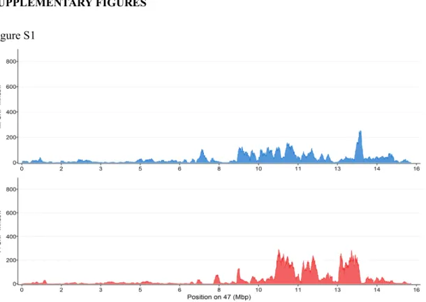
linkage groups (Figure 2A) like for instance on LG47 (Fig. S1) with both male and female sex-171
linked signals. Interestingly, LG47 is paralogous to LG22 stemming from the Cyprinidae whole 172
genome duplication [6]. Indeed, due to this recent common ancestry, these two chromosomes 173
share large homologous and syntenic regions (Fig. S2) that could have resulted in some false 174
remapping of the pool-sequencing reads leading to some of these secondary minor signals. 175
176
Identification of candidate SD genes
Searches for annotated genes by BLAST within the 20 contigs found in our male goldfish draft 178
genome assembly based on the RAD-Seq approach did not return any matches for a candidate 179
SD gene, but mostly transposable elements (Supplementary excel file 3). All genes within the 180
SDR (N= 373) were extracted because they are potential candidates for being SD gene(s) 181
(Supplemental excel file 4). Interestingly, among these genes the anti-Mullerian hormone gene 182
(amh) was found at the beginning of the SDR on LG22 (Fig. 3B). This gene has been reported 183
to be a sex-determining gene in other fish species [14, 15]. However, we did not identify any 184
male-specific SNPs in the coding sequence of goldfish amh. In addition, other male specific 185
alleles within the 5kb promoter region did not show any sex-linkage. 186
187
DISCUSSION
188
Though goldfish is an important economic ornamental fish and a useful model for studying 189
development, evolution, neuroscience, and human disease [3], characterization of goldfish sex-190
specific sequences and potential sex chromosomes have not been reported. In this study, we 191
explored goldfish sex determination using two complementary whole-genome approaches and 192
found that this species has a XX/XY sex determination system as previously described [24] and 193
a large, non-recombining sex determination region on LG22. Although RAD-sequencing or 194
pool-sequencing have been often used separately to explore sex determination in vertebrates 195
[16, 30, 31], we choose to combine these two approaches in goldfish because of the significant 196
female-to-male sex reversal induced by temperature [25] that would have prevented a clear 197
identification of the sex determining region using only a pooled strategy, which mixes genetic 198
XY males and XX males resulting from the sex reversal of genetic females. Because RAD-199
sequencing keeps track of each individual, we were able to identify sex-reversed individuals in 200
goldfish that might have masked sex-linked markers in Pool-seq. 201
202
Sex markers identification is an important step to characterize SD systems [32-38]. Using two 203
complementary whole-genome approaches, we characterized genomic regions containing sex-204
linked markers. In goldfish, these sex-linked markers are genomic DNA variations including 205
gaps, indels and SNPs that present heterozygote polymorphisms in all males and complete 206
homozygosity in all females. This male-specific heterozygosity pattern agrees with a male 207
heterogametic XX/XY system as previously reported using progeny testing of hormonally sex-208
reversed breeders [24]. We found, however, a strong environmental influence leading to a 209
relatively high proportion (around 50%) of female-to-male sex-reversal in the first experimental 210
population that we used for the RAD-Sequencing approach. These animals were actually two-211
year old goldfish raised in an outdoor experimental facility and obtained at different spawning 212
times i.e., from May-June to late September. Some of these animals experienced early 213
development during summer time at potentially higher temperature and others had their early 214
developmental period at lower temperatures. Considering the known effects of high temperature 215
on female-to-male sex reversal in goldfish [25], the fact that some of these fish were exposed 216
to a high summer temperature could explain this relatively high percentage of female-to-male 217
sex-reversed animals. This high percentage was not found in our other experiments in which 218
fish were raised in indoor recirculating system facilities with a tightly controlled low 219
temperature (18 ) maintained throughout the whole early development phase (3 months). This 220
situation indeed confirms earlier findings showing that temperature is probably a major trigger 221
of neomasculinization in goldfish, but we also found that even at this low temperature there 222
was still a small percentage of female-to-male sex-reversal (7.8%), suggesting that other 223
environmental factors, potentially social factors as demonstrated in other species [8, 39], could 224
also play a role on goldfish sex determination. Apart from goldfish, sex determination in other 225
teleost fish, including Tilapia [40], medaka [41] and tongue sole [42] is also regulated by 226
temperature, which overrides the genetic sex determination mechanisms and leads to female-227
to-male sex reversal. By developing genetic sexing tools in goldfish that allows the 228
identification of Y-allele carrying animals, we also brought additional evidence that some of 229
these phenotypic males were indeed sex-reversed XX genetic females. These genetic sexing 230
tools are indeed important for better deciphering genetic and environmental sex determination 231
in goldfish. But these PCR primers could be also used to facilitate the industrial production of 232
commercial goldfish-related hybrid fish in China [43, 44], by helping to identify neomales i.e., 233
XX female-to-male sex reversed animals. 234
235
Sex determination in vertebrates is highly variable with the major exceptions of Eutherian 236
mammals and birds in which XX/XY and ZZ/ZW monofactorial sex determination systems 237
have been conserved over a long evolutionary period [45, 46]. In contrast, fish exhibit much 238
more diverse and dynamic sex determination [9, 10, 47], with monofactorial and polyfactorial 239
[48, 49] genetic systems and frequent switches and turnovers of master sex-determining genes 240
[12, 14, 15, 17, 21, 50]. In goldfish, we identified specific markers and obvious male-241
specific SNPs strongly enriched on LG22. This result confirms that goldfish has an XX-XY 242
system [24] and also indicated that LG22 is the sex chromosome in that species. Evidence is 243
accumulating for the hypothesis that sex chromosomes, in most cases, evolve from autosomes 244
with de novo initial evolution of a new sex determination mechanism that subsequently 245
becomes fixed and extended by the suppression of recombination on the sex chromosome in 246
the vicinity of the initial sex locus, which may increase the size of this non recombining sex 247
determination locus [51]. In goldfish, ~11.7 Mb of LG22 contains numerous male-specific 248
SNPs. A similar large size of the non-recombining region on the sex chromosomes was also 249
found in tilapia including 17.9 Mb in Sarotherodon melanotheron and 10.7 Mb in Oreochromis 250
niloticus [30, 31]. The large non-recombining region on LG22 contains 373 gene models based
251
on the goldfish genome annotation and also a large number of transposable elements (TEs) that 252
were found to be strongly enriched in the male specific contigs identified by our RAD-Sex and 253
our draft genome analysis. Enrichment of TEs around sex loci has been found in other vertebrate 254
species [52] and may play a crucial role for suppression of recombination leading to an 255
expansion of sex chromosome divergence. 256
257
With LG22 being the potential sex chromosome in goldfish, it is reasonable to believe that the 258
non-recombining region that we characterize on LG22 contains the goldfish master sex 259
determining gene. But the only “usual suspect” master sex determining candidate found in this 260
region and the additional non-assembled scaffolds containing sex-linked markers is the anti-261
Mullerian hormone gene (amh) that is located at the beginning of the LG22 non-recombining 262
region. Duplications of amh have been characterized as the master sex determining gene in 263
different fish species [14, 15], making Amh and members of the TGF-beta pathway [17, 19, 20] 264
likely candidates for this sex-determining function. But we have not been able to characterize 265
sex-linked variation neither in the amh coding DNA sequence nor in its 5 kb proximal promoter 266
sequence. Even if we cannot rule out the hypothesis that amh regulation could be affected by 267
sex-specific cis-regulatory elements located very far upstream from amh, our results do not 268
provide any clear and direct evidence that this gene is the goldfish master sex determining gene. 269
Indeed, not all master sex determining genes are classical “usual suspects” known to be 270
involved in the sex-differentiation pathway like TGF-beta members [17, 19, 53], Sox3 [21], or 271
Dmrt1[50, 54]. For instance, the rainbow trout master sex determining gene arose from the 272
duplication / transposition / evolution of an immune-related gene [12]. This finding suggests 273
that goldfish could also have an unusual master sex determining gene, preventing an easy and 274
direct identification just with simple genome-wide analyses and candidate gene approaches. 275
276
The goldfish genome, like the genomes of the common carp and other species of the cyprinid 277
subfamily cyprininae is characterized by a relatively recent whole genome duplication (WGD) 278
that occurred approximately 14 million years ago [6]. This WGD adds an extra complexity to 279
our search for sex-linked regions and sex determining candidate genes because some of these 280
duplicated regions may still retain large blocks of high sequence similarity. The cyprininae 281
genome duplication probably explains why we found an additional sex-biased signal on LG47 282
that stems from the duplication of the same ancestral chromosome that LG22. In addition to the 283
cyprininae WGD, the current goldfish reference genome sequence [6] was assembled from the 284
sequences of an XX gynogenetic animal, meaning that the LG22 sex chromosome sequence is 285
an X chromosome sequence in which potential Y specific regions may be not present. We 286
indeed produced a first draft genome sequence of an XY male but a higher contiguity male 287
genome including long-read technology would be needed to better explore sex-chromosome 288
differences and characterize potential sex-determining candidates. 289
290
CONCLUSIONS
291
Our results confirm that sex determination in goldfish is a complex mix of environmental and 292
genetic factors, and that its genetic sex determination system is male heterogametic (XX/XY). 293
We also characterized a relatively large non-recombining region (~11.7 Mb) on LG22 that is 294
likely to be the goldfish Y chromosome. This large non-recombining region on LG22 contains 295
a single obvious candidate as a potential master sex gene, namely the anti-Mullerian hormone 296
gene (amh). No sex-linked polymorphism, however, was detected in the goldfish amh gene and 297
its 5 kb proximal promoter sequence. Our work provides the foundation required for additional 298
studies that are now required to better characterize sex determination in goldfish and to 299
characterize its master sex-determining gene. 300
301
MATERIALS AND METHODS
302
Experiment fish
303
Fish used for RAD-seq and Pool-seq were reared outdoors and obtained from different 304
spawning times i.e., between May-June and late September. Putative XY and XX males were 305
selected using Y-allele specific primers and these two males were crossed with the same female 306
to produce two goldfish populations that were incubated and reared indoor at 18℃ during three 307
months after fertilization to minimize the chance of sex reversal induced by temperature 308
according to previous research [25]. After these 3 months at 18°C, the rearing temperature was 309
gradually increased to 24℃ over a period of 7 days to avoid suddenly dramatic temperature 310
variation. One-year old fish were euthanized with Tricaine before dissection. Gonads of 311
goldfish were fixed in Bouin’s fixative solution for 24 hours and then embedded gonads were 312
cut serially into 7 µm sections and stained with Hematoxylin to characterize ovarian or testicular 313
features. Fin clips were stored in 90% alcohol for DNA extraction and genotyping. Statistics 314
were applied to test for significant sex ratio differences and genotype/phenotype sex-linkage 315
with a Chi-squared test (p < 0.05). 316
317
DNA extraction and genotyping
318
For genotyping, fin clips were lysed with 5% Chelex and 20 mg Proteinase K at 55℃ for 2 319
hours, and subsequently denatured by Proteinase K at 99℃ for 2 min. Supernatant containing 320
genomic DNA (gDNA) was collected to a new tube after a brief centrifugation. Finally, DNA 321
was diluted to half and stored at -20℃. For genome sequencing, gDNA was extracted with 322
NucleoSpin Kits for Tissue (Macherey-Nagel, Duren, Germany) following the manufacturer’s 323
instructions. gDNA concentration and quality were measured with a NanoDrop ND2000 324
spectrophotometer (Thermo Scientific, Wilmington, DE) and a Qubit3 fluorometer (Invitrogen, 325
Carlsbad, CA). 326
Primers were designed from the sequences of male-biased contigs for sex genotyping and a 327
positive control (Table S1) based on contig flattened_line_0 from our Illumina male genome 328
assembly (Accession number: WSJC00000000) using Primer3 version 0.4.0 329
(http://primer3.ut.ee). PCRs were performed with 0.1 µM of each primer, 50 ng of gDNA 330
adjusted at 50 ng/µl, 100 µM dNTP mixture, and 1 µl of 10× PCR Buffer (Sigma Aldrich) with 331
0.25 units of JumpStart Taq DNA Polymerase (Sigma Aldrich) in a total volume of 25 µl. The 332
PCR thermal cycle procedures were: 94℃ for 30s for denaturing, 58℃ for 30s for annealing 333
and 72℃ for 30s for extending for 35 cycles. Finally, PCR products were electrophoresed on 334
1.5% agarose gels. 335
336
Restriction-site association sequencing (RAD-seq) and male-marker discovery
337
Genomic DNA was extracted from 30 males and 30 females and digest with the restriction 338
enzyme SbfI for constructing a RAD-seq library according to standard protocols [55]. Briefly, 339
for each sample, 1µg of DNA was digested using SbfI. Digested DNA was purified using 340
AMPure PX magnetic beads (Beckman Coulters) and ligated to indexed P1 adapters (one index 341
per sample) using concentrated T4 DNA ligase (NEB). Ligated DNA was purified using 342
AMPure XP magnetic beads. Each sample was quantified using microfluorimetry (Qubit 343
dsDNA HS assay kit, Thermofisher) and all samples were pooled in equal amount. The pool 344
was fragmented on a Biorputor (Diagenode) and purified using a Minelute column (Qiagen). 345
Sonicated DNA was size selected on an 1,5 % agarose cassette aiming for an insert size of 300 346
bp to 500 bp. Size selected DNA was extracted from the gel using the Qiaquick gel extraction 347
kit (Qiagen), repaired using the End-It DNA-end repair kit (Tebu Bio) and adenylated on its 3’ 348
ends using Klenow (exo-) (Tebu-Bio). P2 adapter was ligated using concentrated T4 DNA 349
ligase (NEB) and 50 ng of the ligated product was engaged in a 12 cycles PCR. After AMPure 350
XP beads purification, the resulting library was checked on a Bioanalyzer (Agilent) using the 351
DNA 1000 kit and quantified by qPCR using the KAPA Library quantification kit (Roche, ref. 352
KK4824). The library was sequenced on one lane of Hiseq2500 in single read 100nt mode using 353
the clustering and SBS v3 kit following the manufacturer’s instructions. 354
Raw reads were demultiplexed with the program process_radtags.pl of Stacks with default 355
settings. 135,019,110 (79.1%) reads were kept after this procedure. Demultiplexed reads were 356
subsequently processed by the RADSex software version 2.0.0 357
(http://github.com/RomainFeron/RadSex). The distribution of sequences between male and 358
female were calculated with function distrib with all settings to default. This distribution of 359
sequences was visualized with plot_sex_distribution function of radsex-vis 360
(http://github.com/RomainFeron/RADSex-vis) (Fig 1.A). Sequences significantly associated 361
with sex were extracted using the function signif, which identifies sex-bias tags. 362
Male-biased tags were compared to the male de novo assembly with ncbi-blast+ (version: 2.6.0) 363
setting the e-value cutoff to 1e-20 to identify long, homologous male-biased contigs. Male 364
specific PCR primers were designed from these contigs sequences (see Table S1) using Primer3 365
version 0.4.0 (http://primer3.ut.ee). 366
367
Pooled genome sequencing (Pool-seq) and sex differentiated region identification
368
Genomic DNA extracted from the fin clips of 13 phenotypic females and 13 genotypic males 369
selected from the animals used for the RAD-Seq experiment, were used for the Pool-Seq 370
analysis. The 13 genotypic males were genotyped using the three Y-allele PCR primers 371
described above. Genomic DNA were pooled in equimolar ratio according to sex and Pool-372
seq libraries were generated using the Truseq nano DNA sample prep kit (Illumina, ref. FC-373
121-4001) following the manufacturer’s instructions. Briefly, each pool was sonicated using a 374
Bioruptor (Diagenode). The sonicated pools were repaired, size selected on magnetic beads 375
aiming for a 550 pb insert size and adenylated on their 3’ ends. Adenylated DNA was ligated to 376
Illumina’s specific adapters and, after purification on magnetic beads, was amplified in an 8 377
cycles PCR. Libraries were purified using magnetic beads, checked on a Fragment Analyzer 378
(Agilent) using the HS NGS Fragment kit (DNF-474-33) and quantified by qPCR using the 379
KAPA Library quantification kit (Roche, ref. KK4824). Each library was sequenced on half a 380
lane of a rapid v2 flow cell (Illumina) in paired end 2x250nt mode. 381
Reads from the male and female pools were remapped to a genome sequence coming from a 382
gynogenesis-derived female [QPKE00000000] using BWA mem version 0.7.17 with default 383
parameters. Then, BAM files were sorted and merged with Picard tools version 2.18.2 with 384
default parameters. After that, PCR duplicates were removed with Picard tools. Reads with 385
mapping quality less than 20 and that did not map uniquely were also removed with Samtools 386
version 1.8. Subsequently, the two sex BAM files were used to generate a pileup file using 387
samtools mpileup with per-base alignment quality disabled (-B). A sync file was created using 388
popoolation mpileup2sync version 1.201 (parameters: --min-qual 20), which contains the 389
nucleotide composition of each sex for each position in the reference. Finally, with this sync 390
file, SNPs and coverage between the two sexes of all reference positions were overall calculated 391
with PSASS (version 2.0.0, doi:10.5281/zenodo.2615936). We used a 100kb sliding window 392
with an output point every 500bp to identify sex-specific SNPs enriched regions with PSASS. 393
The PSASS parameters were as follows: minimum depth set to 10 (--min-depth 10), range of 394
heterozygous SNP frequency for a sex-linked locus 0.5±0.2 (--freq-het 0.5, --range-het 0.2), 395
homologous SNP frequency for a sex-linked locus >0.98 (--freq-hom 1, --range-hom 0.02), 396
overlapped sliding window (--window-size 100000, --output-resolution 500). Data 397
visualization was implemented with an R package (
http://github.com/RomainFeron/PSASS-398
vis). 399
Sequencing and de novo assembly of a goldfish male genome
401
One genetic male was selected for de novo assembly using the Y-specific primers described 402
above. Library was generated using the Truseq nano DNA sample prep kit (Illumina, ref. FC-403
121-4001) following the manufacturer’s instructions. Briefly, DNA from a single male 404
individual was sonicated using a Bioruptor (Diagenode). The sonicated DNA was repaired, size 405
selected on magnetic beads aiming for a 550 pb insert size and adenylated on its 3’ ends. 406
Adenylated DNA was ligated to Illumina’s specific adapters and, after purification on magnetic 407
beads, was amplified in an 8 cycles PCR. Library was purified using magnetic beads, checked 408
on a Fragment Analyzer (Agilent) using the HS NGS Fragment kit (DNF-474-33) and 409
quantified by qPCR using the KAPA Library quantification kit (Roche, ref. KK4824). The 410
library was sequenced on one lane of a rapid v2 flow cell (Illumina) in paired end 2*250nt 411
mode. Illumina paired-end reads were assembled using DiscovarDeNovo (reference 412
https://software.broadinstitute.org/software/discovar/blog/) with standard parameters. 413
414
ABBREVIATIONS: RAD-seq: Restriction site-associated DNA sequencing; SNP: Single
415
nucleotide polymorphism; SD: Sex determination; SDR: Sex differentiated region, MSD: 416
master sex determining genes. 417
418
DECLARATIONS
419
Ethics approval: Research involving animal experimentation conformed to the principles for
420
the use and care of laboratory animals, in compliance with French (“National Council for 421
Animal Experimentation” of the French Ministry of Higher Education and Research and the 422
Ministry of Food, Agriculture, and Forest) and European (European Communities Council 423
Directive 2010/63/UE) guidelines on animal welfare. 424
425
Consent for publication: Not applicable
426 427
Availability of data and material: This Whole Genome Shotgun project has been deposited
428
at DDBJ/ENA/GenBank under the accession WSJC00000000. The version described in this 429
paper is version WSJC01000000. Genome sequencing reads of the male genome, the male and 430
female pool-sequencing reads and the RAD-seq demultiplexed sequences have been deposited 431
in the Sequence Read Archive (SRA), under BioProject PRJNA592334. 432
433
Competing interests: The authors declare that they have no competing interests.
434 435
Funding: This project was supported by funds from the “Agence Nationale de la Recherche”
436
and the “Deutsche Forschungsgemeinschaft” (ANR/DFG, PhyloSex project, 2014-2016) to MS 437
and YG. Montpellier Genomics (MGX) facility was supported by France Génomique National 438
infrastructure, funded as part of “Investissement d’avenir” program managed by Agence 439
Nationale pour la Recherche (contract ANR-10-INBS-09). This work was also supported by 440
Grant-in-Aid for Scientific Research (19K22426) to YO, and grants R01OD011116 and 441
5R01GM085318 from the USA National Institutes of Health to JHP. 442
443
Authors' contributions:
444
Conceived and designed the experiments: YG, MW 445
Funding acquisition: YG, MS, JP, LJ 446
Investigation: MW, MP, JG, EJ, AH, CR, HP, SB, YO 447
Bioinformatics analysis: RF, CK, CC, MZ 448
Visualization: MW 449
Wrote the paper: MW, YG 450
451
Acknowledgements: We are grateful to the Genotoul bioinformatics platform Toulouse
Midi-452
Pyrenees (Bioinfo Genotoul) for providing computing and/or storage resources and to the 453
INRA-LPGP experimental facilities for taking care of goldfish experiments. 454
455
Supplementary information
456
Supplementary excel file 1: Sequences of putative Y-allele RAD-tags (N= 32) found in some
457
males but absent from all females. 458
Supplementary excel file 2: Contigs from a goldfish Illumina male genome assembly with
459
homologies with the putative Y-allele RAD-tags. 460
Supplementary excel file 3: Annotation of potential Y chromosome contigs by sequence
461
comparisons to NCBI Non-redundant protein sequence database using blastx. 462
Supplementary excel file 4: Detailed information of annotated genes in the goldfish sex
463
determination regions extracted from the NCBI genome annotation file (accession number 464
QPKE00000000). 465
REFERENCES
1. Blanco AM, Sundarrajan L, Bertucci JI, Unniappan S: Why goldfish? Merits and
challenges in employing goldfish as a model organism in comparative endocrinology research. General and comparative endocrinology 2018, 257:13-28.
2. Popesku JT, Martyniuk CJ, Mennigen J, Xiong H, Zhang D, Xia X, Cossins AR, Trudeau VL: The goldfish (Carassius auratus) as a model for neuroendocrine
signaling. Molecular and cellular endocrinology 2008, 293(1-2):43-56.
3. Omori Y, Kon T: Goldfish: an old and new model system to study vertebrate
development, evolution and human disease. J Biochem 2019, 165(3):209-218.
4. Ota KG, Abe G: Goldfish morphology as a model for evolutionary developmental
biology. Wiley Interdisciplinary Reviews: Developmental Biology 2016, 5(3):272-295.
5. Choe Y, Yu JE, Park J, Park D, Oh J-I, Kim S, Moon KH, Kang HY: Goldfish,
Carassius auratus, as an infection model for studying the pathogenesis of Edwardsiella piscicida. Veterinary research communications 2017, 41(4):289-297.
6. Chen Z, Omori Y, Koren S, Shirokiya T, Kuroda T, Miyamoto A, Wada H, Fujiyama A, Toyoda A, Zhang S: De novo assembly of the goldfish (Carassius auratus) genome
and the evolution of genes after whole-genome duplication. Science Advances 2019, 5(6):eaav0547.
7. Mohammad T, Moulick S, Mukherjee CK: Economic feasibility of goldfish
(Carassius auratus Linn.) recirculating aquaculture system. Aquaculture Research
2018, 49(9):2945-2953.
8. Devlin RH, Nagahama Y: Sex determination and sex differentiation in fish: an
overview of genetic, physiological, and environmental influences. Aquaculture 2002, 208(3-4):191-364.
9. Pan Q, Anderson J, Bertho S, Herpin A, Wilson C, Postlethwait JH, Schartl M, Guiguen Y: Vertebrate sex-determining genes play musical chairs. C R Biol 2016, 339(7-8):258-262.
10. Herpin A, Schartl M: Plasticity of gene-regulatory networks controlling sex
determination: of masters, slaves, usual suspects, newcomers, and usurpators. EMBO reports 2015, 16(10):1260-1274.
11. Matsuda M, Nagahama Y, Shinomiya A, Sato T, Matsuda C, Kobayashi T, Morrey CE, Shibata N, Asakawa S, Shimizu N: DMY is a Y-specific DM-domain gene required
for male development in the medaka fish. Nature 2002, 417(6888):559.
12. Yano A, Guyomard R, Nicol B, Jouanno E, Quillet E, Klopp C, Cabau C, Bouchez O, Fostier A, Guiguen Y: An immune-related gene evolved into the master
sex-determining gene in rainbow trout, Oncorhynchus mykiss. Curr Biol 2012, 22(15):1423-1428.
13. Hattori RS, Murai Y, Oura M, Masuda S, Majhi SK, Sakamoto T, Fernandino JI, Somoza GM, Yokota M, Strüssmann CA: A Y-linked anti-Müllerian hormone
duplication takes over a critical role in sex determination. Proceedings of the National Academy of Sciences 2012, 109(8):2955-2959.
14. Li M, Sun Y, Zhao J, Shi H, Zeng S, Ye K, Jiang D, Zhou L, Sun L, Tao W et al: A
Chromosome Is Essential for Male Sex Determination in Nile Tilapia, Oreochromis niloticus. PLOS Genetics 2015, 11(11):e1005678.
15. Pan Q, Feron R, Yano A, Guyomard R, Jouanno E, Vigouroux E, Wen M, Busnel J-M, Bobe J, Concordet J-P: Identification of the master sex determining gene in
Northern pike (Esox lucius) reveals restricted sex chromosome differentiation. BioRxiv 2019:549527.
16. Feron R, Zahm M, Cabau C, Klopp C, Roques C, Bouchez O, Eche C, Valiere S, Donnadieu C, Haffray P: Characterization of a Y-specific duplication/insertion of
the anti-Mullerian hormone type II receptor gene based on a chromosome-scale genome assembly of yellow perch, Perca flavescens. BioRxiv 2019:717397.
17. Kamiya T, Kai W, Tasumi S, Oka A, Matsunaga T, Mizuno N, Fujita M, Suetake H, Suzuki S, Hosoya S et al: A trans-species missense SNP in Amhr2 is associated with
sex determination in the tiger pufferfish, Takifugu rubripes (fugu). PLoS Genet
2012, 8(7):e1002798.
18. Rondeau EB, Messmer AM, Sanderson DS, Jantzen SG, von Schalburg KR, Minkley DR, Leong JS, Macdonald GM, Davidsen AE, Parker WA: Genomics of sablefish
(Anoplopoma fimbria): expressed genes, mitochondrial phylogeny, linkage map and identification of a putative sex gene. BMC genomics 2013, 14(1):452.
19. Myosho T, Otake H, Masuyama H, Matsuda M, Kuroki Y, Fujiyama A, Naruse K, Hamaguchi S, Sakaizumi M: Tracing the emergence of a novel sex-determining gene
in medaka, Oryzias luzonensis. Genetics 2012, 191(1):163-170.
20. Reichwald K, Petzold A, Koch P, Downie Bryan R, Hartmann N, Pietsch S, Baumgart M, Chalopin D, Felder M, Bens M et al: Insights into Sex Chromosome Evolution
and Aging from the Genome of a Short-Lived Fish. Cell 2015, 163(6):1527-1538.
21. Takehana Y, Matsuda M, Myosho T, Suster ML, Kawakami K, Shin IT, Kohara Y, Kuroki Y, Toyoda A, Fujiyama A et al: Co-option of Sox3 as the male-determining
factor on the Y chromosome in the fish Oryzias dancena. Nat Commun 2014, 5:4157.
22. Matsuda M, Sakaizumi M: Evolution of the sex-determining gene in the teleostean
genus Oryzias. General and comparative endocrinology 2016, 239:80-88.
23. Ospina-Alvarez N, Piferrer F: Temperature-dependent sex determination in fish
revisited: prevalence, a single sex ratio response pattern, and possible effects of climate change. PloS one 2008, 3(7):e2837.
24. Yamamoto TO, Kajishima T: Sex hormone induction of sex reversal in the goldfish
and evidence for male heterogamity(XX/XY). Journal of Experimental Zoology
1968, 168(2):215-221.
25. Goto-Kazeto R, Abe Y, Masai K, Yamaha E, Adachi S, Yamauchi K:
Temperature-dependent sex differentiation in goldfish: Establishing the temperature-sensitive period and effect of constant and fluctuating water temperatures. Aquaculture
2006, 254(1-4):617-624.
26. Chen Z, Omori Y, Koren S, Shirokiya T, Kuroda T, Miyamoto A, Wada H, Fujiyama A, Toyoda A, Zhang S et al: De Novo assembly of the goldfish (Carassius auratus)
genome and the evolution of genes after whole genome duplication. 2018.
27. Anderson JL, Mari AR, Braasch I, Amores A, Hohenlohe P, Batzel P, Postlethwait JH:
revealed by RAD mapping and population genomics. PloS one 2012, 7(7):e40701.
28. Gamble T: Using RAD-seq to recognize sex-specific markers and sex chromosome
systems. Molecular ecology 2016, 25(10):2114-2116.
29. Schlötterer C, Tobler R, Kofler R, Nolte V: Sequencing pools of individuals—mining
genome-wide polymorphism data without big funding. Nature Reviews Genetics
2014, 15(11):749-763.
30. Gammerdinger WJ, Conte MA, Baroiller J-F, D’cotta H, Kocher TD: Comparative
analysis of a sex chromosome from the blackchin tilapia, Sarotherodon melanotheron. BMC genomics 2016, 17(1):808.
31. Gammerdinger WJ, Conte MA, Acquah EA, Roberts RB, Kocher TD: Structure and
decay of a proto-Y region in Tilapia, Oreochromis niloticus. Bmc Genomics 2014, 15(1):975.
32. Kincaid-Smith J, Boissier J, Allienne JF, Oleaga A, Djuikwo-Teukeng F, Toulza E: A
Genome Wide Comparison to Identify Markers to Differentiate the Sex of Larval Stages of Schistosoma haematobium, Schistosoma bovis and their Respective Hybrids. PLoS Negl Trop Dis 2016, 10(11):e0005138.
33. Charlesworth D, Mank JE: The birds and the bees and the flowers and the trees:
lessons from genetic mapping of sex determination in plants and animals. Genetics
2010, 186(1):9-31.
34. Pan ZJ, Li XY, Zhou FJ, Qiang XG, Gui JF: Identification of Sex-Specific Markers
Reveals Male Heterogametic Sex Determination in Pseudobagrus ussuriensis. Mar Biotechnol (NY) 2015, 17(4):441-451.
35. Carmichael SN, Bekaert M, Taggart JB, Christie HR, Bassett DI, Bron JE, Skuce PJ, Gharbi K, Skern-Mauritzen R, Sturm A: Identification of a sex-linked SNP marker
in the salmon louse (Lepeophtheirus salmonis) using RAD sequencing. PLoS One
2013, 8(10):e77832.
36. Kafkas S, Khodaeiaminjan M, Guney M, Kafkas E: Identification of sex-linked SNP
markers using RAD sequencing suggests ZW/ZZ sex determination in Pistacia vera L. BMC Genomics 2015, 16:98.
37. Gamble T, Zarkower D: Identification of sex-specific molecular markers using
restriction site-associated DNA sequencing. Mol Ecol Resour 2014, 14(5):902-913.
38. Fowler BL, Buonaccorsi VP: Genomic characterization of sex-identification
markers in Sebastes carnatus and Sebastes chrysomelas rockfishes. Mol Ecol 2016, 25(10):2165-2175.
39. Kobayashi Y, Nagahama Y, Nakamura M: Diversity and plasticity of sex
determination and differentiation in fishes. Sex Dev 2013, 7(1-3):115-125.
40. Wessels S, Hörstgen-Schwark G: Temperature dependent sex ratios in selected lines
and crosses with a YY-male in Nile tilapia (Oreochromis niloticus). Aquaculture
2011, 318(1-2):79-84.
41. Hattori R, Gould R, Fujioka T, Saito T, Kurita J, Strüssmann C, Yokota M, Watanabe S: Temperature-dependent sex determination in Hd-rR medaka Oryzias latipes:
gender sensitivity, thermal threshold, critical period, and DMRT1 expression profile. Sexual Development 2007, 1(2):138-146.
modification and inheritance in sexual reversal of fish. Genome research 2014, 24(4):604-615.
43. Wen M, Peng L, Hu X, Zhao Y, Liu S, Hong Y: Transcriptional quiescence of
paternal mtDNA in cyprinid fish embryos. Scientific reports 2016, 6:28571.
44. Xu K, Wen M, Duan W, Ren L, Hu F, Xiao J, Wang J, Tao M, Zhang C, Wang J:
Comparative analysis of testis transcriptomes from triploid and fertile diploid cyprinid fish. Biology of reproduction 2015, 92(4):95, 91-12.
45. Wallis M, Waters P, Graves J: Sex determination in mammals—before and after the
evolution of SRY. Cellular and molecular life sciences 2008, 65(20):3182.
46. Ellegren H: Evolution of the avian sex chromosomes and their role in sex
determination. Trends in Ecology & Evolution 2000, 15(5):188-192.
47. Mank JE, Avise JC: Evolutionary Diversity and Turn-Over of Sex Determination
in Teleost Fishes. Sexual Development 2009, 3(2-3):60-67.
48. Roberts NB, Juntti SA, Coyle KP, Dumont BL, Stanley MK, Ryan AQ, Fernald RD, Roberts RB: Polygenic sex determination in the cichlid fish Astatotilapia burtoni.
BMC Genomics 2016, 17:835.
49. Liew WC, Bartfai R, Lim Z, Sreenivasan R, Siegfried KR, Orban L: Polygenic Sex
Determination System in Zebrafish. PLOS ONE 2012, 7(4):e34397.
50. Cui Z, Liu Y, Wang W, Wang Q, Zhang N, Lin F, Wang N, Shao C, Dong Z, Li Y:
Genome editing reveals dmrt1 as an essential male sex-determining gene in Chinese tongue sole (Cynoglossus semilaevis). Scientific reports 2017, 7:42213.
51. Wright AE, Dean R, Zimmer F, Mank JE: How to make a sex chromosome. Nat
Commun 2016, 7:12087.
52. Chalopin D, Volff JN, Galiana D, Anderson JL, Schartl M: Transposable elements
and early evolution of sex chromosomes in fish. Chromosome Res 2015,
23(3):545-560.
53. Rondeau EB, Laurie CV, Johnson SC, Koop BF: A PCR assay detects a male-specific
duplicated copy of Anti-Müllerian hormone (amh) in the lingcod (Ophiodon elongatus). BMC research notes 2016, 9(1):230.
54. Nanda I, Kondo M, Hornung U, Asakawa S, Winkler C, Shimizu A, Shan Z, Haaf T, Shimizu N, Shima A et al: A duplicated copy of DMRT1 in the sex-determining
region of the Y chromosome of the medaka, Oryzias latipes. Proc Natl Acad Sci U S A 2002, 99(18):11778-11783.
55. Baird NA, Etter PD, Atwood TS, Currey MC, Shiver AL, Lewis ZA, Selker EU, Cresko WA, Johnson EA: Rapid SNP discovery and genetic mapping using sequenced RAD
TABLES:
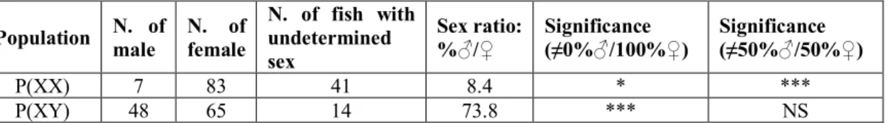
Table 1. Statistics of phenotypic sex in two populations
Population N. of male N. of female
N. of fish with undetermined sex Sex ratio: %♂/♀ Significance (≠0%♂/100%♀) Significance (≠50%♂/50%♀) P(XX) 7 83 41 8.4 * *** P(XY) 48 65 14 73.8 *** NS
P(XX): putative neomale (XX) offspring population; P(XY): putative genetic male (XY) offspring population; NS: Non-significant. Chi-squared test was applied for statistics in R
Table 2. Goldfish Y-allele sex-linkage
Population Male# Female# Undetermined sex# Sex linkage
P(XX) 0 / 7 0 / 83 0 / 41 NS
P(XY) 48 / 48 1 / 65 10 / 14 ***
# Y-allele positive genotyping / total number of samples. P(XX): putative neomale (XX) offspring population; P(XY): putative
26
Table S1. Sequences of the primers used for Y-allele genotyping in goldfish.
Primers PCR
product (bp)
Genome location
names Sequence(5’ - 3’) Male assembly NCBI_genome
Marker 1 Forward: AATACAACATTCCCAGGGAGTGCA Reverse: CATCAAGGGCTATCTGACCAAGA 1169 Flattened_line_394560:620-1788 NW_020523543.1
Marker 2 Forward: GTGCTCAATAGACGACGGATTCTC Reverse: GTCTGTCTGTTAGCCTGTTCTCCA 1189 Flattened_line_270798:2006-3194 NW_020525535.1
Marker 3 Forward: GATGAAGGTCTCGGTCTGTTGTTA Reverse: CCCTGTTATGTTTGTATTGGCTAC 2548 Flattened_line_35862:4409-6956
NC_039250.1 (LG8) Positive control Forward: AAGAGCGCCTCCTAGTGTTT Reverse: GAGACGGAGGAGTGGTATCG 994 Flattened_line_0:6858 -7842 NC_039245.1 (LG3)
Three Y-allele primer pairs (marker 1 to 3) and one autosomal primer pair (positive control) were designed on our XY male genome assembly (male assembly). Name of the contig and nucleotide position (3’-5’) are given in the genome location column.
FIGURES: Figure 1
Figure 1. RAD-sex tags and male-specific markers in goldfish. (A) Haplotypes heatmap in
phenotypic males and females’ goldfish. Each cell in the heatmap represents the number of haplotypes presented in x phenotypic males and y phenotypic females (x: cumulative number of males, y: cumulative number of females). Haplotypes present in more than 12 males and absent in all females were identified as male-specific haplotypes (highlighted by red box). (B) Genotyping of goldfish males and females with three Y-allele primer pairs and one autosomal primer pair used as a positive control. Goldfish are categorized into three groups i.e., putative genetic males (XY), putative XX neomales, and genetic females by combining the results of both Y-allele genotyping and sex phenotyping.
Figure 2
Figure 2. Sex determining regions identified by remapping the Pool-seq male and female reads onto the female genome assembly. SNPs were counted using 100kb sliding window
with an output point every 500bp. (A) Circular plot showing the genome wide metrics of the Pool-seq analysis. All the 50 goldfish linkage groups (LGs) are labelled with their LG number and all unplaced scaffolds are fused together. Outer to inner tracks show respectively: the male-specific SNPs, the female-male-specific SNPs, and the reads depth ratio between males and females. (B) Manhattan plot of the male- and female-specific SNPs showing a strong enrichment of male-specific SNPs on LG22.
Figure 3
Figure 3. Distribution of male-specific SNPs on LG22 and unplaced scaffolds NW_020523543.1 and NW_020523609.1. SNPs were counted using 100kb sliding window
with an output point every 500bp and female- and male-specific SNPs were respectively indicated by red and blue color. (A) A large sex-determination region was identified on LG22, which is highlighted with a black box. The candidate sex-determining gene amh is located on this LG22, but not in the high density, male-specific SNP region. The region from 8Mb to 10Mb containing amh is zoomed in panel (B). (C) The NW_020523543.1 unplaced scaffold exhibits a region around 0.1Mb harboring a small region (200 kb) with a high-density of male-specific SNPs. Meanwhile, sequence comparisons demonstrate that 7 male-biased RAD-tags (colored circles) on a total of 32 map with a high identity onto this scaffold. In contrast, few female-specific SNPs were enriched on this scaffold (red area). (D) The unplaced NW_020523609.1 scaffold is enriched in male-specific SNPs.
SUPPLEMENTARY FIGURES
Figure S1
Figure S1: Distribution of sex-biased SNPs on LG47. SNPs were counted using 100kb
sliding window with an output point every 500bp. The top panel displays the profile of male-specific SNPs (blue area), while the bottom panel displays the profile of female-male-specific SNPs (red area).
Figure S2
Figure S2: Dot plot comparison of LG22 and LG47 showing conserved synteny between these
Figure S3
Figure S3: Sex genotyping with Y-allele primers of the offspring of a putative XX neomale
with a normal XX female. Genotyping was conducted with three Y-allele primers and one autosomal primer used as a gDNA quality control. Phenotypic sex was determined by gonadal histology and males and females are shown using red and yellow color respectively. Female-to-male sex-reversed animals (N= 7) are highlighted by red boxes. Hashes indicate animals with unknown phenotypic sex with undifferentiated gonads based on histology.
Figure S4
Figure S4. Sex genotyping with Y-allele primers of the offspring of a putative XY male with a
normal XX female. Genotyping was conducted with three Y-allele primers and one autosomal primer used as a gDNA quality control. Phenotypic sex was determined by gonadal histology and males and females are shown using red and yellow color respectively. The female-to-male sex-reversed animal (N= 1) is highlighted by a red box. Hashes indicate animals with unknown phenotypic sex with undifferentiated gonads based on histology.