HAL Id: hal-02639452
https://hal.inrae.fr/hal-02639452
Submitted on 28 May 2020
HAL is a multi-disciplinary open access
archive for the deposit and dissemination of
sci-entific research documents, whether they are
pub-lished or not. The documents may come from
teaching and research institutions in France or
abroad, or from public or private research centers.
L’archive ouverte pluridisciplinaire HAL, est
destinée au dépôt et à la diffusion de documents
scientifiques de niveau recherche, publiés ou non,
émanant des établissements d’enseignement et de
recherche français ou étrangers, des laboratoires
publics ou privés.
Distributed under a Creative Commons Attribution - NonCommercial| 4.0 International
License
diseases
Nathalie Vergnolle
To cite this version:
Nathalie Vergnolle. Protease inhibition as new therapeutic strategy for GI diseases. Gut, BMJ
Publishing Group, 2016, 65 (7), pp.1215-1224. �10.1136/gutjnl-2015-309147�. �hal-02639452�
Protease inhibition as new therapeutic strategy
for GI diseases
Nathalie Vergnolle
1,2,3,4,5 1Inserm, U1220, Toulouse, France
2
Université de Toulouse, Université Paul Sabatier, Institut de Recherche en Santé Digestive (IRSD), Toulouse, France
3Inra, U1416, Toulouse, France 4
Ecole Nationale Vétérinaire de Toulouse (ENVT), France
5
Department of Pharmacology and Physiology, University of Calgary, Calgary, Alberta, Canada
Correspondence to Dr Nathalie Vergnolle, INSERM, UMR-1220, IRSD, Place du Dr. Baylac, CHU Purpan, CS 60039, Toulouse 31024, Cedex 3, France; nathalie.vergnolle@inserm.fr Received 3 April 2015 Revised 5 February 2016 Accepted 12 February 2016 Published Online First 12 April 2016
To cite: Vergnolle N. Gut 2016;65:1215–1224.
ABSTRACT
The GI tract is the most exposed organ to proteases, both in physiological and pathophysiological conditions. For digestive purposes, the lumen of the upper GI tract contains large amounts of pancreatic proteases, but studies have also demonstrated increased proteolytic activity into mucosal tissues (both in the upper and lower GI tract), associated with pathological conditions. This review aims at outlining the evidences for dysregulated proteolytic homeostasis in GI diseases and the pathogenic mechanisms of increased proteolytic activity. The therapeutic potential of protease inhibition in GI diseases is discussed, with a particular focus on IBDs, functional GI disorders and colorectal cancer.
INTRODUCTION
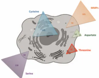
Proteases represent up to 2% of the human genome, with 500–600 different proteases that have been identified. Through the evolution, pro-teases have adapted to the different conditions that characterise complex organisms: pH variation, oxydo-reduction environment, temperature, etc. Proteases specifically cleave proteins at their extremities (N-terminal or C-terminal regions) and are then called exopeptidases, or in the middle of the proteins, being qualified then as endopepti-dases. Depending on their proteolytic mechanism, human proteases are classified as serine, threonine, cysteine, aspartic or metalloproteases (figure 1and table 1). Some of them are secreted and released in the extracellular milieu, while others have intracel-lular functions and exclusively remain inside the cells (figure 1).
PROTEASES AND PROTEASE INHIBITORS OF THE GI TRACT
Proteases
In the GI tract, proteases are heavily present, both in the lumen and deeply into the tissues.1 Pancreatic proteases (trypsins, chymotrypsin, elas-tase, etc) are released into the lumen of the upper GI tract, where they exert digestive functions. The microbiota constitutes also an important source of proteases (figure 2). Bacteria, yeasts and helminths potentially present in the intestinal lumen produce and release proteases.2For some pathogens such as pathogenic forms of Escherichia coli or the entero-toxigenic Bacteroides fragilis, their ability to release proteases is crucial for their pathogenicity. Serine, cysteine, aspartic and metalloproteases are expressed and released by the microbiota (table 2).2 However, it is interesting to note that when the nature of the proteases present in human faeces was investigated, only host proteases were
identified. These findings could question the contri-bution of microbial proteases, to overall luminal proteolytic activity in the gut. Another study has established significant associations between specific bacterial subgroups and faecal protease activity, sug-gesting that microbiota composition could affect intestinal proteolytic homeostasis. More recently, forms of secreted proteases have been identified in intestinal epithelium: mesotrypsin mRNA is found in human intestinal epithelial cells3 and trypsin activity is released by cultures of those cells (Vergnolle, personal communication). Other resi-dent cells of the intestinal mucosa produce and release proteases (figure 2). For instance, the major protein content of mucosal mast cells is proteases. Mast cells release different forms of proteases: tryptase and chymase for the vast majority, and also cathepsin G and granzyme B. Resident macro-phages also produce and/or release different forms of proteases: matrix metalloproteinases (MMPs) (MMP-12 among other MMPs), caspase, cathepsins L and D.1In the inflamed gut, inflammatory cells are another major source of proteases, which they
Open Access Scan to access more
free content
Key messages
▸ Protease inhibition as therapeutic approach in intestinal pathologies: what should we consider?
– Profiles of active proteases have to be performed in pathological tissues in order to define the best molecular targets for therapeutic intervention
– Large spectrum inhibitors might have severe side effects
– Promote the expression or delivery of natural endogenous inhibitors could be a safe therapeutic option
– Local versus systemic delivery would have to be considered
– The use of food-grade bacteria as carrier for the delivery of therapeutic proteins has been proposed
▸ Protease targets for IBD
– MMPs inhibitors have been abandoned – Serine proteases are considered
– Ubiquitin–proteasome system inhibitors are considered
▸ Protease targets for IBS – Trypsin inhibition – Tryptase inhibitors
– Protease-Activated Receptor (PAR1)/PAR2
use to degrade extracellular tissues and intracellular particles, thereby increasing their phagocytic properties.4Upon in flamma-tory cell infiltration and activation, tissue proteolytic activity is considerably increased. Neutrophils in particular release massive amounts of elastase, proteinase-3 and cathepsin G5 (figure 2). Finally, all resident cells of the GI tract express intracellular pro-teases: caspases, which have fundamental roles in cell apoptosis, and autophagins, which are the proteolytic enzymes responsible of autophagy processes6 (table 1). A special case can be made for deubiquitylases, which are crucial regulators of intracellular protein turnover through the proteasome system. These enzymes present in all cell types, are either cysteines or metallo-proteinases and target ubiquitinylated proteins, thereby chan-ging their degradation fate inside the cell.
Although specific proteases can be detected in tissues, the cel-lular origin of most proteases is quite difficult to define, and no study so far has determined the origin of proteases detected in intestinal tissues. The site of action of a given protease is also debated. As of today, one can only specify the possible site of action of a given protease.
Another level of difficulty in studying proteases is that for activity tests, substrates are never fully specific of one protease, neither are their inhibitors. Therefore, the proteolytic activity that is measured is possibly due to a mix of proteases and cannot be attributed to one specific protease.
Protease inhibitors
Protease inhibitors have coevolved with proteases, in order to control their destructive nature. Natural endogenous protease inhibitors are particularly present in the GI tract.1 They are either circulating inhibitors produced at distance from the GI tract (mostly in the liver), or are produced on site, by intestinal epithelial cells or infiltrated inflammatory cells (table 3). Serpins A1, A3, A4, E1 and C1 are circulating protease inhibitors inhi-biting serine proteases such as trypsins, chymase, tryptase, elas-tases, kallikreins and cathepsin G (table 3). Secretory leucocyte protease inhibitor (SLPI) and elafin are produced in situ by intestinal epithelial cells or leucocytes. Both inhibit elastase and proteinase-3, while SLPI also inhibits trypsin, chymotrypsin, cathepsin G, tryptase and chymase7 (table 3). Tissue inhibitors of metalloproteinases (TIMPs) are ubiquitously produced, TIMP-1, TIMP-2, TIMP-3 and TIMP-4 are present in the GI tract, where they inhibit a number of different MMPs8(table 3). The caspase-9 inhibitor, which is a cellular inhibitor of apop-tosis protein-2 (c-IAP2) is also ubiquitously produced by cells present in intestinal tissues.
PROTEASES AND INTESTINAL PHYSIOLOGY
The roles and functions of proteases and their inhibitors under physiological conditions have been poorly investigated. While Figure 1 Representation of human cell proteases according to their
catalytic mechanism and their intracellular or extracellular representation. MMPs, matrix metalloproteinases.
Table 1 Proteases identified in GI tissues and cells, and disease-associated upregulation
Upregulated expression in
Family Proteases Cellular location Possible sites of action CD UC IBS CRC
Serine proteases Elastases Intra/extra L, M, P, I + + + +
Proteinase-3 Intra/extra L, M, P, I + + Chymase Extra L, M, P + + + Kallikreins Extra L, M, P + + + Granzymes Intra/extra L, M, I + + + Tryptase Extra L, M, P + + + + Plasminogen Extra M, P + Activator Trypsins Extra L, M, P + + + Cathepsin G Intra/extra L, M, P, I + + Thrombin L, M, P + +
Factors V and VIII L, M, P +
Matriptase Membrane-bound M, I +
Cysteine proteases Caspases Intra I + +
Cathepsins (B, L) Extra M, P + +
Autophagins Intra I
Calpains Intra I + +
Deubiquitinases Intra I + + + +
Aspartate proteases Cathepsin D Intra I + + +
Renin Intra/extra M, P, I + +
Metalloproteinases MMPs Intra/extra M, P, I + + +
ADAMTS Extra M, P, I = =
Deubiquitinases Intra I
digestive proteases are released into the lumen of the upper GI tract for digestive purposes, intestinal microbes largely inhibit them as they progress down to the tract.9In addition to their physiological role in digestive process, constitutive expression of some proteases seems also to be necessary to intestinal homeo-stasis. Matriptase for example, is a trypsin-like protease that colocalises with E-cadherin in intestinal epithelial cells. Mice deficient for matriptase expression specifically in intestinal epi-thelial cells develop from birth diarrhoea, and then later in life develop megacolon and colitis.10
Proteases from the A Disintegrin And Metalloprotease (ADAM) family also seem to play roles in maintaining intestinal barrier function. ADAM-19 colocalises with the tight junction-associated protein zonula occludens-1,11 ADAM-17 deficiency in human induces bowel dysfunctions.12 Cathepsin K-deficient mice showed a disrupted expression of Occludin, a deposit of type IV collagen at the basement membrane and an increased expression of E-cadherin at the apical junction, all together suggesting barrier dysfunctions.13
Mucus formation and properties also seem to be tightly regu-lated by endogenous proteases. Recently, a study has demon-strated that in contrast to physiological states, mice deficient for the metalloproteinase meprinβ has an attached mucus layer in the small intestine, which can be released by the addition of meprinβ.14In the small intestine, mucus is secreted attached to the goblet cells, and requires a protease meprinβ, to be detached from the epithelium. This example illustrates the importance of some proteases for mucus properties, and mucosal homeostasis. Figure 2 Source of proteases in the
GI tract.
Table 2 Major identified pathogen-associated microbial proteases Protease
category Microbial proteases Examples of pathogens Aspartic Type 4 prepilin
peptidase
Enterohaemorrhagic escherichia coli
Preflagellin Archaeal bacteria Cysteine Sortases Enterococcus faecalis
Gingipains Porphyromonas gingivalis Staphopain Staphylococcus aureus Serine Subtilisin Clostridium difficile
Elastase Pseudomonas aeruginosa Metalloproteinases Fragilysin Enterotoxigenic Bacteroides
fragilis Gelatinase E. faecalis
Elastase P. aeruginosa, Helicobacter pylori
Collagenase Salmonella typhimurium
Table 3 Endogenous protease inhibitors detected in the GI tract
Family
Protease
inhibitor Targeted proteases Source
Possible sites of action Serpins Serpin A1 Trypsin/chymase/tryptase/
elastase/proteinase-3/ cathepsin G/thrombin/ kallikreins Systemic M, P Serpin A3 Chymotrypsin/chymase/ cathepsin G Systemic M, P Serpin A4 Kallikreins Systemic M, P Serpin E1 Plasminogen activator Systemic M, P Serpin C1 Thrombin Systemic M, P Chelonianin SLPI Elastase/cathepsin G/trypsin/
chymotrypsin/tryptase/ chymase
Local L, M, P, I
Elafin Elastase, proteinase-3 Local L, M, P, I TIMPs TIMP-1 MMP-1/MMP-2/MMP-3/ MMP-4/MMP-6/MMP-19/ ADAM-10/ADAM-17 Systemic and local M, P TIMP-2 MMP-1, MMP-2, MMP-14 M, P TIMP-3 Membrane-bound MMPs Local M, P TIMP-4 MMP-1/MMP-2/MMP-3/
MMP-4/MMP-6/MMP-19
Systemic M, P c-IAP2 Caspase-9 Local I
ADAM, A Disintegrin And Metalloprotease; c-IAP2, cellular inhibitor of apoptosis protein-2; I, intracellular; L, lumen; M, matrix; MMP, matrix metalloproteinases; P, plasma; TIMPs, tissue inhibitors of metalloproteinases; SLPI, secretory leucocyte protease inhibitor.
DYSREGULATED PROTEOLYTIC HOMEOSTASIS IN GI DISEASES
Because of the large distribution of proteases in the GI tract, and their tight control by endogenous protease inhibitors, asso-ciation of dysregulated proteolytic homeostasis with GI patholo-gies has often been investigated (table 1).
IBDs including Crohn’s disease and UC were the first diseases to be investigated, initially because of the additional source of proteases represented by infiltrated and activated inflammatory cells. The expression of a very large number of proteases is upregulated in IBD.1Protein or mRNA expressions of proteases from infiltrated immune cells (neutrophil elastase, proteinase-3, cathepsin G, tryptase, chymase or granzymes) are obviously increased in inflamed tissues from patients with IBD (table 1). Being involved in tissue remodelling, a process of major import-ance in IBD, MMPs expression is also significantly increased both in Crohn’s disease and UC, while ADAMTS proteases expression is unchanged.1 15 16 Inappropriate induction of cell death through apoptosis or autophagy is also involved in IBD, and proteases involved in such processes (caspases, autophagins) are upregulated in IBD, particularly in UC.17 Genetic evidence supporting the association of proteases and protease inhibitors genes with IBDs was raised in a systematic review. In that study, 75 genes coding for proteases and 7 genes coding for protease inhibitors were retained for Crohn’s disease, while for UC, 14 proteases and 4 protease inhibitors genes were retained.18 Among the identified genes, proteins of the ubiquitin–prote-asome system were top ranked, and further studies have identi-fied single nucleotide polymorphism in five of those proteins (CYLD, USP40, USP3, DAG1 and APEH) associated with IBD.19 The expression of protease inhibitors in IBD is rather conflicting, reporting increased, decreased or stable levels of expression for serpins,1 elafin or SLPI.20–24 TIMP-1 and TIMP-2 seem to be consistently increased in UC and Crohn’s disease,16 while TIMP-3 is decreased in Crohn’s disease25 26 and c-IAP2 is decreased in UC.17
One major problem with most of the studies that have investi-gated protease expression in colonic tissues of patients is that this does not reflect the function of proteases associated with the disease. Indeed, mRNA or protein expression may be increased, but depending on the presence of endogenous inhibi-tors in tissues, the biological activity of proteases might remain the same. Similarly, investigating mutations on protease genes does not provide answers on the function of the protein. Definitive answers on the role of proteases associated with disease states have to come from studies investigating the in situ net activity of proteases. Elastolytic activity has been investigated in tissues from patients with IBD,22 demonstrating that elastase activity was upregulated, mostly in the mucosa. Surprisingly, elastolytic activity was upregulated both in inflamed tissues from patients with Crohn’s disease or UC and in non-inflamed parts of the colon of those patients, where no inflammatory cell infil-tration was detected. Interestingly, when in situ zymography for elastolytic activity is performed in human colonic tissues of healthy and Crohn’s disease patients, the strongest elastolytic activity is detected on intestinal epithelial cells (figure 3A). These two observations made on tissue proteolytic activity suggest that elastase might not originate exclusively from in fil-trated inflammatory cells, and provide unexpected directions to investigate the role of elastase in the context of IBD. Only few studies have investigated protease activities in IBD. A recent study has shown that increased MMP activity in tissues from patients with IBD was restored to control levels after infliximab treatment.27 Trypsin activity was also increased in tissues from
patients with Crohn’s disease and UC.28 Other studies have investigated the proteolytic activity in stools of patients with IBD, reporting an increased activity, associated with dysbiosis.2 However, depending on the faeces collection and conservation methods, variable results could be observed in faecal proteolytic activity.
To a lesser extent, protease expression has been investigated in tissues from patients with IBS. The expression of specific serine proteases such as tryptase,29elastase,30trypsin28 31or cathepsin G32 were significantly increased in tissues or in the faeces of patients with IBS, compared with healthy controls. Two types of cysteine proteases (calpain-8 and proteases from the proteasome) were also upregulated in tissues from patients with IBS, com-pared with controls.33 34But here again, very few studies have investigated the resultant proteolytic activities in tissues or faeces of patients with IBS. Trypsin-like activity seems to be upregu-lated in tissues from patients with IBS, compared with healthy controls, with a predominant activity in intestinal epithelial cells, as observed by in situ zymography (figure 3B). Faecal protease activity was found upregulated in faeces from patients with IBS30 and association between proteolytic activity and specific intestinal bacterial groups has further been established.35
In colorectal cancer as well, the expression of a number of proteases was upregulated (table 1), but the proteolytic activity associated with colorectal cancer tissues is for the most part unknown. Among the upregulated proteases in colorectal cancer, serine proteases are well represented, but caspases, cathepsins, calpains, deubiquitinases and MMPs are also preva-lent (table 1).
MECHANISMS OF ACTION OF PROTEASES IN GI DISEASES
Proteases present in diseased intestinal tissues dispose of several mechanisms of action to participate in pathogenesis or symp-toms generation. They act by proteolytic processing of other Figure 3 In situ proteolytic activity (elastolytic in A, trypsin-like in B) performed as previously described in ref.20, in human colons of healthy individuals, patients with IBS and patients with Crohn’s disease.
molecules (mediators, receptors), thereby inducing a number of intracellular signals (figure 4).
Receptor activation
In the GI tract, the receptors that have been mostly studied belong to the family of protease-activated receptors (PARs).36 37 These receptors are ubiquitously expressed in the GI tract ( present in intestinal epithelial cells, in neurons, in infiltrated inflammatory cells, in mast cells, in fibroblasts, etc).37 They are activated by the proteolytic cleavage of their extracellular N-terminal domain, which releases a new N-terminal domain that acts as a tethered ligand to induce intracellular signals.38 39 Members of the PAR family (PAR1, PAR2, PAR3and PAR4) can be activated by serine, and cysteine and metalloproteinases.40 Activation of PARs in the GI tract induces a wide array of pro-inflammatory, pronociceptive and proliferative effects (figure 4). In the gut, PAR activation is able to modify a number of physiological functions: ion exchange,41 motility,42 nocicep-tion,43permeability,44 45secretion, etc. The involvement of PAR activation in GI diseases has been proposed for IBD, IBS and colorectal cancer.46 47Because PARs are expressed both on the apical and basolateral sides of intestinal epithelial cells, these receptors might be activated both by lumenal proteases (includ-ing microbial proteases) and by tissue proteases.37
Elastase seems to have receptor-dependent effects involving another type of receptor: the Gram-negative bacteria receptor toll-like receptor-4. Proteolytic activity is necessary to this effect, but the exact mechanism is still unknown.48
Cathepsin G interacts with the G protein-coupled formyl peptide receptor, leading to the activation of mitogen-activated protein kinase (MAPK) pathways.49However, the pathophysio-logical consequences of this activation are still unclear.
Inflammatory mediators processing
Proteases may also modulate the bioactivity of inflammatory mediators. This is the case for cytokines, chemokines and their cognate receptors. Proteolytic cleavage increases the bioactivity of chemokines and cytokines by promoting the processing of an inactive precursor, thereby increasing their pro-inflammatory or
chemotactic properties. For example, this has been shown for CXCL-8 and CXCL-5, which respectively can be cleaved by proteinase-3 and cathepsin G, the truncated forms of these che-mokines having higher chemotactic activity towards neutro-phils.50 51 Proteinase-3 is also known to activate interleukin (IL)-1, IL-18 and tumour necrosis factor (TNF)-α.52–54 However, proteases can also have opposite effect, degrading cytokines: IL-6 is inactivated by cathepsin G and proteinase-3,55 while elastase and cathepsin G both degrade mature TNF.56The net effect of proteolytic modifications of chemokines and cyto-kines, particularly in the context of IBD still has to be clarified. The initial steps of leucocyte recruitments (ie, rolling and adhe-sion events) might also be tightly controlled by proteases. Selectins, which are expressed at the cell surface, where they ini-tiate the rolling signals, are shed by metalloproteinases (ADAM-17), by stromelysin, collagenase and chymotrypsin,57 58 but not by other serine proteases.59 Proteases also regulate the next step of leucocyte recruitment, which involves integrins. Cathepsins are able to cleave members of the integrin family, inhibiting the attachment of migrating cells to extracellular matrix components.60 Here again, the net effect of proteolytic modifications on diapedesis and migration of leucocytes still has to be clarified, but this mechanism of action could play a central role in inflammatory and cancer pathologies.
Apoptosis and anoikis
Caspases and autophagins play essential roles in programmed cell death, which is an important process in chronic in flamma-tory diseases and cancer. Proteases such as thrombin and gran-zymes are also able to induce apoptosis or anoikis.44 61When apoptosis is induced in inflammatory cells, this process favours the resolution of inflammation. Protease-induced neutrophil apoptosis would therefore be protective in the context of chronic intestinal inflammation. In contrast, epithelial cell apop-tosis leads to a decreased barrier function.44 In that case, protease-induced apoptosis would further feed inflammatory response in the gut, by favouring a leaky barrier, and further penetration of luminal content.
Figure 4 Mechanism of action of proteases in GI diseases. PAR, protease-activated receptor.
Tight junction degradation
Proteases have been shown to disrupt cell–cell interactions. Therefore, depending on their cellular target, proteases can potentially influence transmigration and microvascular leakage by acting on endothelial cells, or proteases can influence intes-tinal barrier functions by acting on intesintes-tinal epithelial cells. Some proteases such as chymase are able to alter tight junction proteins (ZO-1, occludins)62 or in the case of elastase-2A, a form of chymase, to directly cleave proteins important in barrier functions.63 Adherent junctions seem also to be the targets of some proteases. This is for example the case for trophil elastase, which upon the transepithelial passage of neu-trophils in inflammatory conditions,64 cleaves the E-cadherin protein. However, neutrophil elastase is unable to cleave tight junction proteins.65 66Other proteases overexpressed in in flam-matory conditions might be able to degrade tight junction pro-teins, although the question of the accessibility of those proteases to tight junction proteins has not really been addressed in vivo. One can question whether proteases could have a direct access to tight junction domain proteins, or whether the effects of proteases on barrier functions are rather mediated by the activation of receptors. Indeed, in the case of thrombin and trypsin, their effects on increased intestinal per-meability are mediated by PAR1and PAR2activation.44 45
Matrix remodelling
The extracellular matrix is a highly dynamic structure, which interacts with cells to regulate proliferation, migration and dif-ferentiation. Cleavage of extracellular matrix components con-stitutes the main regulatory process of these functions. MMPs, ADAMs and ADAMTS are the main enzymes involved in extra-cellular matrix remodelling. Their activities are controlled by TIMPs (table 3). Excessive extracellular matrix degradation, as observed in chronic inflammatory disorders such as IBD or in colorectal cancer, causes tissue destruction, inflammatory cell infiltration, fibrosis and metastasis.8
Mucus cleavage
Mucus is a major component of mucosal barrier. It efficiently protects host tissues from their luminal content. Mucins are large highly glycosylated proteins that constitute the major com-ponent of mucus. Defective mucus layer leads to pathophysio-logical mechanisms including chronic inflammation and infection. Digestive enzymes are usually unable to digest the glycans composing the mucus, thereby leaving mucins intact.67 Probiotic bacteria such as Lactobacillus and Bifidobacterium do not release proteases that can cleave the MUC2 mucin, the mucus core protein,68while others, such as Akkermansia muci-niphila, are able to degrade mucins.69 Proteases from bacterial pathogens such as Porphyromonas gingivalis,70 from parasites such as Entamoeba histolytica71or nematodes such as Trichuris muris72 degrade mucus barrier. Under pathophysiological cir-cumstances such as IBD, where proteolytic activity is largely increased in the mucosa, it is reasonable to think that proteases (microbial or host proteases) modify mucus properties.
Immunoglobulin cleavage
Immunoglobulins are sensitive to proteases. A number of studies have demonstrated that bacterial proteases are able to degrade both IgG and IgA, the immunoglobulins the most present at the intestinal mucosa surface.73–75 Indeed, a specific subclass of microbial proteases called‘IgA proteases’ constitutes a group of extracellular endopeptidases. In pathologies-associated dysbiosis,
microbial proteases might then be able to modify the compos-ition and function of resident immunoglobulins and therefore, to modify intestinal immune response. In vivo degradation of immunoglobulins in the intestinal mucosa has never been demonstrated, and one can only speculate on whether bacterial proteases might act on immunoglobulins from the luminal side or whether they could penetrate the tissues. It is not known yet whether host intestinal proteases are also capable of immuno-globulin degradation in an immune-related pathological context.
PROTEASE INHIBITION AS POSSIBLE TREATMENTS FOR IBD
Overall, considering all their mechanisms of action, proteases associated with IBD exert rather pro-inflammatory properties: they potentiate cytokines and chemokines pro-inflammatory properties, they remodel extracellular matrix to allow leucocyte infiltration, they degrade tight junction proteins inducing plasma extravasation and increased intestinal permeability, they induce apoptosis in intestinal epithelial cells and it is known that activation of PAR1, PAR2 and PAR4 in the colon leads to pro-inflammatory effects.37 40 Taken together, these facts suggest that protease inhibition could have strong therapeutic benefits to treat IBD. However, considering the large number of proteases that have been found upregulated in IBD (table 1), and their diverse functions, it is quite difficult to identify single molecular targets among all those proteases. As previously dis-cussed, one major step would be to define which proteases are overactivated in pathological situation, and to establish the profile of IBD-associated overactivated proteases.
One option could be to consider large spectrum protease inhibitors as new therapeutic approach for IBD. However, large spectrum inhibitors might also bear a number of side effects. From all the families of proteases that are upregulated in IBD, MMPs have raised some interests, mainly due to the fact that synthetic inhibitors have been developed for cancer research. MMP inhibitors demonstrated good anti-inflammatory proper-ties in animal models of colitis, but in human, they appeared to be more efficient at helping mucosal healing and extracellular matrix restructuration. MMPs are important factors of extracel-lular matrix remodelling. Inhibition of proteases implicated in matrix turnover could therefore induce tissuefibrosis. More sur-prisingly, the use of MMP inhibitors has revealed antitumori-genic and anti-inflammatory effects for some MMPs.76 These data identify MMPs as antitargets for inflammation and cancer rather than targets.
Upon active protease identification, studies have identified some interesting targets in IBD. Elastase is one of them, as its activity is dramatically increased in IBD and elastase has demon-strated a large number of pro-inflammatory effects. Trypsin activity might be another interesting proteolytic target as more aggressive disease and rapid progression to surgery was observed in patients with UC bearing a serpin A1 (or α-1-antitrypsin) deficiency.77For both targets, instead of raising synthetic inhibi-tors, which might bear off-target effects, a better option might be to favour the expression of natural endogenous inhibitors of these targeted proteases. Re-equilibrating the protease –antipro-tease balance in the inflamed gut by delivering natural endogen-ous protease inhibitors, which are down-regulated in disease, could constitute a safe and efficient therapeutic option. One challenge though would be to deliver protease inhibitors locally, where they are naturally produced, and where they exert their homeostatic role. Local delivery would also decrease possible side effects of therapeutic intervention. To that aim, the use of genetically modified bacteria could constitute a major advance.
Commensal or probiotic bacteria that colonise the gut can be genetically transformed to express human epithelium-derived protease inhibitors such as elafin or SLPI. Strong anti-inflammatory properties have been described in different animal models for such recombinant bacteria.22 78 Elafin delivered by recombinant lactic acid bacteria after oral administration in mice was detected in the colon lumen, as well as in the mucosal tissues. How this recombinant protein was able to cross the intestinal barrier: through passive diffusion in damaged epithelia or through active transport, is not clear yet. However, its pres-ence was detected both in damaged areas and in areas where the epithelium was intact.22Therefore, one can consider that prote-ase inhibitor delivery through this approach might act both from the lumen and superficial mucosal tissues. Anti-inflamma-tory properties have also been demonstrated in cultured biopsy supernatants from patients with IBD.22Treatments with bacteria recombinant for the expression of protease inhibitors were dras-tically more effective than treatments with bacteria recombinant for anti-inflammatory cytokines such as IL-10 or transforming growth factor-β. This is strongly in favour of targeting proteo-lytic activity for therapeutic options in IBD. However, the use of the recombinant bacteria strategy will have to consider the development of non-disseminating bacteria because of their gen-etically modified nature. Such development has already been described for other recombinant bacteria.79
Other interesting proteolytic targets for IBD treatment are the proteases from the ubiquitin–proteasome system.18 19 Polymorphisms on several genes of this system have been identi-fied in patients with IBD, and pathogenic bacteria modify this system turnover.19 Proteasome inhibitors therapy targeting the ubiquitin–proteasome system, such as the use of bortezomib, which was successfully developed for cancer treatment, could constitute a new option to treat efficiently patients with IBD.
PROTEASE INHIBITION AS POSSIBLE TREATMENT FOR FUNCTIONAL GI DISORDERS
Proteases, through the activation of PARs, modify a number of physiological functions that are dysregulated in IBS. PAR2 acti-vation causes visceral hypersensitivity, modifies intestinal motil-ity and both PAR1 and PAR2 activation increase intestinal epithelial permeability.37 All these functions take an important part in IBS symptoms generation. In addition, increased trypsin-like activity (measured using a preferred trypsin substrate) has been demonstrated in tissues from patients with IBS.28 The increased activity was observed in all patient subgroups: diarrhoea-predominant, constipation-predominant or alternate-predominant, suggesting that protease activity might be a unify-ing feature of IBS. Further, several studies have reported that proteolytic activity released from tissues of patients with IBS provoke an increased permeability, and signal to extrinsic sensory neurons and intrinsic enteric neurons.80–83 This con-firms the prominent effect of IBS-associated proteases on neuron signalling. Taken together, these studies highlight trypsin proteases as important molecular targets for IBS treatment.
Tryptase is another protease that is significantly increased in the mucosa of patients with IBS. Studies have demonstrated that enhanced tryptase activity is responsible for the increased per-meability of rectal mucosa in diarrhoea-predominant patients.84 Tryptase inhibitors have been raised for mast cell-associated pathologies and may be tested in IBS, particularly on visceral hypersensitivity symptoms and increased permeability.
Both trypsin and tryptase have been shown to activate PAR2.85In all animal studies investigating by which mechanisms tryptase, trypsin or IBS patient biopsy supernatants were causing
increased permeability, neuron hyperexcitability or visceral hypersensitivity, proteases and/or PAR2 activation were identi-fied as the principal mechanism of action.86 This suggests that PAR2antagonism could constitute a valid therapeutic option for the treatment of IBS. However, a study investigating the effects of IBS patient biopsy supernatants on human submucosal or myenteric neurons preparations has determined that PAR1 rather than PAR2was activated in human tissues.87 88This sug-gests that in human, PAR1antagonists should be considered for the treatment of IBS symptoms. However, the most recent advances in the pharmacology of PARs has taught us that PARs have several ways to signal other than calcium mobilisation usually measured.89 Adenylyl cyclase, MAPK and ERK signal-ling and β-arrestin recruitment would also have to be investi-gated in PAR2response of human neurons, before ruling out a possible involvement of PAR2. In addition, the most striking effect of PAR2activation was observed on visceral hypersensitiv-ity symptoms and in sensory primary afferents, which might respond differently from submucosal or myenteric neurons. Therefore, for the time being, both PAR1and PAR2antagonism should still be considered as potential therapeutic options for IBS treatment.
Downstream from PAR activation (at least PAR2 and PAR4), mobilisation and potentiation of TRPV4 channel seem to be involved in the context of somatic mechanical hyperalgesia,90 and in the context of IBS.91–93 Most recently, a study has demonstrated that proteases, through the activation of PAR2, were able to induce the release of TRPV4 endogenous agonists, which were found upregulated in tissues from patients with IBS.94 Taken together, these data established the ion channel (TRPV4)-dependent mechanisms by which proteases influence neuronal signalling and visceral hypersensitivity in IBS.
A study that has investigated faecal proteases suggests that in diarrhoea-predominant patients with IBS, most of faecal prote-ase activity is coming from the pancreas and is due to acceler-ated transit.30Lowering transit time could therefore constitute a way in those patients to decrease luminal proteolytic activity and thereby the potential effects of this activity on microbiota composition or intestinal permeability.
PROTEASE INHIBITION AS POSSIBLE TREATMENT FOR COLORECTAL CANCER
As discussed above, numerous proteases are upregulated and potentially play a role in colorectal cancer. The identification of proteases that favour normal physiological functions instead of helping oncogenesis or tumour growth had most important clin-ical implications. The fact that proteases might have opposite effects in cancer might explain the failure of clinical trials that have used large spectrum protease inhibitors for treating patients with cancer.95 Furthermore, a significant number of proteases, and in particular intracellular proteases, have been defined as tumour suppression natural agents. Therefore, extreme caution is now associated with any antiprotease thera-peutic strategy for cancer, and the inhibitory profile of antipro-tease therapy is carefully evaluated according to the characteristics of the enzyme to be targeted, and its cellular source.
The ubiquitin–proteasome system is however the most protease-targeted system for cancer treatment. A number of bio-active compounds targeting E1, E2 enzymes and E3 ligase are now available for therapeutic tests96 and are currently under investigation.
THE SPECIAL CASE OF COELIAC DISEASE
Coeliac disease is an autoimmune disorder of the small intestine, which involves an immune reaction to gluten non-degraded pep-tides such as gliadin. Strict and life-long gluten-free diet consti-tutes an effective treatment. However, therapies based on protease or antiprotease therapies have recently been suggested. First, the idea that assisted digestion to detoxify gluten by using microbial endopeptidases has been proposed.97 The use of microbial peptidase is necessary because no human enzyme exists to cleave at proline and glutamine sites, which are the most prominent sites in toxic gliadin peptides. This approach has numerous drawbacks, and in particular the fact that most of the enzymes used are inactivated in the stomach by pepsin and acidic pH. Rather than enabling patients to have a full gluten diet, protease therapy can protect patient with severe disease from unwanted or hidden exposure to gluten. In that case, pro-teases, but not protease inhibition, are considered as a thera-peutic approach.
In contrast, a recent study proposes to use a protease inhibitor for coeliac disease treatment. In that study, the authors described that coeliac disease patients express lower amounts of the natural endogenous elastase inhibitor elafin.98 They further demonstrated that elafin inhibited the transformation of gliadin peptide into its immunogenic form. Finally, they demonstrated in a mouse model of coeliac disease that elafin delivery decreased inflammatory symptoms and enhanced barrier func-tion. This study thus highlights the possible use of the protease inhibitor elafin as a therapeutic option for coeliac disease.
BENEFICIAL EFFECTS OF PROTEASES IN GUT PATHOLOGIES
Intestinal tissues demonstrate basal proteolytic activity in physio-logical conditions. Although very low compared with the activ-ity detected in pathological tissues, the presence of low proteolytic activity in healthy tissues suggests that proteases can exert physiological functions in intestinal tissues and may even be protective. As discussed above, this has been clearly estab-lished for some MMPs that demonstrated antitumorigenic and anti-inflammatory properties.76 Surprisingly, some proteases such as chymotrypsin and neutrophil elastase seem to foster intestinal barrier function at least in vitro, increasing transe-pithelial resistance of intestinal etranse-pithelial cell monolayers.99The authors demonstrated that this effect was independent of PAR activation. Thesefindings could indeed suggest a protective role for some proteases in intestinal pathologies associated with a loss of intestinal barrier integrity. Additional anti-inflammatory effects for host or microbial proteases have been described along with their ability to degrade pro-inflammatory cytokines and chemokines.100–103 MMPs, microbial serine protease such as lactocepin or even cysteine proteases such as cathepsin B are among the proteases exerting such effect, which therefore pro-tects from chronic inflammatory insults.100 101 103Clearly, the physiological functions of proteases and their potential protect-ive effects in gut pathologies have to be considered and taken into account, especially for therapeutic initiatives that would propose the use of protease inhibitors. However, more studies are necessary to define the spectrum of protective proteases and above all, the concentrations and conditions at which they might exert their protective effect.
CONCLUSION
Protease inhibition has definitively been raised in the recent years to the rank of‘hot-topic’ for therapeutic strategies to treat
GI diseases. Initially considered for cancer treatment, protease inhibition strategy has considerably evolved from strategies tar-geting large spectrum proteases, to strategies now tartar-geting spe-cific proteases. The evolution has also considered other indications than cancer. A very large amount of work has been performed in the domain of IBD and IBS, identifying new pro-teolytic targets (mostly extracellular proteases). New approaches, based on natural protease inhibitor delivery, and re-equilibration of specific proteolytic homeostasis have also been proposed and are considered as the most promising strat-egies in the near future. In the long term, there is a need to characterise the proteolytic profiles associated with each intes-tinal disease, or even within a same pathology, the proteolytic profile of patient’s subgroups. Such definition will have to take into account only active proteases. To a given proteolytic profile, an adapted therapeutic strategy could then be proposed, targeting one or several proteases.
Acknowledgements Thanks to Dr Celine Deraison and to Claire Rolland-Fourcade for realising and providing the in situ zymography pictures infigure 3.
Collaborators Celine Deraison, Claire Rolland-Fourcade.
Funding This work was supported by the Agence Nationale de la Recherche (R12177BB), the Region Midi-Pyrénées, by the European Research Council (ERC-2012-StG-20111109), the AFA (Association Francois Aupetit) and the AFER. Competing interests None declared.
Provenance and peer review Commissioned; externally peer reviewed. Open Access This is an Open Access article distributed in accordance with the Creative Commons Attribution Non Commercial (CC BY-NC 4.0) license, which permits others to distribute, remix, adapt, build upon this work non-commercially, and license their derivative works on different terms, provided the original work is properly cited and the use is non-commercial. See: http://creativecommons.org/ licenses/by-nc/4.0/
REFERENCES
1 Motta JP, Martin L, Vergnolle N. Proteases/antiproteases in inflammatory bowel diseases. In: Vergnolle N, Chignard M, eds. Proteases and their receptors in inflammation. Basel: Springer, 2011:173–215.
2 Carroll IM, Maharshak N. Enteric bacterial proteases in inflammatory bowel disease—pathophysiology and clinical implications.World J Gastroenterol 2013;19:7531–43.
3 Knecht W, Cottrell GS, Amadesi S, et al. Trypsin IV or mesotrypsin and p23 cleave protease-activated receptors 1 and 2 to induce inflammation and hyperalgesia. J Biol Chem2007;282:26089–100.
4 Pederzoli-Ribeil M, Gabillet J, Witko-Sarsat V. Proteases from inflammatory cells: regulation of inflammatory response. In: Vergnolle N, Chignard M, eds. Proteases and their receptors in inflammation. Basel: Springer, 2011:73–100.
5 Segel GB, Halterman MW, Lichtman MA. The paradox of the neutrophil’s role in tissue injury.J Leukoc Biol2011;89:359–72.
6 Fernández ÁF, López-Otín C. The functional and pathologic relevance of autophagy proteases.J Clin Invest2015;125:33–41.
7 Scott A, Weldon S, Taggart CC. SLPI and elafin: multifunctional antiproteases of the WFDC family.Biochem Soc Trans2011;39:1437–40.
8 Bonnans C, Chou J, Werb Z. Remodelling the extracellular matrix in development and disease.Nat Rev Mol Cell Biol2014;15:786–801.
9 Ramare F, Hautefort I, Verhe F, et al. Inactivation of tryptic activity by a human-derived strain of Bacteroides distasonis in the large intestines of gnotobiotic rats and mice. Appl Environ Microbiol 1996;62:1434–6. 10 Netzel-Arnett S, Buzza MS, Shea-Donohue T, et al. Matriptase protects against
experimental colitis and promotes intestinal barrier recovery.Inflamm Bowel Dis 2012;18:1303–14.
11 Franzè E, Caruso R, Stolfi C, et al. High expression of the “A Disintegrin And Metalloprotease” 19 (ADAM19), a sheddase for TNF-α in the mucosa of patients with inflammatory bowel diseases.Inflamm Bowel Dis2013;19:501–11. 12 Blaydon DC, Biancheri P, Di WL, et al. Inflammatory skin and bowel disease linked
to ADAM17 deletion.N Engl J Med2011;365:1502–8.
13 Arampatzidou M, Schütte A, Hansson GC, et al. Effects of cathepsin K deficiency on intercellular junction proteins, luminal mucus layers, and extracellular matrix constituents in the mouse colon.Biol Chem2012;393:1391–403.
14 Schütte A, Ermund A, Becker-Pauly C, et al. Microbial-induced meprinβ cleavage in MUC2 mucin and a functional CFTR channel are required to release anchored small intestinal mucus.Proc Natl Acad Sci USA2014;111:12396–401.
15 Biancheri P, Di Sabatino A, Corazza GR, et al. Proteases and the gut barrier. Cell Tissue Res2013;351:269–80.
16 Lakatos G, Hritz I, Varga MZ, et al. The impact of matrix metalloproteinases and their tissue inhibitors in inflammatory bowel diseases.Dig Dis2012;30:289–95. 17 Seidelin JB, Nielsen OH. Expression profiling of apoptosis-related genes in
enterocytes isolated from patients with ulcerative colitis.APMIS 2006;114:508–17.
18 Cleynen I, Jüni P, Bekkering GE, et al. Genetic evidence supporting the association of protease and protease inhibitor genes with inflammatory bowel disease: a systematic review.PLoS ONE2011;6:e24106.
19 Cleynen I, Vazeille E, Artieda M, et al. Genetic and microbial factors modulating the ubiquitin proteasome system in inflammatory bowel disease.Gut 2014;63:1265–74.
20 Schmid M, Fellermann K, Fritz P, et al. Attenuated induction of epithelial and leukocyte serine antiproteases elafin and secretory leukocyte protease inhibitor in Crohn’s disease. J Leukoc Biol 2007;81:907–15.
21 Motta JP, Magne L, Descamps D, et al. Modifying the protease, antiprotease pattern by elafin overexpression protects mice from colitis.Gastroenterology 2011;140:1272–82.
22 Motta JP, Bermudez-Humaran LG, Deraison C, et al. Food-grade bacteria expressing elafin protect against inflammation and restore colon homeostasis. Sci Transl Med2012;4:158ra44.
23 Ho S, Pothoulakis C, Koon HW. Antimicrobial peptides and colitis. Curr Pharm Des 2013;19:40–7.
24 Wehkamp J, Schmid M, Stange EF. Defensins and other antimicrobial peptides in inflammatory bowel disease.Curr Opin Gastroenterol2007;23:370–8. 25 Monteleone I, Federici M, Sarra M, et al. Tissue inhibitor of metalloproteinase-3
regulates inflammation in human and mouse intestine.Gastroenterology 2012;143:1277–87.e1-4.
26 Cesaro A, Abakar-Mahamat A, Brest P, et al. Differential expression and regulation of ADAM17 and TIMP3 in acute inflamed intestinal epithelia.Am J Physiol Gastrointest Liver Physiol2009;296:G1332–43.
27 de Bruyn M, Arijs I, Wollants WJ, et al. Neutrophil gelatinase B-associated lipocalin and matrix metalloproteinase-9 complex as a surrogate serum marker of mucosal healing in ulcerative colitis.Inflamm Bowel Dis2014;20:1198–207. 28 Cenac N, Andrews CN, Holzhausen M, et al. Role for protease activity in visceral
pain in irritable bowel syndrome.J Clin Invest2007;117:636–47. 29 Barbara G, Stanghellini V, De GR, et al. Activated mast cells in proximity to
colonic nerves correlate with abdominal pain in irritable bowel syndrome. Gastroenterology2004;126:693–702.
30 Tooth D, Garsed K, Singh G, et al. Characterisation of faecal protease activity in irritable bowel syndrome with diarrhoea: origin and effect of gut transit.Gut 2014;63:753–60.
31 Kerckhoffs AP, Ter Linde JJ, Akkermans LM, et al. Trypsinogen IV, serotonin transporter transcript levels and serotonin content are increased in small intestine of irritable bowel syndrome patients.Neurogastroenterol Motil2008;20:900–7. 32 Annaházi A, Gecse K, Dabek M, et al. Fecal proteases from diarrheic-IBS and
ulcerative colitis patients exert opposite effect on visceral sensitivity in mice.Pain 2009;144:209–17.
33 Swan C, Duroudier NP, Campbell E, et al. Identifying and testing candidate genetic polymorphisms in the irritable bowel syndrome (IBS): association with TNFSF15 and TNFα.Gut2013;62:985–94.
34 Coëffier M, Gloro R, Boukhettala N, et al. Increased proteasome-mediated degradation of occludin in irritable bowel syndrome.Am J Gastroenterol 2010;105:1181–8.
35 Carroll IM, Ringel-Kulka T, Ferrier L, et al. Fecal protease activity is associated with compositional alterations in the intestinal microbiota.PLoS ONE2013;8:e78017. 36 Vergnolle N. Review article: proteinase-activated receptors-novel signals for
gastrointestinal pathophysiology. Aliment Pharmacol Ther 2000;14:257–66. 37 Vergnolle N. Clinical relevance of proteinase-activated receptors in the gut.Gut
2005;54:867–74.
38 Hollenberg MD. Protease-mediated signalling: new paradigms for cell regulation and drug development.Trends Pharmacol Sci1996;17:3–6.
39 Ramachandran R, Hollenberg MD. Proteinases and signalling: pathophysiological and therapeutic implications via PARs and more. Br J Pharmacol 2008;153(Suppl 1):S263–82.
40 Vergnolle N. Proteinase-activated receptors (PARs) in infection and inflammation in the gut.Int J Biochem Cell Biol2008;40:1219–27.
41 Vergnolle N, Macnaughton WK, Al-Ani B, et al. Proteinase-activated receptor 2 (PAR2)-activating peptides: identification of a receptor distinct from PAR2 that regulates intestinal transport.Proc Natl Acad Sci USA1998;95:7766–71. 42 Cattaruzza F, Cenac N, Barocelli E, et al. Protective effect of proteinase-activated
receptor 2 activation on motility impairment and tissue damage induced by intestinal ischemia/reperfusion in rodents.Am J Pathol2006;169:177–88. 43 Coelho AM, Vergnolle N, Guiard B, et al. Proteinases and proteinase-activated
receptor 2: a possible role to promote visceral hyperalgesia in rats. Gastroenterology2002;122:1035–47.
44 Chin AC, Vergnolle N, MacNaughton WK, et al. Proteinase-activated receptor 1 activation induces epithelial apoptosis and increases intestinal permeability.Proc Natl Acad Sci USA2003;100:11104–9.
45 Cenac N, Garcia-Villar R, Ferrier L, et al. Proteinase-activated receptor-2-induced colonic inflammation in mice: possible involvement of afferent neurons, nitric oxide, and paracellular permeability.J Immunol2003;170:4296–300. 46 Darmoul D, Gratio V, Devaud H, et al. Protease-activated receptor 2 in colon
cancer: trypsin-induced MAPK phosphorylation and cell proliferation are mediated by epidermal growth factor receptor transactivation.J Biol Chem
2004;279:20927–34.
47 Darmoul D, Gratio V, Devaud H, et al. Activation of proteinase-activated receptor 1 promotes human colon cancer cell proliferation through epidermal growth factor receptor transactivation. Mol Cancer Res 2004;2:514–22.
48 Devaney JM, Greene CM, Taggart CC, et al. Neutrophil elastase up-regulates interleukin-8 via toll-like receptor 4.FEBS Lett2003;544:129–32. 49 Sun R, Iribarren P, Zhang N, et al. Identification of neutrophil granule protein
cathepsin G as a novel chemotactic agonist for the G protein-coupled formyl peptide receptor.J Immunol2004;173:428–36.
50 Padrines M, Wolf M, Walz A, et al. Interleukin-8 processing by neutrophil elastase, cathepsin G and proteinase-3.FEBS Lett1994;352:231–5.
51 Nufer O, Corbett M, Walz A. Amino-terminal processing of chemokine ENA-78 regulates biological activity.Biochemistry1999;38:636–42.
52 Sugawara S, Uehara A, Nochi T, et al. Neutrophil proteinase 3-mediated induction of bioactive IL-18 secretion by human oral epithelial cells.J Immunol
2001;167:6568–75.
53 Coeshott C, Ohnemus C, Pilyavskaya A, et al. Converting enzyme-independent release of tumor necrosis factor alpha and IL-1beta from a stimulated human monocytic cell line in the presence of activated neutrophils or purified proteinase 3.Proc Natl Acad Sci USA1999;96:6261–6.
54 Robache-Gallea S, Morand V, Bruneau JM, et al. In vitro processing of human tumor necrosis factor-alpha.J Biol Chem1995;270:23688–92.
55 Bank U, Kupper B, Reinhold D, et al. Evidence for a crucial role of
neutrophil-derived serine proteases in the inactivation of interleukin-6 at sites of inflammation.FEBS Lett1999;461:235–40.
56 Scuderi P, Nez PA, Duerr ML, et al. Cathepsin-G and leukocyte elastase inactivate human tumor necrosis factor and lymphotoxin.Cell Immunol 1991;135:299–313.
57 Murphy G. The ADAMs: signalling scissors in the tumour microenvironment. Nat Rev Cancer2008;8:929–41.
58 Preece G, Murphy G, Ager A. Metalloproteinase-mediated regulation of L-selectin levels on leucocytes.J Biol Chem1996;271:11634–40.
59 Bazil V, Strominger JL. Metalloprotease and serine protease are involved in cleavage of CD43, CD44, and CD16 from stimulated human granulocytes. Induction of cleavage of L-selectin via CD16. Journal of Immunology 1994;152:1314–22.
60 Lechner AM, Assfalg-Machleidt I, Zahler S, et al. RGD-dependent binding of procathepsin X to integrin alphavbeta3 mediates cell-adhesive properties.J Biol Chem2006;281:39588–97.
61 Laforge M, Bidere N, Carmona S, et al. Apoptotic death concurrent with CD3 stimulation in primary human CD8+ T lymphocytes: a role for endogenous granzyme B.J Immunol2006;176:3966–77.
62 Scudamore CL, Jepson MA, Hirst BH, et al. The rat mucosal mast cell chymase, RMCP-II, alters epithelial cell monolayer permeability in association with altered distribution of the tight junction proteins ZO-1 and occludin.Eur J Cell Biol 1998;75:321–30.
63 Bonnart C, Deraison C, Lacroix M, et al. Elastase 2 is expressed in human and mouse epidermis and impairs skin barrier function in Netherton syndrome through filaggrin and lipid misprocessing.J Clin Invest2010;120:871–82.
64 Chin AC, Lee WY, Nusrat A, et al. Neutrophil-mediated activation of epithelial protease-activated receptors-1 and -2 regulates barrier function and transepithelial migration.J Immunol2008;181:5702–10.
65 Ginzberg HH, Cherapanov V, Dong Q, et al. Neutrophil-mediated epithelial injury during transmigration: role of elastase. Am J Physiol Gastrointest Liver Physiol 2001;281:G705–17.
66 Nava P, Kamekura R, Nusrat A. Cleavage of transmembrane junction proteins and their role in regulating epithelial homeostasis.Tissue Barriers2013;1:e24783. 67 Johansson ME, Sjövall H, Hansson GC. The gastrointestinal mucus system in health
and disease.Nat Rev Gastroenterol Hepatol2013;10:352–61.
68 Subramani DB, Johansson ME, Dahlén G, et al. Lactobacillus and Bifidobacterium species do not secrete protease that cleaves the MUC2 mucin which organises the colon mucus.Benef Microbes2010;1:343–50.
69 Derrien M, Collado MC, Ben-Amor K, et al. The Mucin degrader Akkermansia muciniphila is an abundant resident of the human intestinal tract.Appl Environ Microbiol2008;74:1646–8.
70 van der Post S, Subramani DB, Backström M, et al. Site-specific O-glycosylation on the MUC2 mucin protein inhibits cleavage by the Porphyromonas gingivalis secreted cysteine protease (RgpB).J Biol Chem2013;288:14636–46.
71 Lidell ME, Moncada DM, Chadee K, et al. Entamoeba histolytica cysteine proteases cleave the MUC2 mucin in its C-terminal domain and dissolve the protective colonic mucus gel.Proc Natl Acad Sci USA2006;103:9298–303. 72 Hasnain SZ, McGuckin MA, Grencis RK, et al. Serine protease(s) secreted by the
nematode Trichuris muris degrade the mucus barrier.PLoS Negl Trop Dis2012;6: e1856.
73 Brezski RJ, Vafa O, Petrone D, et al. Tumor-associated and microbial proteases compromise host IgG effector functions by a single cleavage proximal to the hinge.Proc Natl Acad Sci USA2009;106:17864–9.
74 Guentsch A, Hirsch C, Pfister W, et al. Cleavage of IgG1 in gingival crevicular fluid is associated with the presence of Porphyromonas gingivalis.J Periodontal Res 2013;48:458–65.
75 Rao MB, Tanksale AM, Ghatge MS, et al. Molecular and biotechnological aspects of microbial proteases. Microbiol Mol Biol Rev 1998;62:597–635.
76 Dufour A, Overall CM. Missing the target: matrix metalloproteinase antitargets in inflammation and cancer.Trends Pharmacol Sci2013;34:233–42.
77 Yang P, Tremaine WJ, Meyer RL, et al. Alpha1-antitrypsin deficiency and inflammatory bowel diseases.Mayo Clin Proc2000;75:450–5.
78 Bermúdez-Humarán LG, Aubry C, Motta JP, et al. Engineering lactococci and lactobacilli for human health.Curr Opin Microbiol2013;16:278–83.
79 Steidler L, Neirynck S, Huyghebaert N, et al. Biological containment of genetically modified Lactococcus lactis for intestinal delivery of human interleukin 10.Nat Biotechnol2003;21:785–9.
80 Valdez-Morales EE, Overington J, Guerrero-Alba R, et al. Sensitization of peripheral sensory nerves by mediators from colonic biopsies of diarrhea-predominant irritable bowel syndrome patients: a role for PAR2.Am J Gastroenterol
2013;108:1634–43.
81 Ibeakanma C, Vanner S. TNFalpha is a key mediator of the pronociceptive effects of mucosal supernatant from human ulcerative colitis on colonic DRG neurons.Gut 2010;59:612–21.
82 Buhner S, Li Q, Vignali S, et al. Activation of human enteric neurons by supernatants of colonic biopsy specimens from patients with irritable bowel syndrome.Gastroenterology2009;137:1425–34.
83 Piche T, Barbara G, Aubert P, et al. Impaired intestinal barrier integrity in the colon of patients with irritable bowel syndrome: involvement of soluble mediators. Gut2009;58:196–201.
84 Lee JW, Park JH, Park DI, et al. Subjects with diarrhea-predominant IBS have increased rectal permeability responsive to tryptase.Dig Dis Sci2010;55:2922–8. 85 Vergnolle N. Protease-activated receptors as drug targets in inflammation and
pain.Pharmacol Ther2009;123:292–309.
86 Nasser Y, Boeckxstaens GE, Wouters MM, et al. Using human intestinal biopsies to study the pathogenesis of irritable bowel syndrome.Neurogastroenterol Motil 2014;26:455–69.
87 Kugler EM, Mazzuoli G, Demir IE, et al. Activity of protease-activated receptors in primary cultured human myenteric neurons.Front Neurosci2012;6:133. 88 Mueller K, Michel K, Krueger D, et al. Activity of protease-activated receptors in
the human submucous plexus.Gastroenterology2011;141:2088–97.e1. 89 Zhao P, Lieu T, Barlow N, et al. Cathepsin S causes inflammatory pain via biased
agonism of PAR2 and TRPV4.J Biol Chem2014;289:27215–34.
90 Grant AD, Cottrell GS, Amadesi S, et al. Protease-activated receptor 2 sensitizes the transient receptor potential vanilloid 4 ion channel to cause mechanical hyperalgesia in mice.J Physiol (Lond)2007;578:715–33.
91 Brierley SM, Page AJ, Hughes PA, et al. Selective role for TRPV4 ion channels in visceral sensory pathways.Gastroenterology2008;134:2059–69.
92 Cenac N, Altier C, Chapman K, et al. Transient receptor potential vanilloid-4 has a major role in visceral hypersensitivity symptoms.Gastroenterology
2008;135:937–46, 946.e1-2.
93 Augé C, Balz-Hara D, Steinhoff M, et al. Protease-activated receptor-4 (PAR 4): a role as inhibitor of visceral pain and hypersensitivity.Neurogastroenterol Motil 2009;21:1189–e107.
94 Cenac N, Bautzova T, Le Faouder P, et al. Quantification and potential functions of endogenous agonists of transient receptor potential channels in patients with irritable bowel syndrome.Gastroenterology2015;149:433–44.e7.
95 López-Otin C, Matrisian LM. Emerging roles of proteases in tumour suppression. Nat Rev Cancer2007;7:800–8.
96 Liu J, Shaik S, Dai X, et al. Targeting the ubiquitin pathway for cancer treatment. Biochim Biophys Acta2015;1855:50–60.
97 Makharia GK. Current and emerging therapy for celiac disease.Front Med (Lausanne)2014;1:6.
98 Galipeau HJ, Wiepjes M, Motta JP, et al. Novel role of the serine protease inhibitor elafin in gluten-related disorders.Am J Gastroenterol2014;109:748–56. 99 Swystun VA, Renaux B, Moreau F, et al. Serine proteases decrease intestinal
epithelial ion permeability by activation of protein kinase Czeta.Am J Physiol Gastrointest Liver Physiol2009;297:G60–70.
100 Dufour A. Degradomics of matrix metalloproteinases in inflammatory diseases. Front Biosci (Schol Ed)2015;7:150–67.
101 Cotton JA, Motta JP, Schenck LP, et al. Giardia duodenalis infection reduces granulocyte infiltration in an in vivo model of bacterial toxin-induced colitis and attenuates inflammation in human intestinal tissue.PLoS ONE2014;9: e109087.
102 Cotton JA, Bhargava A, Ferraz JG, et al. Giardia duodenalis cathepsin B proteases degrade intestinal epithelial interleukin-8 and attenuate interleukin-8-induced neutrophil chemotaxis.Infect Immun2014;82:2772–87.
103 von Schillde MA, Hörmannsperger G, Weiher M, et al. Lactocepin secreted by Lactobacillus exerts anti-inflammatory effects by selectively degrading proinflammatory chemokines.Cell Host Microbe2012;11:387–96.