HAL Id: inserm-00331816
https://www.hal.inserm.fr/inserm-00331816
Submitted on 17 Nov 2008HAL is a multi-disciplinary open access archive for the deposit and dissemination of sci-entific research documents, whether they are pub-lished or not. The documents may come from teaching and research institutions in France or abroad, or from public or private research centers.
L’archive ouverte pluridisciplinaire HAL, est destinée au dépôt et à la diffusion de documents scientifiques de niveau recherche, publiés ou non, émanant des établissements d’enseignement et de recherche français ou étrangers, des laboratoires publics ou privés.
Ontology-Based Annotation of Brain MRI Images
Ammar Mechouche, Christine Golbreich, Xavier Morandi, Bernard Gibaud
To cite this version:
Ammar Mechouche, Christine Golbreich, Xavier Morandi, Bernard Gibaud. Ontology-Based Annota-tion of Brain MRI Images. AMIA - American Medical Informatics AssociaAnnota-tion, Nov 2008, Washington, United States. �inserm-00331816�
Ontology-Based Annotation of Brain MRI Images
Ammar Mechouche
1, Christine Golbreich
2 3, Xavier Morandi
1and Bernard Gibaud
1 1Unit/Project VisAGeS U746, INSERM/INRIA/CNRS/ Univ. of Rennes 1, Rennes, France
2Univ. of Versailles Saint-Quentin, Versailles -
3LIRMM UMR 5506, Montpellier, France
Abstract
This paper describes a hybrid system for annotating anatomical structures in brain Magnetic Resonance Images. The system involves both numerical knowledge from an atlas and symbolic knowledge represented in a rule-extended ontology, written in standard web languages, and symbolic constraints. The system combines this knowledge with graphical data automatically extracted from the images. The annotations of the parts of sulci and of gyri located in a region of interest selected by the user are obtained with a reasoning based on a Constraint Satisfaction Problem solving combined with Description Logics inference services. The first results obtained with both normal and pathological data are promising.
Introduction
The segmentation of images has always been an important topic in medical image processing. However relating segmented data to explicit semantic names (e.g. of anatomical structures) was given little attention so far, and in most image processing labs, such references are mostly either not explicit, or implemented in trivial ways through e.g. local file naming conventions. This situation is changing as a result of new needs arising, e.g. from translational research aiming at facilitating the exploitation of experimental data across several disciplines and scales.1 Such needs call for semantic annotation of
images, based on consensual and application-independent reference knowledge vocabularies. However, this is a very challenging topic, due to the difficulty of defining such vocabularies, on the one hand, and to use them in image annotation tools, on the other hand.
The application that we address concerns the preparation of surgical procedures in neurosurgery. Specifically, we try to assist the user in labeling the cortical gyri and sulci (i.e. ridges and valleys on the cortex surface) in the region surrounding the lesion, whose resection is the primary objective of the procedure. This is important because they provide useful anatomical landmarks for surgery, especially in eloquent cortex.2 Moreover, this is interesting in
terms of documentation of the clinical cases, and with respect to the ability of retrieving similar cases for decision support concerning future procedures.
Our approach consists in first extracting graphical primitives from a brain MRI image to delineate parts of sulci and parts of gyri (called patches in the following of the paper) on the cortical surface. Then, we use symbolic knowledge of an ontology to label them that is, to create instances of the ontology’s classes, based on the mereo-topological knowledge represented in the ontology, while taking into account the labeling suggestions provided by the user. However, due to the very large number of possible combinations we adopted a hybrid approach, consisting in previously selecting a reasonable number of initial hypotheses for the labels of patches, relying on a numeric atlas, and next selecting some valid combinations among them, based on existing prior knowledge about the spatial arrangement of the gyri and parts of gyri in the brain. The paper is organized as follows: the method section provides further details on the knowledge sources and reasoning techniques involved in the labeling of patches and sulci; the results section reports on first experiments achieved with both normal and pathological data; the discussion section compares our approach to the existing methods, identifies its present limitations and future possible improvements.
Method
First, some elementary definitions of used terms are necessary for the easy comprehension of the paper:
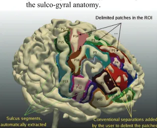
• A segment (figure 1) is a part of an external trace of a sulcus. The segments are organized in a graph describing the connections between them.
• A conventional separation (figure 1) is a fictitious line added by the user in order to connect two segments separated by a gyrus.
• A patch (figure 1) is a subset of the brain surface, corresponding to a part of a gyrus, and delimited by a set of continuous segments and conventional separations.
• An interpretation consists of a set of labels associated to the patches and segments of a Region Of Interest (ROI); each patch and each segment has only one label.
• A consistent interpretation is a set of annotations, where all patch labels and segment labels are pairwise compatible
given our prior ontology knowledge about the sulco-gyral anatomy.
System Overview
The complete labeling process is depicted in figure 2. (1) The brain is segmented from a T1-MRI scan, and the external traces of the sulci are automatically extracted; (2) the ROI is selected by the user, the patches are delimited, and described; (3) the patches are pre-labelledby matching the sub-graph of sulcus
segments belonging to the ROI with an atlas (SPAMs); 3 (4) the consistent interpretations for the
patches are inferred by a Constraint Satisfaction Problem solver, based on our prior knowledge about the spatial arrangement of the gyri and parts of gyri in the brain; (5)the best interpretation for the patches is determined interactively using information supplied by the user; (6) the sulcus segments are classified exploiting the best interpretation computed for the patches, the spatial relations between the segments and the patches, and the logical definitions of the sulci in the ontology. The final annotations are produced and represented in OWL (the Ontology Web Language), in order to facilitate their sharing and exploitation.
Knowledge involved in the Labeling System Numerical Knowledge: The system uses a numerical atlas, in order to initialize the reasoning with a set of hypotheses for the patches. The SPAMs 3 (Statistical
Probability Anatomy Maps) are used by the system to reach this goal. A SPAM is a 3D probabilistic map associated to a particular anatomical structure. The value at each voxel position represents the probability of belonging to this structure at that location. The SPAMs were derived from a database of 305 normal subjects, after re-alignment of MRI data into a common reference system (called stereotaxic space).
Figure 1: Example of a Region of Interest.
Symbolic Knowledge: It consists of an OWL DL (OWL based on Description Logics) ontology modelling the mereo-topological features about the sulci and the gyri. The resources used to model this knowledge were: (1) previous work of Dameron 4
about brain anatomy; (2) the Foundational Model of Anatomy (FMA1); (3) the Ono atlas of cerebral sulci;
5 (4) and the expertise of a neuroanatomist. For more
details on our brain ontology see 6. The logical
definitions of the sulci in the ontology serve to classify sulcus segments with a Description Logics classifier. The ontology knowledge was also used to
manually specify the definitions of the spatial constraints between gyri parts, in order to be used by the Constraint Satisfaction Problem solver when labeling the patches. This takes into account both the respective orientation and adjacency relationships between the gyri and gyri parts. So, we defined six spatial constraints corresponding to the six orientations, each represented by a set of tuples, such as (RightPreCentralGyrus, RightPostCentralGyrus), one of the tuples defining the anteriorTo constraint, which
expresses that any part of the right precentral gyrus must be anterior to any part of the right postcentral gyrus. Some tuples may be in the form
(RightSuperiorFrontalGyrus, RightSuperiorFrontalGyrus),
Figure 2: Complete Labeling Process:
Bent corner squares: Knowledge, Squares: treatments, Circles: input-output data. The numbers (1...6) on the squares correspond to the different treatments referred to in the System Overview section.
expressing that some part of the right superior frontal gyrus may be anterior to some other part of the same gyrus. As opposed to the orientation constraints, the adjacency constraints between gyri and parts of gyri are of utmost importance in the system, because they have the advantage to be independent of the coordinate system and the numerical computation programs, which may make some erroneous decisions. So the adjacency constraints are privileged in our system.
Patch Labeling
Patch Extraction: The system first extracts the sub-graph of the sulci segments corresponding to the ROI selected by the user. Then, the latter defines a set of contiguous patches by introducing a number of conventional separations (figure 1). Then, the system computes the topological relations and orientations between neighbouring patches, and between the patches and the segments forming them (figure 2–a). The description of these spatial relations is represented in OWL (file#1).
Atlas-Based Pre-Labeling of the Patches: The segments of the sub-graph are realigned into the SPAMs space (stereotaxic space) thanks to the registration matrix produced during the segmentation process (figure 2-b). A program analyzes the position of each segment with respect to each SPAM, and determines whether it bounds it or not, and with which orientation. This information is also represented in OWL (we name it file#2). The
matching of information from file#1 and file#2 in case of normal subjects is done by rules of the following form: Bounds(x,y) ^ SulcusPart(x) ^ Patch(y) ^ anteriorTo(x,y) ^ Bounds(x,z) ^ Gyrus(z) ^ anteriorTo(x,z) → partOf(y, z). This rule infers that a patch y is a part of a
particular gyrus z of the ontology, if y and the SPAM corresponding to z are both bounded by a segment x with the same orientation anteriorTo. Six similar rules are defined in our system for normal subjects, they correspond to the six spatial orientations (anteriorTo, posteriorTo, superiorTo, inferiorTo, lateralTo, and medialTo). Slightly different rules are defined for pathological cases. They do not take in consideration the orientations between the parts of sulci and the SPAMs, because they might lead to erroneous decisions due to displacements related to some
pathology. These rules allow for assigning to each patch a set of labels viewed as initial hypotheses. The correct label is assumed to belong to this set.
Constraint Reasoning for Determining Consistent Interpretations for the Patches: A Constraint Satisfaction Problem (CSP) consists of a number of variables and a number of constraints. A variable is defined by its domain, i.e. the set of values that can be assigned to this variable. A constraint relates several variables and restricts the involved variables values to legal assignments. Constraint reasoning is the process of computing a solution to the given CSP, i.e. an assignment of values to the variables that satisfy all the given constraints on the variables. 7 The
adaptation of our problem to a CSP was easy (figure 2-b). Indeed, the patches represent the variables, the hypotheses computed for the patches represent the domains of the variables, and the spatial relations between the patches represent the constraints. The CSP solver provides all possible consistent interpretations for the patches with respect to our knowledge about the spatial arrangement of the gyri and parts of gyri in the brain.
User-Assisted Determination of the Best Interpretation: The goal is to determine, from a user perspective, which is the best interpretation, among those returned by the CSP solver. To reach this goal, the user is invited to assign a label to a patch exhibiting different labels in different interpretations. Then, the system eliminates all the proposed
interpretations that are not consistent with the user choices (figure 2-b). The interactions are repeated until only one interpretation remains for the patches. Figure 3: Logical definition of the RightCentralSulcusPart concept
Illustrative example: Consider the simple example depicted in figure 4 exhibiting three patches (P1, P2,
P3) delimited in the ROI (figure 4–a). The goal is to
find the correct labels for the three patches. From the patch description we have the following spatial relations: posteriorTo(P1, P3), adjacentTo(P1, P3),
anteriorTo(P3, P2), adjacentTo(P3, P2), superiorTo(P1, P2),
and adjacentTo(P1, P2). We suppose that the following hypotheses are inferred for the patches from the SPAMs matching rules above: P1(R-PreCG, R-PostCG),
R-IntFG, R-PostCG). The graphical representation is transformed into a CSP representation (figure 4–b), and its resolution returns three possible interpretations for the patches (I1, I2, and I3) (figure 4-c). Now, the system asks the user to validate one label for the patch having the highest number of possible labels in the different interpretations (P3 in the example, since it has three possible labels). If the user validates P3(R-PreCG), then the system eliminates the previous interpretations where the label of P3
differs from R-PreCG. So the best interpretation in this case is I3 (figure 4-d).
Sulcus segments Labeling
The sulcus segments labeling relies on a Description Logics reasoning (figure 2–c). The system uses the best interpretation computed for the patches, the topological relations and the orientations calculated between the segments and the patches, and the logical definitions of the sulci defined in the ontology. Let us consider the segment S1 in figure 4–a. The best interpretation computed above was I3. From the patch description, it is known that S1 bounds P1 with an anterior orientation and P3 with a posterior orientation. The logical definition, in the ontology, of the right central sulcus part shown on figure 3, expresses that a part of the central sulcus of the right hemisphere bounds a part of the right postcentral
gyrus with an anterior orientation, and a part of the precentral gyrus with a posterior orientation, and it
does not bound other gyri. Consequently, the segment
S1 will be identified as a RightCentralSulcusPart
instance.
Experiments and material
Preliminary experiments were made using T1-MRI images obtained with a 3T scanner (Philips Achieva) from three normal subjects and two patients. In the two pathological cases the lesion was located in the right frontal lobe. The brain segmentation and the
extraction of the external traces of the sulci were done with Brainvisa2 tools and Vistal3, respectively. We used 44 SPAMs corresponding to the gyri. The system is implemented in C++ and Java, the connection between C++ and Java programs is done thanks to JNI (Java Native Interface). The ontology is edited and created with the Protégé4 software. The rules are edited and created with the SWRL5 Plugin. The results for the patches were obtained with the Java Constraint Library JCL6 (a CSP solver), and the segments were classified with the KAON27 reasoner (an inference engine for rule-extended ontologies).
Figure 4: Illustrative example of patch labeling
Results
The evaluation was done on ROIs defined by one expert neuroanatomist. They include the superior frontal gyrus, the middle frontal gyrus, the inferior frontal gyrus, the precentral gyrus, the postcentral gyrus, the central sulcus, the superior precentral sulcus, the intermediate precentral sulcus, the inferior precentral sulcus, the superior frontal sulcus, and the inferior frontal sulcus regions. We compared the results provided by the system to the labels assigned by the expert, considered as a gold standard. The same procedure was applied to the five MRI datasets except for the matching rules, since orientations were not considered in case of pathological data.
The following results were obtained: the number of patches extracted from the selected ROIs was around 16 (mean: 16.4, std. dev: 1.14), the number of consistent interpretations inferred for the patches was about 5 (mean: 5.2, std. dev: 1.64), and the best interpretation for the patches was reached after about 3 interactions with the user (mean 2.6, std. dev: 0.55). The accuracy of patch labels (defined as the ratio of the accurately labeled patches over the total number of patches in the ROI) was optimal (100%) for all cases, and the accuracy for the sulci segments (defined as the ratio of the accurately labeled sulci segments over the total number of sulci segments in the ROI) was acceptable (mean: 92.08 %, std. dev: 1.61); it was not perfect due to insufficient accuracy in the computation of the orientations between the patches and the segments. It is important to notice that the system performance was not decreased in pathological cases in spite of significant structures displacement according to the expert.
2http://brainvisa.info/index_f.html 3http://www.irisa.fr/visages/software/VIsTAL/ 4http://protege.stanford.edu 5http://www.w3.org/Submission/SWRL/ 6http://liawww.epfl.ch 7 http://kaon2.semanticweb.org
Discussion
Our approach differs in many ways from classical methods used for segmenting the cortex in MRI images. 8 Those are generally global, i.e. they try to
label the whole cortex, have limited precision (i.e. consider the major gyri and sulci only), refer to simple term lists, are entirely automatic, mostly based on numerical priors (i.e. atlases), and not robust in presence of pathology. In contrast, our approach is local (i.e. focuses on a particular region in the image), may provide labels with high anatomical precision, involves a user participation, is based on symbolic prior knowledge (provided by the ontology), and may be relevant for pathological cases as well as normal ones. It also differs from similar works on image annotation using semantic web technologies, such as the method of Dashmahapatra: 9 in particular, our
identification process involves reasoning on topological properties of the entities to be labelled, based on the graphical information extracted from the image. Regarding the ontology, we used both Dameron’s work4 and FMA, since FMA did not
provide the topological knowledge concerning the sulci.
Our first experiments using both normal and pathological data are very promising. We plan to still improve the system in the near future with regards to several aspects. The definition of the conventional separations between gyri is defined manually in the current implementation, and could be automated. The use of atlases as a means to produce the initial hypotheses could be optimized, e.g. by using different types of numerical atlases, more adapted to the particular case under study. The orientations’ computation could be improved, e.g. based on the method of Yann. 10 Finally, the ontology could be
refined, in order to include more fine-grained gyri and sulci, and this knowledge could be directly used to derive automatically the orientations’ definitions in the CSP problem. Moreover, the dependence of the system performance on initial processing steps (especially the definition of conventional separations) should be further investigated.
Conclusion
We have presented a hybrid and interactive system developed to semi-automatically label brain MRI images with semantic annotations and the first results obtained so far. The presented approach is novel with respect to several aspects: (1) the use of a CSP solver to select consistent interpretations of the gyri, (2) the easy generation of semantically-rich annotations of gyral/sulcal structures, (3) the use of explicit prior knowledge described in a formal ontology, (4) its
representation in OWL, the Ontology Web Language, to facilitate knowledge sharing. Our first results with both normal and pathological data are very promising and we will still improve the system in several well-identified aspects in the near future.
Acknowledgments: We are grateful to Louis Collins of the Montreal Neurological Institute for providing us with the SPAMs database and to the Regional Council of Brittany for supporting this project.
References
1. Ruttenberg A, Clark T, Bug W, Samwald M, Bodenreider O, Chen H, Doherty D, Forsberg K, Gao Y, Kashyap V, Kinoshita J, Luciano J, Marshall M, Ogbuji C, Rees J, Stephens S, Wong G, Wu E, Zaccagnini D, Hongsermeier T, Neumann E, Herman I, Cheung K. Advancing translational research with the Semantic Web. BMC Bioinformatics. 2007; 8(Suppl 3):2. 2. Jannin P, Morandi X, Fleig OJ, Le Rumeur E,
Toulouse P, Gibaud B, Scarabin JM. Integration of sulcal and functional information for multimodal neuronavigation. Journal of Neurosurgery. 2002; 96(4):713-23.
3. Collins DL, Zijdenbos AP, Baare WFC, Evans AC. Improved cortical structure segmentation. IPMI. 1999; LNCS 1613, 210-223.
4. Dameron O, Gibaud B, Burgun A, Morandi X. Towards a sharable numeric and symbolic knowledge base on cerebral cortex anatomy: lessons from a prototype. AMIA. 2002; 185-189. 5. Ono M, Kubik S, Abernathey C: Atlas of the
Cerebral Sulci. Thieme Med Publishers. 1990. 6. Mechouche A, Golbreich C, Gibaud B. Towards
a hybrid system using an ontology enriched by rules for the semantic annotation of brain MRI images. RR. 2007; LNCS 4524, 219-228.
7. Krzysztof A. Principles of Constraint Programming. Cambridge Univ. Press. 2003. 8. Cachia A, Mangin JF, Rivière D,
Papadopoulos-Orfanos D, Kherif F, Bloch I, Régis J. A generic framework for parcellation of the cortical surface into gyri using geodesic Voronoï diagrams. Medical Image Analysis. 2003; 7(4):403-416. 9. Dashmahapatra S, Dupplaw D, Hu B, Lewis H,
Lewis P, Shadbolt N. Facilitating multi-disciplinary knowledge-based support for breast cancer screening. J. of Healthcare Technology and Management. 2006 7(5), 403-420.
10. Yann H, Aline D. Qualitative spatial relationships for image interpretation by using semantic graph. In Escolano. GbRPR. 2007; LNCS 4538, 240-250.