Nephrol Dial Transplant (2007) 22: 2426–2429 doi:10.1093/ndt/gfm321
Advance Access publication 7 June 2007
High salt intake: a cause of blood pressure-independent left
ventricular hypertrophy?
Michel Burnier, Olivier Phan and Qing Wang
Division of Nephrology, Department of Medicine and University of Lausanne, Lausanne, Switzerland
Keywords: blood pressure; cardiac hypertrophy; potassium; sodium
Introduction
Left ventricular hypertrophy (LVH) is a frequent and well-recognized consequence of a chronically elevated blood pressure (BP). Several studies have demon-strated that LVH is an independent cardiovascular risk factor, associated with an increased likelihood to develop cardiovascular complications such as conges-tive heart failure, sudden cardiac death, coronary heart disease and stroke [1,2]. Conversely, an aggres-sive treatment of hypertension can prevent and reverse LVH and reduce the incidence of cardiovascular events [3,4].
Non-haemodynamic factors leading to LVH
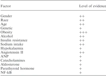
If BP is definitively the major trigger for the develop-ment of cardiac hypertrophy in hypertension, left ventricular mass may be affected by several other non-haemodynamic factors which may also play an important role in the pathophysiology of LVH, as listed in Table 1. Factors such as gender, age, race, obesity, alcohol consumption, catecholamines, ANP, aldosterone and angiotensin II and genetic factors (polymorphism of ACE) have all been considered as BP-independent determinants of left ventricular struc-ture. However, in clinical conditions, it is often difficult to demonstrate that the effect of these factors is really independent of BP, because small but long-standing changes in BP, which may contribute to the develop-ment of LVH may be missed, such as for example, nighttime increases in BP. Indeed, the discrimination between normotension and hypertension is arbitrary
and a loss of the normal nocturnal decline in BP has been associated with an increased incidence of LVH [5]. Similarly, an excessive rise in BP may induce LVH in subjects who participate in vigorous but unsustained exercises.
Experimental studies in transgenic mice have some-times provided good evidence that some of these factors induce cardiac hypertrophy, independently of changes in systemic BP. For example, the over-expression of angiotensinogen in cardiomyocytes leads to an increased production of angiotensin II limited to the heart with normal plasma levels [6]. Interestingly, these transgenic mice develop a marked cardiac hypertrophy in the absence of systemic hypertension and the increase in cardiac mass could be prevented by the administration of a blocker of the angiotensin II AT1 receptor [6]. In another transgenic mice model, inactivation of the guanylyl-cyclase-A gene selectively in the heart has been found to exhibit a mild cardiac hypertrophy, a marked increase in mRNA expression of cardiac hypertrophy markers, but a 7– 10 mmHg lower BP than control mice [7]. In some experiments, a regression of LVH was found without a decrease in BP [8,9]. For example, non-selective blockade of endothelin receptors with bosentan has been found to lower cardiac hypertrophy without lowering BP [8]. A similar observation was made with the inhibition of the transcription nuclear factor (NF)-kB in spontaneously hypertensive rats [9]. Taken together, these experimental data suggest that there are definite mechanisms which contribute to the development of cardiac hypertrophy independently of BP, but their role in humans remains to be demonstrated.
Does sodium have a role in the development of LVH? There is also increasing evidence that a high salt intake may have deleterious effects on the cardiovascular system and lead to the development of cardiac and vascular hypertrophy, independently or in addition to its effect on BP [10–12]. However, once again, it is difficult to ascertain whether the impact of the high sodium intake on cardiac mass or vascular
Correspondence and offprint requests to: M. Burnier, Division of Nephrology and Hypertension Consultation, Centre Hospitalier Universitaire Vaudois, Rue du Bugnon 17 CH-1011 Lausanne, Switzerland. Email: [email protected]
hypertrophy really is independent of BP, unless this latter is measured carefully over 24 h, which was not the case in these studies. Indeed, BP during sleeping hours may be crucial for the development of LVH even in animals [13].
In rats, increasing the sodium content of the diet often increases BP but there are some reports suggest-ing that sodium may induce LVH even in normo-tensive animals [14]. We recently investigated the combined effects of sodium and mineralocorticoids on BP and cardiac and renal hypertrophy in two strains of mice, one carrying one renin gene (the Ren-1c gene) and a second one carrying two renin genes, i.e. the Ren-1d and the Ren-2 genes [15]. The major difference between these two strains is that the two-renin gene mice exhibit a 10-fold higher plasma renin activity and a 100-fold higher plasma renin
concentration, which are not suppressed by a high sodium intake. Two-renin gene mice are salt sensitive and have a higher baseline BP. When these mice are uninephrectomized and receive a 1% sodium diet together with deoxycorticosterone for several weeks, their BP increases markedly and they develop a significant cardiac hypertrophy and an increase in the mass of the remaining kidney. To our surprise, mice with only one renin gene exhibited no increase in BP when receiving DOCA/salt. However, these animals also develop cardiac and renal hypertrophy. These data therefore suggested that sodium, when added to a high dose of mineralocorticoids, could contribute to induce cardiac and renal hypertrophy, even in the absence of BP changes [15]. To further investigate the respective role of mineralocorticoids and sodium in the development of cardiac hypertrophy, the same one-renin gene mice were treated for 4 weeks with DOCA and either a low or a high sodium diet [16]. Interestingly, mice receiving DOCA and a low sodium diet develop all the signs of hypermineralo-corticism but no significant increase in cardiac mass (Figure 1). These data indicate that an excess of miner-alocorticoid per se is not sufficient to induce cardiac and renal hypertrophy and the presence of a high sodium intake is a necessary condition for the development of mineralocorticoid-induced cardiac and renal damages.
To further explore the role of mineralocorticoids and sodium in the development of cardiac hypertrophy, we examined the direct consequences of chronically elevated plasma aldosterone levels on cardiac mass in a mouse model (a epithelium Na channel -/- Tg), which is normotensive under a normal sodium diet but exhibits chronic hyperaldosteronism [17]. After birth, these mice develop a pseudohypoaldosteronism type 1 (PHA-1) characterized by a metabolic acidosis, urinary salt wasting and growth retardation. Within 2 weeks after birth 50% of these mice die, but the survivors develop a compensated state of PHA-1 with a normal acid-base status, a compensated salt wasting and 6-fold higher plasma aldosterone levels than control. These mice and their control were examined after 16 months of life. We found that despite chronically elevated aldosterone levels and a low BP, these mice had no evidence of cardiac hypertrophy, remodelling or fibrosis [17]. Therefore, this observation suggests that hypermineralocorticism does not induce cardiac hyper-trophy in mice with a urinary salt wasting phenotype. This further emphasizes the importance of sodium as an important cofactor for the development of cardiac hypertrophy and that a hypermineralocorticism per se has little if any effect on the heart, unless animals receive a high sodium intake.
In more recent studies, we have shown that metabolic factors other than sodium may contribute to the development of cardiac hypertrophy in the DOCA/salt mice model. Indeed, the DOCA/salt model is characterized by the development of hypoka-laemia and metabolic alkalosis. We demonstrated that these two factors may also participate in the
Table 1. Non-haemodynamic factors and the development of left ventricular hypertrophy
Factor Level of evidence
Gender þþ Race þþ Age þþ Genetic þ Obesity þþþ Alcohol þþ Insulin resistance þþ Sodium intake þþ Hypokalaemia þ Angiotensin II þþ ANP þ Catecholamines þ Aldosterone þ Parathyroid hormone þ NF-kB þ
þþþ: Very strong evidence, þþ strong evidence, þ evidence that needs confirmation. The evidence is based on clinical and experi-mental data.
Control High salt (HS) Low salt (LS) DOCA/LS DOCA/HS 0 1 2 3 4 5 6 P<0.01 P<0.01
Cardiac weight index(mg/g)
Fig. 1. Effect of a high and low sodium diet on cardiac weight index in deoxycorticosterone-treated mice. BP was normal and comparable in all groups. Cardiac hypertrophy was found in one-renin gene mice receiving DOCA and a high salt intake (1% NaCl in the drinking water). No cardiac hypertrophy was observed on a low sodium diet (0.5 mg Na/g food). Adapted from references 14 and 15.
pathophysiology of cardiac hypertrophy, as correction of the hypokalaemia can prevent the development of cardiac hypertrophy without affecting BP [18]. These data suggest that there might be an interaction between the high-salt intake and the mineralocorticoid-induced loss of potassium to trigger hypertrophic factors leading to cardiac hypertrophy. Yet, other factors may contribute to the development of LVH in the mineralocorticoid-salt-induced model, including the alkalosis itself and the development of hypomagnesae-mia, which has also been reported to accompany mineralocorticoid excess and to interact negatively on heart function with the development of hypo-kalaemia [19].
What is the normal BP in mice with cardiac hypertrophy?
The development of cardiac hypertrophy in the presence of a so-called ‘normal’ BP might raise the question of the normality of BP. One could postulate that there is a resetting of the BP/cardiac hypertrophic-response relationship and that in this situation a lower BP could perhaps prevent the occurrence of cardiac hypertrophy. To test this hypothesis, we recently treated normotensive DOCA/salt mice with a calcium channel blocker, in order to lower their ‘normal’ BP and to assess the impact of lower BP on cardiac hypertrophy. To our surprise, reducing BP in these mice was associated with a reduction in cardiac mass, as shown in Figure 2 [20]. These results would thus suggest that there is indeed a resetting and that in the presence of LVH, one should perhaps aim at a lower BP.
Conclusions
LVH is an independent risk factor for the develop-ment of cardiovascular complications in hypertensive
patients. Although BP is the major trigger for the development of LVH, several non-haemodynamic factors should also be taken into account, including sodium and potassium intake. Recent animal data have indeed suggested that a high sodium intake is a crucial cofactor contributing to the patho-physiology of cardiac hypertrophy, particularly in situations of mineralocorticoid excess. These observa-tions further emphasize the importance of non-pharmacological recommendations in the management of hypertensive patients, in order to prevent target organ damages.
Conflict of interest statement. None declared.
References
1. Levy D, Garrison RJ, Savage DD et al. Prognostic implica-tions of echocardiographically determined left ventricular mass in the Framingham Heart Study. New Engl J Med 1990; 322: 1561–1566
2. Verdecchia P, Schillaci G, Borgioni C et al. Prognostic significance of serial changes in left ventricular mass in essential hypertension. Circulation 1998; 97: 48–54
3. Dahlof B, Pennert K, Hansson L. Reversal of left ventricular hypertrophy in hypertensive patients: A meta-analysis of 109 treatment studies. Am J Hypertens 1992; 5: 95–110
4. Dahlof B, Devereux RB, Kjeldsen SE et al. and the LIFE (Losartan Intervention for Endpoint Reduction) Study group. Cardiovascular morbidity and mortality in the Losartan Intervention For Endpoint reduction in hypertension study (LIFE): a randomised trial against atenolol. Lancet 2002; 359: 995–1003
5. Verdecchia P, Schillaci G, Guerrieri M et al. Circadian blood pressure changes and left ventricular hypertrophy in essential hypertension. Circulation 1990; 81: 528–536
6. Mazzolai L, Pedrazzini T, Nicoud F, Gabbiani G, Brunner HR, Nussberger J. Increased cardiac angiotensin II levels induce right and left ventricular hypertrophy in normotensive mice. Hypertension2000; 35: 985–991 DOCA/salt DOCA/salt+ Nifedipine P<0.001 130 120 110 100 90 80 15:00 18:00 21:00 00:00 03:00 06:00 09:00 12:00 24 h MeanBP (mmHg) DOCA/salt DOCA/salt+ Nifedipine DOCA/salt DOCA/salt + Nifedipine 5.0 4.8 4.6 4.4 4.2 4.0
Cardiac weight index (mg/g)
Time of the day
Fig. 2. Effect of lowering BP on cardiac hypertrophy in normotensive mice. DOCA/salt normotensive one-renin gene mice were receiving nifedipine (30 mg/kg) or a placebo for 4 weeks. BP measured by telemetry was significantly lowered (left panel) and cardiac hypertrophy was prevented (right panel).
7. Holtwick R, van Eickels M, Skryabin BV et al. Pressure-independent cardiac hypertrophy in mice with cardiomyocyte-restricted inactivation of the atrial natriuretic peptide receptor guanylyl cyclase-A. J Clin Invest 2003; 111: 1399–1407 8. Dvorak P, Kramer HJ, Backer A et al. Blockade of endothelin receptors attenuates end-organ damage in homozygous hyper-tensive ren-2 transgenic rats. Kidney Blood Press Res 2004; 27: 248–258
9. Gupta S, Young D, Sen S. Inhibition of NF-kB induces regression of cardiac hypertrophy, independent of blood pressure control, in spontaneously hypertensive rats. Am J Physiol Heart Circ Physiol2005; 289: H20–H29
10. Schmieder RE, Messerli FH, Garavaglia GE, Nunez BD. Dietary salt intake. A determinant of cardiac involvement in essential hypertension. Circulation 1988; 78: 951–956
11. Kupari M, Koskinen P, Virolainen J. Correlates of left ventricular mass in a population sample aged 36 to 37 years. Focus on lifestyle and salt intake. Circulation 1994; 89: 1041–1050
12. Safar ME, Thuilliez C, Richard V, Benetos A. Pressure-independent contribution of sodium on large artery structure and function in hypertension. Cardiovasc Res 2000; 46: 269–276 13. Morgan T, Brunner H, Aubert J et al. Cardiac hypertrophy depends upon sleep blood pressure: A study in rats. J Hypertens 2000; 18: 445–451
14. Yuan B, Leenen F. Dietary sodium intake and left ventricular hypertrophy in nomotensive rats. Am J Physiol 1991; 261: H1397–H1401
15. Wang Q, Hummler E, Nussberger J et al. Blood pressure, cardiac and renal responses to salt and DOCA in mice: role of renin genes. J Am Soc Nephrol 2002; 13: 1509–1516
16. Wang Q, Burnier M. Sodium modulates the mineralocorticoid-induced cardiac and renal hypertrophy independently of blood pressure in one-renin gene mice. J Hypertension 2004; 22 [Suppl. 1]: S10 (abstract)
17. Wang Q, Clement S, Gabbiani G et al. Chronic hyperaldoster-onism in a transgenic mouse model fails to induce cardiac remodelling and fibrosis under normal salt diet. Am J Physiol 2004; 286: F1178–F1184
18. Wang Q, Domenighetti A, Pedrazzini T, Burnier M. Potassium supplementation reduces cardiac and renal hypertrophy inde-pendent of blood pressure in DOCA/salt mice. Hypertension 2005; 46: 547–554
19. Evans SJ, Levi AJ, Jones JV. Low magnesium enhances the pro-arrhythmic effect of low potassium in the hypertrophied rat heart but not in the normal rat heart. J Hypertens 1996; 14: 635–44
20. Wang Q, Burnier M. What is the normal blood pressure in one-renin gene deoxycorticosterone – acetate/salt mice with cardiac hypertrophy? J Hypertens 2007, in press (abstract)
Received for publication: 17.3.07 Accepted in revised form: 29.4.07
Nephrol Dial Transplant (2007) 22: 2429–2431 doi:10.1093/ndt/gfm287
Advance Access publication 7 June 2007
A primer on iron therapy
Liliana Schaefer
1and Roland M. Schaefer
21
Institute of Pharmacology and Toxicology, University of Frankfurt, Theodor-Stern Kai 7, 60590 Frankfurt and
2Department of Medicine, University of Muenster, Albert-Schweitzer-Str 33, 48149 Muenster, Germany
Keywords: Chronic kidney disease; renal anaemia; erythropoiesis-stimulating agents (ESA); intravenous iron therapy; anaphylactoid reactions
Introduction
Iron deficiency is a frequent complication in patients with chronic kidney disease (CKD). Major causes are reduced dietary intake, impaired absorption, chronic blood loss, inflammatory or infectious comorbidity and increased requirements during correction of renal anaemia with erythropoiesis-stimulating agents (ESA).
Inadaequate iron stores with reduced availability of iron to the bone marrow are the main cause of hyporesponsiveness to ESA treatment. Thus, in the vast majority of haemodialysis patients, intravenous iron is used in combination with ESA to treat renal anaemia. Optimal iron management, therefore, includes the monitoring of iron status and the supplementation of adaequate amounts of iron, to maintain haemoglobin levels at target in a cost-effective manner [1].
Monitoring iron status
Ferritin
Serum ferritin is a parameter of iron storage in the reticuloendothelial system. In subjects without kidney disease, a value below 15 ng/ml indicates absolute iron deficiency [2]. In CKD patients, absolute iron
Correspondence and offprint requests to: Roland M. Schaefer, MD, Department of Medicine, University of Muenster, Albert-Schweitzer-Str. 33, 48149 Muenster, Germany.
Email: [email protected]