(Diptera: Culicidae): Activity and Distribution of Trypsin,
Aminopeptidase, and a-Glucosidase in the Midgut
PETER F. BILLINGSLEY1 AND HERMANN HECKER Swiss Tropical Institute, Basel, Switzerland
J. Med. Entomol. 28(6): 865-871 (1991)
ABSTRACT The activities of trypsin, aminopeptidase, and a-glucosidase were studied in the whole midgut, anterior and posterior midgut, and posterior midgut lumen and epithelium of the mosquito Anopheles stephensi Liston. Trypsin activity was restricted entirely to the posterior midgut lumen. No trypsin activity was found before the blood meal, but activity increased continuously up to 30 h after feeding, and subsequently returned to baseline levels by 60 h. Aminopeptidase was active in anterior and posterior midgut regions before and after feeding. In whole midguts, activity rose from a baseline of aS enzyme units (EU) per midgut to a maximum of 12 EU at 30 h after the blood meal, subsequently falling to baseline levels by 60 h. A similar cycle of activity was observed in the posterior midgut and posterior midgut lumen, whereas aminopeptidase in the posterior midgut epithelium decreased in activity during digestion. Aminopeptidase in the anterior midgut was maintained at a constant low level, showing no significant variation with time after feeding, a-glucosidase was active in anterior and posterior midguts before and at all times after feeding. In whole midgut homogenates, a-glucosidase activity increased slowly up to 18 h after the blood meal, then rose rapidly to a maximum at 30 h after the blood meal, whereas the subsequent decline in activity was less predictable. All posterior midgut activity was restricted to the posterior midgut lumen. Depending upon the time after feeding, >25% of the total midgut activity of a-glucosidase was located in the anterior midgut. The enzyme distributions are consistent with described structural models for digestion in mosquitoes. After blood meal ingestion, proteases are active only in the posterior midgut. Trypsin is the major primary hydrolytic protease and is secreted into the posterior midgut lumen without activation in the posterior midgut epithelium. Aminopeptidase activity is also luminal in the posterior midgut, but cellular aminopeptidases are required for peptide processing in both anterior and posterior midguts. a-glucosidase activity is elevated in the posterior midgut after feeding in response to the blood meal, whereas activity in the anterior midgut is consistent with a nectar-processing role for this midgut region.
KEY WORDS Insecta, Anopheles stephensi, bloodmeal digestion, midgut
INSECT-BORNE DISEASE ORGANISMS usually have an ciparum infection (Feldmann et al. 1990). The initial, often prolonged stay in the midgut of the presence of several midgut-associated glycosidases vector, where they develop or multiply in what (P.F.B. & H.H., unpublished data) in An. stephensi may become a hostile environment (Gass 1977). demonstrate that there are still many aspects of Knowledge of physiological events taking place in midgut enzymology yet to be understood, the vector midgut is important in understanding In Ae. aegypti, inactive trypsin is released from vector-parasite interactions necessary for disease small secretory vesicles within midgut epithelial transmission. Although trypsin is the major primary cells into the posterior midgut lumen, where the hydrolytic protease in the mosquito midgut (Brie- enzyme is activated (Graf et al. 1986). Other studies gel & Lea 1975), aminopeptidases in midguts of have demonstrated the spatial distribution of en-Anopheles stephensi Liston (Billingsley 1990b) and zymes across the mosquito midgut and have sug-Aedes aegypti L. (Graf & Briegel 1982) also play gested that the peritrophic membrane may serve important roles in the normal processes of blood as a molecular filter for bloodmeal proteins, as a digestion. Furthermore, differences in aminopep- layer separating hydrolytic events in the lumen, tidases, rather than trypsin, occurring between and as an important layer in separating enzymes strains of An. stephensi may be partly responsible and inhibitors from one another (Borovsky 1986, for the degree of susceptibility to Plasmodium fal- Van Handel & Romoser 1987). However, the most complete and integrated data for digestion in mos-quitoes is based upon ultrastructural observations 'Correspondence and current address: Cellular and Molecular c o m ple m e n t e d with morphometric analyses Parasitology Research Group, Department ot Biology, Imperial . \ , - . „ „ r> j . „ TT I T r ^ n r>-n- l
College of Science, Technology and Medicine, Prince Consort (Hecker 1977, Rudin & Hecker 1979, Billingsley Road, London SW7 2BB, U.K. 1990a). Such studies were used to develop a tem-0022-2585/91/0865-0871$02.00/0 © 1991 Entomological Society of America
poral and spatial model for digestion in mosquitoes, but complementary biochemical data necessary to correlate structure and function (e.g., Billingsley 1988; Billingsley & Downe 1985, 1988) have been lacking, especially in Anopheles spp.
As part of a study on blood digestion and malaria infectivity, the current report provides an overview of activities of three major digestive enzymes in the mosquito An. stephensi. These enzymes have been characterized previously in this species (Ber-ner et al. 1983, Billingsley 1990b, unpublished data), and were examined here in several defined phys-iological compartments of the midgut over the complete digestion period. Qualitative correlation of the results with previously published ultrastruc-tural data is used to develop an integrated model for digestion in this important mosquito species.
Materials and Methods
Insect Rearing and Treatment. Anopheles ste-phensi Liston (all stages), originally obtained from the London School of Hygiene and Tropical Med-icine in 1971, were reared in a 12:12 (L:D) pho-toperiod at 27 ± 2°C. Eggs were hatched in tap water containing a small amount of ascorbic acid, and larvae were fed ground hamster food. Adults were maintained at 75-80% relative humidity, pro-vided with 10% (wt/vol) sucrose ad libitum, and were fed periodically on denbrinated pig blood (Berner et al. 1983).
For experiments, 3-4-d-old female mosquitoes were deprived of sucrose for 12-18 h and then offered a meal of warmed, defibrinated pig blood through a stretched Parafilm membrane. Fully en-gorged females were separated in a cold room and used for further study. At selected intervals after feeding, blood fed mosquitoes were anesthetized by cooling in a deep freeze for a few minutes and then kept on ice until dissection.
Preparation of Midgut Homogenates. Whole, anterior, and posterior midguts were dissected in cold Aedes Ringer's solution (Hayes 1953). All non-midgut tissue was removed during dissection. To separate posterior midgut epithelium from the con-tents of the lumen, the posterior midgut was cut longitudinally and the contents were washed out onto a microscope slide. The contents were then placed into a 1.5-ml Eppendorf tube, while the intact posterior midgut epithelia were washed twice in Ringer's solution to remove any residual luminal material. Originally, we intended to separate ma-terial from each side of the peritrophic membrane, but this layer is not completely formed in An. ste-phensi until after >50% of the blood meal is di-gested (Berner et al. 1983). The delicacy of the peritrophic membrane during dissection prevented the preparation of uncontaminated material from each side, so the endo- and ectoperitrophic spaces were not assayed separately.
Pooled tissue was homogenized in 0.15 M NaCl in a tight fitting Teflon-Glass homogenizer, then
centrifuged at 10,000 x g for 10 min at 4°C. The homogenate supernatant was used in all subsequent enzyme and protein assays, and could be stored at —80°C for several months with no significant loss of activity for the three enzymes studied. The amount of tissue per homogenate was varied ac-cording to the time after feeding and the region being assayed: unfed and from 42 h after the blood meal whole midgut, posterior midgut and posterior midgut lumen—10 mosquito equivalents per 1.0 ml saline; 2-36 h after the blood meal whole mid-gut, posterior midgut and posterior midgut lu-men—2 mosquito equivalents per 1.0 ml saline; anterior midgut and posterior midgut epitheli-um—4-8 mosquito equivalents in 0.6 ml saline.
Enzyme and Protein Assays. Trypsin and ami-nopeptidase were assayed using the substrates ben-zoyl-DL-arginine-p-nitroanilide (BApNA) and leucine-p-nitroanilide (LpNA) respectively (Houseman & Downe 1986, Billingsley 1990b). Calculations of enzyme activities were made using an extinction coefficient of 8,800 nM cm"1 for both substrates at 410 nm (Erlanger et al. 1961). This method of tissue preparation did not detect mem-brane-associated aminopeptidases in the midgut (Billingsley 1990b).
a-glucosidase activity was determined by mod-ifications to the method of Ribeiro & Perreira (1984) (Billingsley & Hecker, unpublished) using the sub-strate p-nitrophenol-a-glycoside (pNaG) in 0.1 M TrisHCl buffer, pH 6.0. Absorbance was mea-sured at 405 nm and activity was calculated from a standard curve of p-nitrophenol made under the same conditions.
Assays were carried out in duplicate, with a min-imum of four samples (number of midguts or mid-gut compartments per sample as described above) for each time point after feeding unless otherwise stated. The results presented in all graphs (except Fig. 5) represent the means ± SE of 4-6 paired samples. Spontaneous breakdown of substrates and endogenous absorbance of samples were controlled by incubating paired tubes containing substrate and buffer, then adding the homogenate supernatant after the stopping agent. Absorbance readings were made on a Beckman (Fullerton, Calif.) 25 or a Cecil (Cambridge, U.K.) CE 292 spectrophotometer.
Protein contents of homogenate supernatants were determined using the trichloroacetic acid pre-cipitation procedure in the macro-assays (2-42 h after the blood meal; whole midgut, posterior mid-gut, and posterior midgut lumen) or microassays (all other samples) described by Peterson (1977). Bovine serum albumin (fraction 5, Sigma Chemical Company, St. Louis, Mo.) was used as the standard. Results were expressed as either relative activi-ty—i.e., total activity in enzyme units (EU) per midgut equivalent or midgut compartment equiv-alent, or as specific activity—the enzyme activity per microgram protein per midgut equivalent de-termined for each sample. The latter value was used to examine the relationship between enzyme
1000i
« 200
0 12 24 36 48 Time after feeding (h)
Fig. 1. Protein content of whole midgut homogenate supernatants of Anopheles stephensi after a blood meal.
10 i
12 24 36 48 Time after feeding (h)
60
activity and protein content of the midgut over the digestion period. One EU is described as one mil-limole of substrate (BApNA, LpNA, or pNaG) hy-drolysed per minute.
Results
Protein Content of the Midgut. The midgut in unfed insects contained 2.5-10 ng protein per whole midgut (Fig. 1). Immediately after feeding, the whole midgut contained «550 ng protein, which remained constant for at least 9 h. The blood meal was digested from 12 h onwards, resulting in a rapid decline in midgut protein until 48 h after feeding. By 60 h the protein per midgut had re-turned to the original amounts (Fig. 1). Throughout most of the digestive period, the bulk of midgut protein was from the blood meal. The protein con-tent of midgut epithelium did not alter significantly during the 60 h period after feeding (data not
201
12 24 36 48
Time after feeding (h)
60
Fig. 2. Relative activities of trypsin (•), aminopep-tidase (•), and a-glucosidase (A) in whole midgut ho-mogenate supernatants of Anopheles stephensi.
10" Trypsin Aminopeptidase alpha-glucosidase 100 200 300 Specific activity
Fig. 3. The relationship between protein and trypsin (•), aminopeptidase (•), and a-glucosidase (A) activities in the midgut of Anopheles stephensi. a. Specific activ-ities plotted on a log scale demonstrate similar trends until 30-42 h. b. Linear regression analysis of mean spe-cific activities and mean relative activities for each en-zyme. Only trypsin shows a clear positive correlation. All mean data points were used (taken from Fig. 2 and 3a) for the correlation calculations, but extreme data points are not shown on the graph.
shown), therefore protein in the posterior midgut epithelium and anterior midgut was not assayed.
Post-Feeding Activity and Distribution of Tryp-sin. Trypsin activity increased from a baseline of no activity in unfed insects to maximum activity at 30 h after the blood meal, and then dropped to no activity by 60 h (Fig. 2). Specific activity, plotted on a log scale, showed a similar pattern with max-imal activity at 30 and 42 h after the blood meal, before decreasing at 54 h (Fig. 3a). When the mean specific and relative activities of trypsin were plot-ted in a linear regression, there was a positive cor-relation between the two (r = 0.622; df = 12; P = 0.02) (Fig. 3b). All detectable trypsin activity was
15-
10-
5-12 24 36 48 Time after feeding (h)
60
Fig. 4. Activities of trypsin (•) and aminopeptidase (•) in homogenate supernatants of the posterior midgut of Anopheles stephensi. 0 . 5 1 0.4-0.3" E 0.2-0.1" UJ 0.0 12 24 36 48 Time after feeding (h)
60
Fig. 5- Post-feeding activity of aminopeptidase in the posterior midgut epithelium homogenate superna-tants of Anopheles stephensi. These results are the means of only two independent determinations.
restricted to the posterior midgut (Fig. 4), and all this activity was luminal. No activity could be de-tected in any other midgut compartment (anterior midgut or posterior midgut epithelium) at any time before or after the blood meal, even after pro-longed incubation with samples containing greater amounts of tissue (up to 25 posterior midgut epi-thelia per 1.0 ml saline dissected at 30 h after the blood meal and incubated for over 3 h).
Post-Feeding Activity and Distribution of Ami-nopeptidase. Aminopeptidase activity was present in all midgut regions before and at all times after feeding. In whole midguts, activity rose from a baseline of « 3 EU per midgut before feeding to a maximum of 12 EU at 30 h after the blood meal (Fig. 2). Activity then declined to baseline amounts by 60 h after the blood meal. The specific activity of aminopeptidase in the whole midgut was char-acterized by a delayed peak in activity at 48 h after the blood meal (Fig. 3a) compared to 30 h after the blood meal for total activity per whole midgut. The resulting correlation between mean relative activity and mean specific activity was poor (r =
-0.032; df = 12; P > 0.1) (Fig. 3b).
In the anterior midgut, aminopeptidase activity was maintained at a very low, almost constant level that represented only 0-0.5% of total midgut ami-nopeptidase activity. Anterior midgut activity therefore showed no clear response to the blood meal and could not be correlated with the major aminopeptidase cycle occurring in the posterior midgut.
More than 99% of the total relative activity was always present in the posterior midgut, and activity in this region (Fig. 4) predictably demonstrated a trend similar to that in the whole midgut (Fig. 2). Aminopeptidase was distributed unevenly between the epithelium and the lumen, ranging from 12.6% at 3 h to 0.8% at 30 h after feeding. The total relative activity in the posterior midgut epithelium
decreased from 0.5 EU per posterior midgut epi-thelium at 3 h to 0.05 EU at 36 h after the blood meal (Fig. 5), the converse trend to luminal activ-ity. Total activity in the epithelium then showed signs of recovery but had not returned to original amounts at 60 h after the blood meal (Fig. 5).
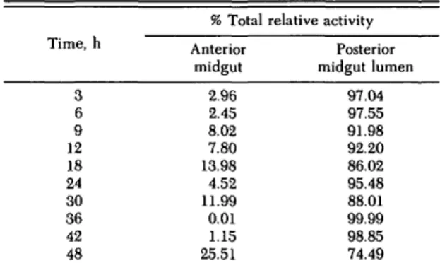
Post-Feeding Activity and Distribution of o-Glucosidase. a-glucosidase was active in both anterior and posterior midgut regions before and at all times after feeding. In whole midgut ho-mogenates, a-glucosidase activity increased slowly up to 18 h after the blood meal, then rose rapidly to a maximum at 30 h after the blood meal, whereas the subsequent decline in activity was less pre-dictable than that for the proteases (Fig. 2). The posterior midgut activity exhibited an overall in-crease after feeding but with no clear trend. All posterior midgut activity was restricted to the pos-terior midgut lumen (Table 1). The specific activity of a-glucosidase in the whole midgut showed a continuous rise from 3 h after the blood meal until 60 h (Fig. 3a), because of elevated activity late in
Table 1. Distribution of a-glucosidase in midgut com-partments of An. stephensi during a 48-h post-feeding period Time, h 3 6 9 12 18 24 30 36 42 48 % Total Anterior midgut 2.96 2.45 8.02 7.80 13.98 4.52 11.99 0.01 1.15 25.51 relative activity Posterior midgut lumen 97.04 97.55 91.98 92.20 86.02 95.48 88.01 99.99 98.85 74.49
t;
1.0- 0.2-0.0 +
48 60
Time after feeding
Fig. 6. Activity of a-glucosidase in anterior midgut homogenate supernatants of Anopheles stephensi.
the digestion period when very little protein was present in the whole midgut. Correlation between the mean relative activity and mean specific activ-ity was poor (r = 0.17; df = 12; F > 0.5) (Fig. 3b). More than 25% of the total midgut activity of a-glucosidase could be located in the anterior mid-gut depending upon the time after feeding (Table 1). The anterior midgut a-glucosidase activity showed a clear specific response to the presence of a blood meal, with maximum activity detectable at 24 h (Fig. 6). Activity then declined to a very low baseline and showed no correlation with other blood digestion processes in the posterior midgut.
Discussion
Our study provides a clear picture of the timing and sites of activity of three major enzymes, all of which have different functions in blood meal di-gestion, in the midgut of An. stephensi. Trypsin is the major primary hydrolytic protease in the mos-quito midgut (Briegel & Lea 1975) and is respon-sible for the initial breakdown of proteins and large peptides. In Ae. aegypti, trypsin is synthesized de novo after blood feeding (Gooding 1973) in two cellular stages (Felix et al. 1991)—immediate trans-lation from trypsin mRNA after feeding, followed by transcription of new trypsin mRNA several hours after the blood meal (Fuchs & Fong 1976, Hecker & Rudin 1979). Although several trypsin molecules can be detected in Ae. aegypti midgut (Graf & Briegel 1985, Kunz 1978), active trypsin is restrict-ed to the posterior midgut lumen; trypsin-immu-noreactive molecules that have been localized in secretory vesicles of posterior midgut epithelium are probably inactive precursors (Graf et al. 1986). Immunolocalization of trypsin clearly shows that the bulk of reactivity is restricted to the posterior midgut lumen of Ae. aegypti (Graf et al. 1986), where it freely crosses the peritrophic membrane to digest the blood meal. This is also the case with
An. stephensi, where all active trypsin was re-stricted to the posterior midgut lumen.
The quantity of trypsin produced is directly pro-portional to the size of the blood meal (Briegel & Lea 1975), and more specifically to soluble peptides in the blood serum (Felix et al. 1991). Consequent-ly, trypsin and midgut protein show clear and pre-dictable correlations (Houseman & Downe 1986). In An. stephensi this is reflected in the specific activity curve, which showed a clear post-feeding trend almost identical to that of total activity per whole midgut. Of the three enzymes examined, only trypsin exhibited such a correlation between relative and specific activities, supporting the the-ory for secretogogue control of trypsin in the mos-quito midgut (Briegel & Lea 1975, Houseman & Downe 1986).
In An. stephensi, three aminopeptidases are re-sponsible for the posttryptic digestion of peptides throughout the midgut (Billingsley 1990b). Low levels of activity in the anterior midgut probably represent a cytosolic soluble aminopeptidase, which would be required for routine intracellular peptide processing; the absence of any influence by the blood meal on anterior midgut aminopeptidase ac-tivity demonstrates its lack of involvement in di-gestion. Conversely, although posterior midgut ep-ithelium would still require a similar cytosolic enzyme, some epithelial activity in the posterior midgut also may represent a processing or secretory step for the luminal enzyme. The decrease after feeding in the epithelium-associated aminopepti-dase was a clear response to the presence of the blood meal in the lumen.
Most of the soluble aminopeptidase was secreted into the posterior midgut lumen, where its activity is probably concentrated in the ectoperitrophic space (Graf & Briegel 1982). Membrane-bound aminopeptidases constitute «50% of total midgut activity in An. stephensi (Billingsley 1990b), but there may still be a transport role for the soluble form of the enzyme. Being active close to the mid-gut wall, the soluble aminopeptidase is in an ideal site to drive amino acid transport from the lumen into the epithelium (and on to the hemolymph) in a fashion similar to alcohol dehydrogenase driving and directing NADH transport under experimental conditions (Vincent et al. 1988).
The specific activity of aminopeptidase peaked later compared with total relative activity or with any trypsin activity, and there was no correlation between relative and specific aminopeptidase ac-tivities. Although trypsin is responsible for primary proteolytic events in the midgut, secondary diges-tion of peptides is brought about by (amino- and carboxy-) peptidases, so aminopeptidase activity should correlate with peptides rather than proteins in the midgut. The peak of specific activity rep-resents, therefore, a poor correlation with proteins, and is delayed probably to coincide with the ap-pearance of peptides after trypsin hydrolysis of the blood meal.
The glycosidases represent a major nonprotein-ase group of enzymes in the mosquito midgut (un-published data). Up to six different glycosidases are active in the midgut of An. stephensi, and probably occupy two major sites—the midgut lumen and the lysosomes of midgut epithelium, a-glucosidase is the major midgut glycosidase in the An. ste-phensi midgut and therefore was chosen for this study. Compared to both proteases, a-glucosidase showed a different distribution and activity after feeding. Like trypsin, a-glucosidase was active only in a soluble form and was restricted to the lumen in the posterior midgut. Like aminopeptidase, a-glucosidase was active before and after feeding, and was found in both anterior and posterior mid-gut regions, a-glucosidase clearly is not membrane associated and is secreted into the lumen in re-sponse to blood feeding. Because the lumen and epithelium of the anterior midgut were not ex-amined separately, it is assumed that a-glucosidase has similar properties and distributions in both an-terior and posan-terior midgut regions.
The pronounced increase of a-glucosidase activ-ity in posterior midgut lumen occurred later in comparison to the proteases. The specific activity never reached a defined peak but continued to rise throughout the digestion period, in complete dis-association from other digestive events, a-glucosi-dase is presumably produced in response to suitable glycoside substrates in the midgut. Although some of these may be free in the serum, many more would be released by the proteolytic degradation of blood meal glycoproteins, especially those in the erythrocyte membranes (Harris & Kellermeyer 1970). There is no evidence to suggest whether a-glucosidase is active in the posterior midgut lu-men in the endo- or ectoperitrophic space, but delay in onset of activity also may influence postse-cretory structural modifications to the peritrophic membrane, which is highly glycosylated (Berner et al. 1983, Rudin & Hecker 1989).
By feeding radiolabelled glucose with blood to An. stephensi, Schneider et al. (1987) demonstrat-ed that, even after peritrophic membrane forma-tion is complete (18 h after feeding), sugar com-ponents of the blood meal still are absorbed in the anterior midgut. The small, but significant, in-crease in a-glucosidase in the anterior midgut after feeding indicates that this region may be involved in blood meal digestion, probably before the plug of peritrophic membrane at the anterior-posterior midgut junction is formed completely. The absence of any proteases in significant quantities in the anterior midgut suggests that the a-glucosidase found in this region does not originate from the posterior midgut.
Ultrastructural observations were used to devel-op a model for digestion in mosquitoes (Hecker 1977,1978; Billingsley 1990a), and the current data on enzyme distribution can be used to support this model. The quantities of synthetic and secretory organelles (ribosomes, rough endoplasmic
reticu-lum, and Golgi) in the midgut cells increase from 2 to 30 h after feeding. This clearly correlates with synthesis and secretion of all three enzymes de-scribed here, particularly in the posterior midgut. It is not possible to distinguish which organellar modifications are responsible for enzyme changes, because all enzyme activities follow a similar trend (Billingsley 1988). The absence of such organellar changes in the anterior midgut supports the model that protein digestion (at least) does not occur in the anterior midgut. The sugar digestive role also may be related to sugar feeding in mosquitoes, because the anterior midgut is thought to receive nectar passed to it from the gut diverticula (Hecker 1977, 1978; Billingsley 1990a).
Acknowledgment
P.F.B. was supported within the European Science Kxchange Programme by a postdoctoral research fellow-ship from the Royal Society, London (England). We are grateful to W. Rudin and B. Betschart for advice and discussions during this work. We also thank S. Marti, A. Habliitzel, and F. Grimm for mosquito rearing.
References Cited
Berner, R., W. Rudin & H. Hecker. 1983. Peritrophic membranes and protease activity in the midgut of the malaria mosquito, Anopheles stephensi (Liston) (Insecta: Diptera) under normal and experimental conditions. J. Ultrastr. Res. 83: 195-204.
Billingsley, P. F. 1988. Morphometric analysis of Rhodnius prolixus Stal (Hemiptera: Reduviidae) midgut cells during blood digestion. Tissue Cell 20: 291-301.
1990a. The midgut ultrastructure of haematophagous insects. Annu. Rev. Entomol. 35: 219-248.
1990b. Blood digestion in the mosquito, Anopheles stephensi Liston (Diptera: Culicidae): partial char-acterization and post-feeding activity of midgut ami-nopeptidases. Arch. Insect Biochem. Physiol. 15:149-163.
Billingsley, P. F. & A.E.R. Downe. 1985. Cellular localisation of aminopeptidase in the midgut of Rhod-nius prolixus Stal (Hemiptera: Reduviidae) during blood digestion. Cell Tissue Res. 241: 421-428. 1988. Ultrastructural localisation of cathepsin B in the
midgut of Rhodnius prolixus Stal (Hemiptera: Redu-viidae) during blood digestion. Int. J. Insect Morphol. Embryol. 17: 295-302.
Borovsky, D. 1986. Proteolytic enzymes and blood digestion in the mosquito, Culex nigripalpus. Arch. Insect Biochem. Physiol. 3: 147-160.
Briegel, H. & A. O. Lea. 1975. Relationship between protein and proteolytic activity in the midgut of mos-quitoes. J. Insect Physiol. 21:1597-1604.
Erlanger, B. F., N. Kokowsky & W. Cohen. 1961. The preparation of two new chromogenic substrates of trypsin. Arch. Biochem. Biophys. 95: 271-278. Feldmann, A. M., P. F. Billingsley & E. Savelkoul.
1990. Bloodmeal digestion by strains of Anopheles stephensi Liston (Diptera: Culicidae) of differing sus-ceptibility to Plasmodium falciparum. Parasitology 101: 193-200.
Felix, C. R., B. Betschart, P. F. Billingsley & T. A. Freyvogel. 1991. Post-feeding induction of trypsin in the midgut of Aedes aegypti L. (Diptera: Culici-dae) is separable into two cellular phases. Insect Bio-chem. 21: 197-203.
Fuchs, M. S. & W. F. Fong. 1976. Inhibition of blood digestion by alpha-amanitin and actinomycin D and its effect on ovarian development in Aedes aegypti. J. Insect Physiol. 22: 465-471.
Cass, R. F. 1977. Influences of blood digestion on the development of Plasmodium gallinaceum (Brumpt) in the midgut of Aedes aegypti (L.). Acta Tropica 34: 127-140.
Gooding, R. H. 1973. The digestive processes of hae-matophagous insects. IV. Secretion of trypsin by Ae-des aegypti. (Diptera: Culicidae). Can. Ent. 105: 599-603.
Craf, R. & H. Briegel. 1982. Comparison between aminopeptidase and trypsin activity in blood-fed fe-males, of Aedes aegypti. Rev. suisse Zool. 89: 845-850.
1985. Isolation of trypsin isozymes from the mosquito
Aedes aegypti. Insect Biochem. 15: 611-618.
Craf, R., A. S. Raikhel, M. R. Brown, A. O. Lea & H. Briegel. 1986. Mosquito trypsin: immunocyto-chemical localization in the midgut of blood-fed Ae-des aegypti (L.). Cell Tissue Res. 245: 19-27.
Harris, J. W. & R. W. Kellermeyer. 1970. The red cell. Production, metabolism, destruction: normal and abnormal. Harvard University Press, Cambridge. Hayes, R. O. 1953. Determination of a physiological
saline solution for Aedes aegypti L. J. Econ. Entomol. 46: 624-627.
Hecker, H. 1977. Structure and function of midgut epithelial cells in Culicidae mosquitoes (Insecta, Dip-tera). Cell Tissue Res. 184: 321-341.
1978. Intracellular distribution of ribosomes in mid-gut cells of the malaria mosquito, Anopheles ste-phensi (Liston) (Insecta: Diptera) in response to feed-ing. Int. J. Insect Morphol. Embryol. 7: 267-272. Hecker, H. &. W. Rudin. 1979. Normal versus
a-amanitin induced cellular dynamics of the midgut
epithelium in female Aedes aegypti L. (Insecta, Dip-tera) in response to blood feeding. Europ. J. Cell Biol. 19: 160-167.
Houseman, J. C. & A.E.R. Downe. 1986. Methods of measuring blood meal size and proteinase activity for determining the effects of mated state on digestive processes of female Aedes aegypti (L.) (Diptera: Cu-licidae). Can. Entomol. 118: 241-248.
Kunz, P. A. 1978. Resolution and properties of the proteinases in adult Aedes aegypti (L.). Insect Bio-chem. 8: 169-175.
Peterson, G. L. 1977. A simplification of the protein assay method of Lowry et al. which is more generally applicable. Anal. Biochem. 83: 346-356.
Ribeiro, J.M.C. & M.E.A. Perreira. 1984. Midgut glycosidases of Rhodnius prolixus. Insect Biochem. 14: 103-108.
Rudin, W. & H. Hecker. 1979. Functional morphol-ogy of the midgut of Aedes aegypti L. (Insecta, Dip-tera) during blood digestion. Cell Tissue Res. 200: 193-203.
1989. Lectin binding-sites in the midgut of the mos-quitoes Anopheles stephensi Liston and Aedes ae-gypti L. (Diptera: Culicidae). Parasitol. Res. 75: 268-279.
Schneider, M., W. Rudin & H. Hecker. 1987. Ab-sorption and transport of radioactive tracers in the midgut of the malaria mosquito, Anopheles stephen-si. J. Ultrastr. Molec. Struct. Res. 97: 50-63.
Van Handel, E. & W. S. Romoser. 1987. Proteolytic activity in the ectoperitrophic fluid of blood-fed Cu-lex nigripalpus. Med. Vet. Entomol. 1: 251-255.
Vincent, J.-C, S. Alexandre & M. Thellier. 1988. How a soluble enzyme can be forced to work as a transport system: description of an experimental design. Arch. Biochem. Biophys. 261: 405-408.
Received for publication 22 January 1991; accepted 1 July 1991.