Positive and negative mutant selection in the human histone hairpin-binding protein using the yeast three-hybrid system
10
0
0
Texte intégral
(2) Nucleic Acids Research, 2000, Vol. 28, No. 7 1595. multifunctional protein controlling most or all important aspects of histone mRNA metabolism. It contains a central domain of ~73 amino acids that is conserved between the human, mouse, Xenopus laevis and C.elegans proteins (5,6,12,24). This region was defined by deletion analysis of the human protein to represent a minimal RNA-binding domain (RBD) (5). Neither the protein as a whole nor the RBD are similar to any known RNA-binding proteins, so that HBP/SLBP appears to represent a new type of RNA–protein interaction. We have used the yeast three-hybrid system to select for mutations in human HBP that allow it to bind to an RNA containing a non-canonical hairpin as well as for mutations that abolish binding to the natural hairpin. All gain-of-function mutations lie in the N- and C-terminal regions flanking the RBD, suggesting that these regions contribute to the full substrate specificity. Consistent with this, the human RBD shows a relaxed substrate specificity both when tested in the yeast three-hybrid system and in direct RNA-binding assays. In contrast, the loss-of-function mutations either belong to a nested set of C-terminal truncations that help to define the C-terminal border of the RBD or contain point substitutions in the RBD that reveal critical residues for RNA binding. MATERIALS AND METHODS Nucleic acids The hairpin RNAs wtHP (5′-GGACAAAAGGCCCUUUUCAGGGCCACCC), cgHP (5′-GGACAAAACCCCCUUUUCAGGGGGACCC; changes underlined), U11G (5′-GGACAAAAGGCCCUGUUCAGGGCCACCC), U13A (5′-GGACAAAAGGCCCUUUACAGGGCCACCC), G5U/U13A (5′-GGACAAAAUGCCCUUUACAGGGCCACCC) and mutHP (5′-GGACAAAACCGGAAAGCCUUCCGGACCC) were transcribed from oligonucleotides by T7 RNA polymerase (25), including [α-32P]UTP in the transcription reaction, purified by denaturing polyacrylamide electrophoresis and stored in H2O. Strains Plasmids were amplified in Escherichia coli strain XL1-blue. The randomly mutated library for the second positive selection was obtained by amplification in the mutagenic E.coli strain XL1 red [endA, gyrA96, thi-1, hsdR17, supE44, relA1, lac, mutD5, mutS, mutT, Tn10(Tetr)] (Stratagene). For protein expression, strain BL21 [F–, ompT, rB–, mB–] (DE3) plysS (Pharmacia) was used. The three-hybrid system procedures were carried out in the Saccharomyces cerevisiae strain L40-coat (4). Plasmids Plasmid pET-15B was used for protein expression in E.coli. The shuttle vector pIII/MS2.2 was used for expression of the hybrid RNA molecules in yeast (4). pIII/wt/MS2 designates a plasmid encoding a hybrid RNA containing, 5′→3′, the RNase P leader sequence, the wild-type histone RNA hairpin and the MS2 binding sites (6). Five additional plasmids expressing mutant hairpin sequences were made by insertion of doublestranded oligonucleotides into the SmaI site of the pIII/MS2.2 plasmid. Oligonucleotides used were: 5′-GGAGCTCAACAAAAGGCCCTGTTCAGGGCCACCC (pIII/U11G/MS2); 5′-GGAGCTCAACAAAAGGCCCTTTACAGGGCCACCC (pIII/U13A/MS2); 5′-GGAGCTCAACAAAATGCCCTTTA-. CAGGGCCACCC (pIII/G5U/U13A/MS2); 5′-GGAGCTCAACAAAACCCCCTTTTCAGGGCCACCC (pIII/cg/MS2); 5′-GGAGCTCAACAAAACCGGAAAGCCTTCCGGACCC (pIII/mut/MS2); only one strand is shown and deviations from the wild-type sequence are underlined. HBPs were expressed as HBP–Gal4 activation domain (AD) fusion proteins in yeast. Mutant HBPs are designated by the change of amino acid at the position within the HBP open reading frame. Mutagenesis and screening procedure A pAct-HBP plasmid library produced by hydroxylamine treatment (26) was introduced into S.cerevisiae L40-coat cells containing the plasmid pIII/cg/MS2 as previously described (6,27). Transformants were grown on synthetic medium (YNB) lacking uracil, histidine and leucine for selection of the URA3, HIS3 and LEU2 marker genes, respectively. To favour the selection of HBP variants binding with high affinity to the mutated cg hairpin, 25 mM 3-amino-1,2,4-triazole (3AT) was included in the medium. Colonies appeared 5–6 days later and were analysed further for lacZ expression by plating on medium lacking uracil and leucine and supplemented with 80 mg/l 5-bromo-4-chloro-3-indolyl-β-D-galactopyranoside (X-Gal). pAct-HBP mutant plasmid DNA from growing strains co-expressing pIII/cg/MS2 RNA was isolated and subjected to further mutagenesis by amplification in E.coli XL1 red before being subjected to a second screening in L40-coat cells expressing cgHP RNA. Colonies growing after 3 days in the presence of 25 mM 3AT were then characterised as described above. To confirm that the changes in marker gene activation described above were caused by mutations in the HBP coding sequence and not by mutations introduced elsewhere in the plasmids, we subcloned the coding regions of the mutant proteins into non-mutagenised pAct plasmids and re-tested activation of the HIS3 marker gene in combination with the various hairpin elements. The results obtained with these constructs were identical to those shown in Figure 2 (data not shown), indicating that these experiments reflect a change in RNA-binding specificity of HBP. To select for loss-of-function mutations, the same pAct-HBP plasmid library produced by hydroxylamine treatment was introduced into S.cerevisiae L40-coat cells containing the plasmid pIII/wt/MS2. Transformants were plated on synthetic medium (YNB) lacking uracil and leucine. Growing colonies were replica plated on YNB medium lacking uracil and leucine supplemented with X-Gal. White colonies appearing among the vast majority of blue ones were further analysed. After confirming the white phenotype by isolating the pAct-HBP plasmid and re-transforming it into S.cerevisiae L40-coat cells containing plasmid pIII/wt/MS2, the HBP cDNA insert of the positive clones was sequenced. Testing of reporter gene activation in the yeast three-hybrid system To determine the level of lacZ expression, β-galactosidase activity was measured (28) in extracts from L40-coat cells expressing both HBP and RNA prepared as previously described (6). Measurements were done with extracts prepared from three independent transformants at two dilutions of extract. Alternatively, drops (3–4 µl) of yeast double transformants grown to OD600 = 0.1 were applied to plates prepared.
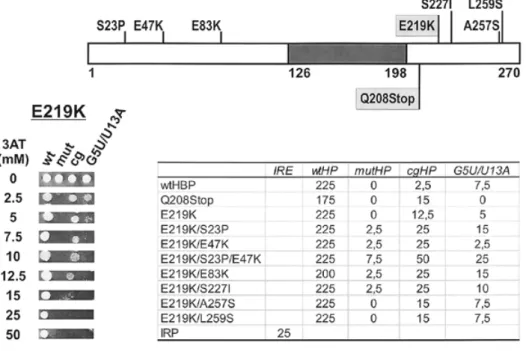
(3) 1596 Nucleic Acids Research, 2000, Vol. 28, No. 7. Figure 1. (A) Sequence variants of the histone RNA hairpin used in this work. The wild-type hairpin (wtHP) from the mouse histone H4-12 gene (49) is shown with base substitutions introduced either singly or in combination in different mutants. Only 4 nt preceding and following the hairpin are shown, although the RNAs used in vitro or in the three-hybrid system contained additional nucleotides (see Materials and Methods). The hairpin structures of mutHP and cgHP are shown below; flanking nucleotides were identical to the wtHP RNA. (B) Schematic drawing of the yeast three-hybrid system and (C) activation of the HIS3 and lacZ genes. Hybrid RNA molecules with binding sites for MS2 coat protein (X) and wild-type or mutant hairpin elements (Y) as indicated were expressed in yeast strain L40-coat along with a fusion between the RBD of MS2 coat protein and the LexA DNA-binding domain that binds to an upstream activating sequence (UAS) in front of the HIS3 and lacZ reporter genes (4) and with the cDNA encoding human HBP fused to the Gal4 AD (6). Expression of the reporter genes was assayed by scoring for growth on His selective plates supplemented with 25 mM 3AT and by β-galactosidase assay of yeast extracts (see Materials and Methods).. with YNB medium lacking uracil and leucine but supplemented with 80 µg/ml X-Gal. Incubation was for 3 days at 30°C. To test for HIS3 activation, drops (3–4 µl) of yeast double transformants grown to OD600 = 0.1 were applied to plates prepared with YNB medium lacking uracil, leucine and histidine but supplemented with different concentrations (0–400 mM) of the competitive His3p inhibitor 3AT (29). Levels of HIS3 expression were defined by the highest 3AT concentration allowing for growth. Production of recombinant proteins Recombinant His-tagged HBP and E219K/E47K/S23P were produced with an N-terminal M(H)6LEA tag in SF21 insect cells using a modified version of the Bac-to-Bac baculovirus expression system (Life Technologies) for protein expression. The RBD was expressed as an N-terminal MGSS(H)6SSGLVPRGSHMLE-tagged fusion protein from plasmid pET-15B in E.coli BL21. Proteins were purified by Ni affinity column chromatography using Ni–NTA resin (Qiagen). The RBD was further purified by phosphocellulose and Superose 12 column chromatography. Binding assays Recombinant HBP or RBD (6 or 2 pmol, respectively) and 55 fmol 32P-labelled RNA were incubated for 20 min on ice in. 10 µl of 10 mM Tris–HCl, pH 7.5, 0.3 M KCl, 10% glycerol, 1 mg/ml BSA, 1 U/µl RNAsin (Promega). Analysis was by native 5% polyacrylamide gel electrophoresis for reactions with HBP and by 8% polyacrylamide gel electrophoresis for reactions with RBD, using 50 mM Tris, 50 mM glycine as buffer system, and products were visualised by autoradiography or using a PhosphorImager. DNA sequencing DNA sequencing was done with the oligonucleotides 5′-TGCTCTACTCTGCGCTCT (located in the Gal4 activation domain) and 5′-GAGAGAGAAAATCATCATCAGGAA (located between nt 421 and 445 in the HBP coding sequence) labelled at their 5′-end with IRD41, using the SequiTherm Long-Read Cycle Sequencing Kit (Epicentre Technologies). Sequences were analysed on an automatic sequencer (Licor 4200). RESULTS Interaction of wild-type HBP with non-canonical hairpins We tested a series of hairpin mutants for their binding to wildtype HBP in the yeast three-hybrid system. For this purpose, plasmid pAct-HBP, previously selected from a human cDNA.
(4) Nucleic Acids Research, 2000, Vol. 28, No. 7 1597. Figure 2. Positive selection of HBP mutants interacting with cgHP. (Top) Positions of mutations. HBP is depicted as a bar with the minimal RBD shown in grey. Numbers indicate amino acid positions. The isolated mutants are indicated. E219K and Q208Stop (indicated by light grey boxes) were isolated in the first round of selection. The other changes were isolated in the second round of selection starting with mutagenised E219K (see text). (Bottom left) Drop tests of E219K. Yeast strain L40-coat expressing the indicated hairpin RNAs as MS2 fusions along with the E219K AD fusion were tested for growth on His selective plates containing different concentrations of 3AT as indicated. (Bottom right) Summary of the drop tests done with yeast expressing wtHBP, wtHP or mutants thereof as indicated. The iron regulatory protein (IRP) and iron response element (IRE) were used as controls (4,37). Numbers indicate the maximal 3AT concentrations allowing for growth; 0 means no growth at 2.5 mM 3AT, which was the lowest 3AT concentration tested.. library (6), was introduced into the L40-coat yeast strain that was already expressing a fusion RNA combining the MS2 site with either the wild-type histone hairpin or one of seven mutant hairpins (see Materials and Methods). Expression of the two reporter genes, HIS3 and lacZ, was determined by growth on plates lacking histidine and supplemented with 25 mM 3AT, which increases selectivity for the HIS3 marker, and by β-galactosidase assay of yeast cell extracts (see Materials and Methods). Three hairpins containing mutations in the loop (U11G, U13A and U11G/U13A) and a single mutation in the stem (C19A) showed reduced binding to wild-type HBP as determined by β-galactosidase activity but were still able to activate HIS3 expression at a high enough level to permit growth of the yeast cells on selective medium containing 25 mM 3AT (Fig. 1); these were therefore not deemed suitable to select for compensating mutations. In contrast, mutant hairpins G5U/U13A, cg and mut were completely unable to promote growth on His selective medium (Fig. 1), making them interesting candidates for three-hybrid selection for compensating mutations in the HBP gene. Isolation of gain-of-function mutations for the cg hairpin We used the G5U/U13A and cg hairpins as baits to select for compensating mutations in the HBP cDNA. Plasmid pActHBP was subjected to random mutagenesis using hydroxylamine, amplified in E.coli and then used for the transformation of yeast strain L40-coat co-expressing either G5U/U13A or cgHP fusion RNA. For each of the hairpins, ~450 000 pAct-HBP transformants were screened by growth on selective media. lacking histidine, uracil and leucine and supplemented with 25 mM 3AT. This allowed the isolation of two mutant HBP–AD fusion proteins able to interact with the cg hairpin, but none for G5U/U13A. Both of these mutations, E219K and Q208Stop, were found to interact only very weakly with the cg hairpin (see below). To obtain mutants interacting more strongly and also to increase the spectrum of possible mutation types, these two plasmids were grown in the mutagenic E.coli strain XL1 red, re-isolated and used in a second round of screening. This allowed the isolation of seven further compensating mutations from the original mutant E219K but none from Q208Stop. Surprisingly, all the compensating mutations from both screens were located outside the previously defined RBD (Fig. 2, top), indicating that the N- and C-terminal regions of the protein might play a role in discriminating between natural and divergent hairpins. Interactions of the isolated HBP mutants with various hairpins were determined by growth at different 3AT concentrations in drop test assays (Fig. 2, bottom; see Materials and Methods). This assay gave highly reproducible results and the values shown indicate the highest concentration of 3AT at which growth was possible. Already the mutations isolated in the first round of screening showed a higher resistance to 3AT than wild-type HBP. The apparent discrepancy that E219K and Q208Stop ceased to grow at 3AT concentrations over 12.5 and 15 mM, respectively, whereas 25 mM had been used for their initial selection can be explained by the fact that the single colonies appeared in the selection only after prolonged incubation (5–6 days) whereas the drop tests, containing a lot of seeded.
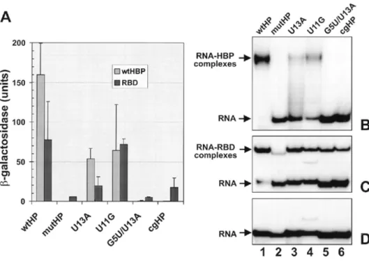
(5) 1598 Nucleic Acids Research, 2000, Vol. 28, No. 7. cells, were interpreted after 3 days. Interestingly, Q208Stop had lost the ability to interact with the G5U/U13A hairpin and showed a slightly reduced 3AT resistance in combination with the wild-type hairpin, whereas these interactions were not significantly affected for E219K. The interaction with the other mutant hairpins, U11G, U13A, U11G/U13A and C19A, was not affected for either mutant (data not shown). Two of the mutants isolated in the second round of screening showed only a slight increase in 3AT resistance (E219K/A257S and E219/ L259S) and also interacted similarly to the original E219K in combination with the other hairpins tested. In contrast, the other four double mutants isolated in the second round showed a marked increase in binding to the cg hairpin. The triple mutant E219K/S23P/E47K showed the strongest interaction with the cg hairpin. Since each of its additional amino acid exchanges was also obtained in one of the double mutants, both of which displayed an intermediate 3AT resistance, we conclude that these substitutions had additive effects. Similar differences between the abilities of these mutants to interact with the cg hairpin were also observed when blue colour on X-gal plates was used as an indicator of lacZ reporter gene expression (data not shown). With the exception of E219K/E83K, none of the mutants isolated in the second screening showed a reduced interaction with the wild-type hairpin. However, all mutant HBPs that strongly activated HIS3 gene expression with the cg hairpin also showed some increase for the other two mutant hairpins, G5U/U13A and mutHP. Again, the triple mutant showed the strongest interaction with these other hairpins. Thus it appears that some of the selected mutants did not have an altered, but rather a relaxed, specificity for hairpin structures (see Discussion). We also tried to test whether the increased interaction of the triple mutant HBP to the cg hairpin could be demonstrated by in vitro RNA-binding assays. For this purpose, we purified recombinant His-tagged wild-type HBP and E219K/S23P/E47K from insect cells infected with recombinant baculovirus to >90% homogeneity using Ni affinity column chromatography and used these proteins in binding studies. In electrophoretic mobility shift assays (EMSA) we detected binding of both proteins to wtHP RNA, but an interaction with cgHP RNA could not be reproducibly demonstrated, suggesting that such an interaction, if it occurred, was too weak to withstand the electrophoresis conditions (data not shown). A close interaction of both wild-type HBP and E219K/S23P/E47K with cgHP RNA could, however, be demonstrated by UV crosslinking (data not shown). For both proteins, the formation of this UV adduct was much weaker than with the wild-type hairpin. Unfortunately, however, the formation of this UV adduct could not be used as a quantitative measure for binding of the two proteins to cgHP RNA, since ‘freezing’ a protein–RNA complex by crosslinking does not reflect the binding affinity. The minimal RBD has a relaxed RNA-binding specificity The fact that all of the above mutations were located in the Nand C-terminal regions of the protein indicated that these regions play a role in discriminating between natural and noncanonical RNA hairpins. It therefore seemed likely that the minimal RBD previously defined by gel mobility shift analysis of in vitro translated truncated proteins (amino acids 126–198) (5) might have a relaxed substrate specificity. To test this, we inserted a HBP fragment spanning amino acids 121–203,. i.e. encompassing the minimal RBD into the pAct vector and analysed its interaction with different hairpin RNAs by its ability to activate the lacZ reporter gene in the yeast threehybrid system (Fig. 3A). The data for HBP–AD have already been presented and described in Figure 1 above, but are shown again for the sake of comparison. Measurements of β-galactosidase activity were done with three independent transformants and at two different concentrations of extract. The data presented in Figure 3A show the mean ± SD obtained for each combination. Interestingly, lacZ activation in cells expressing the RBD–AD fusion and wtHP RNA was only ~50% of that of the corresponding HBP–AD strain (Fig. 3A). However, in contrast to the observations with HBP–AD strains, near identical activation of the lacZ gene was observed in cells expressing RBD–AD in the presence of wtHP and U11G RNAs, indicating a change in binding selectivity of the RBD caused by loss of the flanking sequences. This was further supported by a small but significant lacZ activation in cells expressing cgHP, mutHP and G5U/U13A RNA. Only U13A RNA showed a reduction in β-galactosidase activity that was proportional to that observed in the corresponding HBP–AD cells. To confirm that these differences in relative lacZ activation reflected a change in RNA-binding selectivity, we compared the binding of recombinant HBP and RBD, purified to >90% homogeneity, to short synthetic 32P-labelled RNA molecules derived from the hairpin RNAs used in the three-hybrid system. Figure 3B and C shows the results of a representative in vitro binding experiment in which the protein concentrations had been adjusted to just allow for maximal binding of wtHP RNA. Confirming the observations made with the three-hybrid system, we detected binding of HBP to wtHP (Fig. 3B, lane 1) and more weakly to U13A and U11G RNA (lanes 3 and 4) but not to either mutHP, G5U/U13A or cgHP RNA (lanes 2, 5 and 6). The RBD bound to all RNA substrates used (Fig. 3C, lanes 1–6), but more weakly to the mutant hairpins than to wtHP RNA, confirming the RNA–RBD interactions observed in the three-hybrid system. However, there were quantitative differences between the three-hybrid and in vitro systems. For example, marker gene activation was greater with U11G than with U13A, whereas the opposite was true for RNA binding in vitro (Fig. 3C, lanes 3 and 4). A similarly inverted order was also observed between G5U/U13A and cgHP (lanes 5 and 6). Summarising these results, despite some quantitative differences between the yeast three-hybrid and in vitro RNA-binding assays, the original assumption that removal of N- and C-terminal sequences leads to a relaxed RNA-binding specificity in the RBD could be confirmed in both systems (see Discussion). Isolation of loss-of-function mutations by negative three-hybrid selection We next tried to isolate HBP mutants with reduced ability to interact with wild-type hairpin RNA in the yeast three-hybrid system. For this purpose, L40-coat yeast cells expressing the wild-type hairpin fusion RNA were transformed with mutagenised pAct-HBP and plated on selective medium lacking uracil and leucine. Replicas of these plates were then transferred onto similar plates additionally containing X-Gal. The white colonies among a majority of blue ones appearing on these plates were further analysed for HIS3 expression at different 3AT concentrations and characterised by DNA sequencing of the cDNA.
(6) Nucleic Acids Research, 2000, Vol. 28, No. 7 1599. Figure 3. Binding of full-length human HBP (wtHBP) and its minimal RBD to different hairpin structures. (A) Activation of the lacZ gene in the yeast three-hybrid system. Either wtHBP (6) or a fragment encompassing the RBD (amino acid residues 121–203) were expressed in the yeast three-hybrid system as AD–HBP fusions. β-Galactosidase activities were measured with extracts prepared from three independent transformants at two dilutions of extract (see Materials and Methods). Standard deviations are indicated with error bars. The values for wtHBP are the same as presented in Figure 1. (B and C) EMSA with wtHBP (B) or with the RBD (C) and short synthetic hairpin RNAs. wtHBP and a fragment encompassing the RBD (amino acid residues 121–203) were expressed in insect cells and E.coli, respectively. Both proteins contained an N-terminal histidine tag and were purified to >90% homogeneity. Protein concentrations (HBP 0.6 µM; RBD 0.2 µM) were chosen to give near complete binding of wtHP RNA. The proteins were incubated with the indicated 32P-labelled hairpin RNAs and analysed by non-denaturing gel electrophoresis and autoradiography as described in Materials and Methods. (D) Gel analysis of the free hairpin RNAs used for EMSA in (B) and (C). The additional band in the U11G RNA preparation (lane 4) that is also visible in the binding reactions is most likely due to an alternative folding of the RNA.. insert (see Materials and Methods). A total of 42 white colonies were isolated from ~30 000 transformants and 12 of these had point mutations in the HBP cDNA. Six of the mutants created nonsense codons at positions Q12, Q139, Q141, R163, W183 and W190, respectively (Fig. 4, top). These were unable to grow on His selective medium, independent of the 3AT concentration, indicating that they had completely lost the ability to interact with the wild-type hairpin. The other six clones had single amino acid substitutions at four different positions within the RBD; E157, T171, R181 and D184. Interestingly, three of the mutants affected the same residue, R181, with a Cys substitution being isolated twice and a Gln once. The 3AT titrations indicate that D184N had the strongest effect on interaction with the wildtype hairpin (growth only at 2.5 mM 3AT; Fig. 4, bottom) and E157K had the most moderate effect (100 mM 3AT), with the other four mutants being able to grow at up to 12.5–50 mM 3AT. Thus selection for loss-of-function mutations by the yeast three-hybrid system allowed identification of critical residues for RNA binding and helped to define the C-terminal border of the RBD (see Discussion). Two mutants constructed by site-directed mutagenesis were included in this analysis (indicated by asterisks in Fig. 4). One of these had two Arg at positions 137/138 replaced by Ala, resulting in a complete loss of RNA binding in vitro (data not shown) and a complete inability to grow on His selective plates even at the lowest 3AT concentration of 2.5 mM. (Fig. 4, bottom). The other had an Ala exchange of the same T171 residue for which we isolated a loss-of-binding mutant in the present selection, but showed near normal binding to wildtype hairpin RNA in vitro (J.Link and B.Müller, unpublished observations). In contrast to the selected mutant T171I, the constructed T171A showed only a moderate reduction in 3AT resistance in the yeast three-hybrid system, indicating that, for this residue, not only the position but also the nature of the amino acid change was important. DISCUSSION The yeast three-hybrid system as a positive and negative selection system In this paper we have used the yeast three-hybrid system to select for point mutations in human HBP with altered RNAbinding properties. The three-hybrid system proved to be amenable to both positive and negative selection schemes. This ‘classic genetics’ approach to select altered RNA-binding phenotypes from a random pool of mutants is an important complement to the popular ‘reverse genetics’ strategy and can provide valuable information on functionally important residues in new types of RNA-binding proteins such as HBP. Selection schemes based on the expression of RNA-binding proteins in bacteria have been described (30–34), but the yeast three-hybrid system(s) (3,4) or other eukaryotic selection.
(7) 1600 Nucleic Acids Research, 2000, Vol. 28, No. 7. Figure 4. Negative selection of HBP mutants with reduced binding to wild-type histone hairpin RNA. (Top) Positions of mutations. HBP is depicted as a bar with the minimal RBD shown in grey. Numbers indicate amino acid positions. The isolated mutants are indicated. Two identical mutants (R181C) were isolated independently. RR137/8AA and T171A were derived by site-directed mutagenesis and are marked with an asterisk. (Bottom) Summary of drop tests done with yeast expressing each of the mutant proteins together with wtHP as indicated. The bars indicate the maximal 3AT concentrations allowing for growth; 0 means no growth at 2.5 mM 3AT, which was the lowest 3AT concentration tested.. systems based on different principles (35–37) have the potential advantage that RNA–protein interactions are analysed in a eukaryotic environment. The yeast three-hybrid system has previously been used to quantitate RNA–protein interactions (reviewed in 38) and in positive selection schemes to isolate cDNAs encoding specific RNA-binding proteins (3,5–8) or RNA ligands for a given RNA-binding protein (9). The system has the advantage that two marker genes are available for selection and that the selection stringency for one of them, HIS3, can be varied over a wide range using different concentrations of the drug 3AT. This is particularly useful for a very strong RNA-binding protein such as HBP that shows growth on His selective medium at 3AT concentrations of up to 225 mM (Fig. 2). We exploited the proven suitability of the three-hybrid system for positive selection by searching for compensating mutations of HBP that would allow it to bind to mutant RNA hairpins. The selection criterion was growth on His selective medium at 25 mM 3AT, i.e. the concentration that had been used for the original isolation of HBP cDNA (6). Three of the mutant hairpins analysed, cgHP, G5U/U13A and mutHP, did not allow growth in combination with wild-type HBP–AD fusion protein under these conditions and showed no lacZ activity (Fig. 1), and cgHP and G5U/U13A were then used for selection. However, compensating mutants were only successfully. selected with cgHP RNA as the bait, and not with G5U/U13A RNA. This could be because the cg mutation modified the hairpin element only at the bottom of the stem, whereas G5U/U13A had modifications in both the stem and the loop. Therefore, more extensive mutations may have been needed to allow HBP to bind to this very different RNA structure. Other mutant hairpins had residual lacZ activity and still promoted growth at 25 mM 3AT (Fig. 1). However, considering the extremely high 3AT resistance (225 mM) observed for the wild-type HBP/hairpin combination, it is likely that compensating mutations for these other mutant hairpins could also have been isolated by applying more stringent selection conditions. Although most of the mutants binding more strongly to cgHP displayed a relaxed binding specificity, this does not mean that mutants with a more selectively altered specificity could not be isolated using a similar approach. However, the creation of true altered specificity mutants could require more than one or a few amino acid changes, because the network of interactions in the RBD may be too complex. In this case, it might be necessary to apply an in vivo evolution scheme consisting of multiple rounds of mutagenesis and selection. We know from other work that the RBD of the C.elegans HBP discriminates between hairpins differing only in the first position of the loop and that variants of the human RBD with similar sequence specificity can be obtained by multiple sitedirected amino acid substitutions (F.Michel, D.Schümperli and B.Müller, submitted for publication). The problem of isolating relaxed specificity mutants in the larger N- and C-terminal regions might also be circumvented by targeting the mutagenesis more specifically to the RBD. Finally, other sequence-specific RNA-binding proteins may not be so prone to relaxation of the RNA-binding specificity as human HBP. The isolation of loss-of-binding mutations in HBP represents the first successful case of negative screening using the yeast three-hybrid system. To select the mutants, we made use of the blue/white colour phenotype on X-gal plates. Not unexpectedly, the procedure yielded a larger proportion of false positive colonies (30 of 42) than positive selection, since reporter gene expression can also be lost when other features of the HBP–AD expression plasmid are mutated (e.g. the yeast replicon, the promoter or the activation domain of the fusion gene). All the mutants located in the HBP coding sequence were very informative, with about half of them representing premature stop codons and the other half being missense mutations. If one wanted to avoid full characterisation of the nonsense mutations, one could select for conditional phenotypes (e.g. temperaturesensitive mutations) or screen the mutant colonies by western blot for production of full-length protein. Relationship between reporter gene activities in yeast and in vitro RNA-binding results An interesting aspect from our work bears on the relationship between reporter gene activities in the yeast three-hybrid system and the physical strength of the protein–RNA interaction. For wild-type HBP, hairpins U13A and U11G, which yielded ~50–60 U of β-galactosidase, produced very weakly detectable protein–RNA complexes by EMSA (Fig. 3). In contrast, the RBD formed readily detectable protein–RNA complexes with the cgHP and G5U/U13A RNAs although lacZ activity was only 17.5 and 5.3 U, respectively. For the HIS3 marker, our results with the triple mutant protein E219K/S23P/E47K and.
(8) Nucleic Acids Research, 2000, Vol. 28, No. 7 1601. cgHP RNA indicate that a 3AT resistance over 50 mM is required for in vitro binding of full-length HBP. In contrast, the RBD formed complexes in vitro with cgHP RNA which allowed growth at only 2.5 mM 3AT. This implies that the relationship between reporter gene activation and the biochemical strength of an interaction varies between different protein–RNA pairs. Presumably, the levels of reporter gene activation in the three-hybrid system can be affected in many ways, among others through different folding in combination with the Gal4 AD, oligomerisation or additional interactions with the transcription machinery. Thus, it is important that data from the three-hybrid system be interpreted in terms of relative RNA binding strength only when studying closely related variants in the RNA or protein of a given interaction pair. For minor variants of such a pair, e.g. when comparing the interactions with different hairpin mutants for either full-length HBP (Figs 1 and 3) or for the RBD (Fig. 3), the β-galactosidase assays and the binding observed in vitro by EMSA are in good general agreement, although minor variations have also been observed (see description of Fig. 3 in Results). However, even between these assays the relationship is not necessarily linear. For example, the relative affinities of hairpins with the U11G, U13A or U11G/U13A mutation were previously reported to be 15, 20 and 5% of that of wtHP RNA (39), respectively, and yet the β-galactosidase activity was only reduced ~2.5-fold (Fig. 1). For other mutations, such as mutHP and cgHP, no three-hybrid interaction was observed, in full agreement with earlier in vitro binding studies (6,39,40) and with data from this paper (Fig. 3). For the other assay used in this work, scoring growth on His selective plates with different 3AT concentrations, non-linearity between 3AT resistance and HIS3 expression is even to be expected. Specificity of RNA recognition by HBP: interplay between the N- and C-terminal regions and the minimal RBD Interestingly, all the compensating mutations isolated in the positive selection were located in the N- and C-terminal regions, with none in the RBD (Fig. 2). Moreover, one mutation, Q208Stop, removed most of the C-terminal part of the protein. Several of the double mutations and the triple mutation not only increased the interaction with cgHP but also with mutHP and G5U/U13A RNAs (Fig. 2). Furthermore, the RBD of human HBP was found to have a relaxed specificity compared to full-length wtHBP (Fig. 3). All these findings indicate that the flanking regions contribute to the full binding specificity of HBP, although the RBD by itself is sufficient for binding. There are two possible explanations for these findings which are not mutually exclusive. First, the N- and C-terminal regions might contribute to the full binding specificity by imposing structural constraints on the RBD. Their mutation or removal might render the RBD more flexible, allowing it to accommodate non-canonical hairpins. However, extrapolating from allosterically regulated enzymes, one would expect a loss of substrate binding rather than a reduced binding specificity as a result of such a structural relaxation. Second, it is possible that residues in the N- and C-terminal domains are truly involved in discrimination of non-cognate substrates and that we have identified critical anti-determinants preventing the binding of non-cognate hairpins. Such antideterminants have been identified in the interactions between. aminoacyl-tRNA synthetases and their cognate tRNAs (41–43) and for E.coli initiation factor IF3 (44). Acidic residues (Asp and Glu) play a direct role in this discrimination of non-cognate RNA substrates. In this context it is interesting to note that three of the eight mutations isolated by us were substitutions of Glu residues by Lys (E47K, E83K and E219K). However, as the side chain of Lys is two carbon atoms longer than the Glu side chain, it is also possible that these mutations had a steric effect and changed the structure of the protein, thus interfering with RNA binding. The mutation S227I led to the replacement of a hydrophilic by a hydrophobic amino acid, whereas S23P replaced Ser by Pro, perhaps involving a change in HBP secondary structure. Finally, mutations A257S and L259S had only moderate effects on binding to cgHP RNA, so these changes are presumably not very important. It is possible, however, that the increased binding to the mutHP and G5U/U13A RNAs does not really reflect a loss of binding specificity. We selected for HBP mutants able to bind to cgHP RNA, which has two C-G base pairs at the bottom of the stem. The same C-G base pairs were present in mutHP (along with other changes), while in G5U/U13A RNA the bottom base pair was destabilised by the introduction of another pyrimidine, uridine, on the 5′-side. It is therefore also possible that one or more pyrimidines at the beginning of a double-stranded RNA region where bases are more accessible than within a narrow RNA A-helix (1) is the common structure of these three hairpin RNAs that the HBP mutants have ‘learned’ to recognise. Whichever of the above explanations is correct, it is clear that the N- and C-terminal regions flanking the RBD contribute significantly to the full sequence specificity of the human HBP. Moreover, as we isolated two double mutants (E219K/S23P and E219K/E47K) containing each of the additional mutations present in the triple mutant (E219K/S23P/E47K) and these activated the yeast reporter genes to intermediate levels between that of the single mutant E219K and the triple mutant, it appears that the effect of the three mutations in changing or reducing the binding specificity was roughly additive. In this work we have isolated a total of seven nonsense mutations. One, Q208Stop, retained binding to wtHP RNA and showed increased binding to cgHP RNA (Fig. 2). All the other six mutations with stop codons at Q12, Q139, Q141, R163, W183 and W190 completely lost the ability to interact with wtHP RNA. This allows us to position the C-terminal border of the RBD between amino acids 190 and 208. Previous work using in vitro translated truncated proteins defined the C-terminal border of the RBD as between residues 180 (non-functional) and 198 (functional) (5). Our new data thus narrows this margin to residues 190–198. It is not very surprising that six of the seven nonsense mutations occurred at Q and W residues, since the mutagenic agent used (hydroxylamine) primarily produces C→T or A→G mutations (45). Thus CAA or CAG codons (Asn) can readily be converted to TAA or TAG and TGG (Trp) can be converted to TAG. In our negative screen, we have isolated stop mutations in three of six Asn residues and in two of six Trp codons located in the N-terminal region and the RBD, indicating that the negative screen did not fully saturate the cDNA for mutations. All the missense mutations isolated in the negative selection were located within the previously defined RBD. This region is highly conserved among the five HBPs that have been.
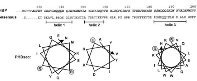
(9) 1602 Nucleic Acids Research, 2000, Vol. 28, No. 7. Figure 5. Structure of the minimal RBD of human HBP. The minimal RBD defined by Marzluff and co-workers (5) is shown in bold italic. The fragment designated RBD in this study corresponds to amino acid residues 121–203. The consensus sequence was derived from a comparison of human, mouse and C.elegans HBPs (5,6) as well as of the two related Xenopus proteins xSLBP1 and xSLBP2 (24). The residues shown are present in at least three of the five proteins. Putative α-helical regions predicted by the PHDsec program are shown below (46,47). Amino acid residues mutated in loss-of-binding mutants are underlined in the human HBP sequence and indicated by circles in the α-helical projections.. characterised (human, mouse, C.elegans and two proteins named xSLBP1 and xSLBP2 from X.laevis) (5,6,24). Comparison of these five proteins within the RBD yields the consensus sequence shown in Figure 5. Moreover, by computer alignment using the PHDsec program (46,47) three α-helical stretches are predicted to occur within this region (Fig. 5). All but one of the loss-of-binding mutations are located within these putative α-helices: the in vitro constructed RR137/8AA in helix 1, E157K in helix 2 and R181Q, R181C and D184N in helix 3. Only T171I is located in the linker region between the predicted helices 2 and 3. All of the mutated residues except for E157 are absolutely conserved between the five proteins; E157 (which had the weakest effect on binding) is altered to A and Q in xSLBP1 and xSLBP2, respectively, but is conserved in mouse and C.elegans. Although the selection did not saturate HBP for mutations (see above), these mutants identify a first set of amino acid residues that are critical for RNA binding. Three of the mutations, RR137/8AA, R181Q and R181C, replaced a positively charged amino acid by an uncharged one and had severe effects on RNA binding. This suggests that these residues may make important contacts with the negatively charged RNA backbone. In particular, R181, for which a Cys substitution was isolated twice and an Asn substitution once, seems to be a very important residue. After having isolated T171I as a loss-of-binding mutation, we also tested an Ala substitution of the same residue that we had obtained by site-directed mutagenesis. Interestingly, the Ala substitution had only a minor effect on binding of HBP to wtHP RNA (Fig. 4), indicating that not only the position but also the nature of the change was very critical. Because of the size difference between Ala and Ile it is likely that the T171I mutation poses a steric problem for RNA binding. The sequence TPNK is a consensus sequence for cdc2/cyclin B kinase, but evidence to be reported elsewhere suggests that this site is not a substrate for phosphorylation by cdc2/cyclin B kinase (J.Link and B.Müller, unpublished observations).. A very interesting mutation is D184N, since the substitution of an Asp by an Asn is a very minor change in terms of steric effect, implying that this residue might be involved in crucial interactions for RNA binding. The negatively charged Asp might, for example, interact directly with one of the bases in the RNA hairpin. In summary, this analysis has allowed us to identify critical residues for RNA binding within the RBD of human HBP. It is interesting to note that HBP is a multifunctional protein. It is certainly involved in histone RNA 3′ processing and presumably also in nucleo-cytoplasmic transport and translation of histone mRNA as well as in the regulation of its cytoplasmic half-life (12). The minimal region required for histone RNA 3′ processing in vitro has been mapped to the RBD plus 20 amino acids beyond its C-terminal border (48). Therefore, some of the residues of the RBD are likely to be important for RNA processing and perhaps also for some other functions of HBP. It will be interesting to analyse in detail whether the residues identified here are also involved in these functions. Moreover, the information gathered here can be used as a starting point for modelling the structure of the complex between the RBD and the histone RNA hairpin. ACKNOWLEDGEMENTS We are grateful to Marvin Wickens for supplying us with the yeast three-hybrid system. The HBP mutants RR137/8AA and T171A were obtained by site-directed mutagenesis by Roger Wiprächtiger and Julia Link, respectively. Ramesh S. Pillai contributed RNA-binding assays that were not included in the final version of the paper. This work was supported by the State of Bern and by Swiss National Science Foundation Grant 31-52619.97 to D.S. and B.M. REFERENCES 1. Cusack,S. (1999) Curr. Opin. Struct. Biol., 9, 66–73. 2. Draper,D.E. (1999) J. Mol. Biol., 293, 255–270..
(10) Nucleic Acids Research, 2000, Vol. 28, No. 7 1603. 3. Putz,U., Skehel,P. and Kuhl,D. (1996) Nucleic Acids Res., 24, 4838–4840. 4. Sengupta,D.J., Zhang,B.L., Kraemer,B., Pochart,P., Fields,S. and Wickens,M. (1996) Proc. Natl Acad. Sci. USA, 93, 8496–8501. 5. Wang,Z.F., Whitfield,M.L., Ingledue,T.C., Dominski,Z. and Marzluff,W.F. (1996) Genes Dev., 10, 3028–3040. 6. Martin,F., Schaller,A., Eglite,S., Schümperli,D. and Müller,B. (1997) EMBO J., 16, 769–778. 7. Zhang,B., Gallegos,M., Puoti,A., Durkin,E., Fields,S., Kimble,J. and Wickens,M.P. (1997) Nature, 390, 477–484. 8. Dahanukar,A., Walker,J.A. and Wharton,R.P. (1999) Mol. Cell, 4, 209–218. 9. Sengupta,D.J., Wickens,M. and Fields,S. (1999) RNA, 5, 596–601. 10. Birnstiel,M.L. and Schaufele,F.J. (1988) In Birnstiel,M.L. (ed.), Structure and Function of Major and Minor Small Nuclear Ribonucleoprotein Particles. Springer Verlag, New York, NY, pp. 155–182. 11. Marzluff,W.F. (1992) Gene Expr., 2, 93–97. 12. Müller,B. and Schümperli,D. (1997) Semin. Cell Dev. Biol., 8, 567–576. 13. Eckner,R., Ellmeier,W. and Birnstiel,M.L. (1991) EMBO J., 10, 3513–3522. 14. Williams,A.S., Ingledue,T.C., Kay,B.K. and Marzluff,W.F. (1994) Nucleic Acids Res., 22, 4660–4666. 15. Sun,J., Pilch,D.R. and Marzluff,W.F. (1992) Nucleic Acids Res., 20, 6057–6066. 16. Gallie,D.R., Lewis,N.J. and Marzluff,W.F. (1996) Nucleic Acids Res., 24, 1954–1962. 17. Pandey,N.B. and Marzluff,W.F. (1987) Mol. Cell. Biol., 7, 4557–4559. 18. Harris,M.E., Böhni,R., Schneiderman,M.H., Ramamurthy,L., Schümperli,D. and Marzluff,W.F. (1991) Mol. Cell. Biol., 11, 2416–2424. 19. Mowry,K.L., Oh,R. and Steitz,J.A. (1989) Mol. Cell. Biol., 9, 3105–3108. 20. Vasserot,A.P., Schaufele,F.J. and Birnstiel,M.L. (1989) Proc. Natl Acad. Sci. USA, 86, 4345–4349. 21. Pandey,N.B., Sun,J. and Marzluff,W.F. (1991) Nucleic Acids Res., 19, 5653–5659. 22. Hanson,R.J., Sun,J.H., Willis,D.G. and Marzluff,W.F. (1996) Biochemistry, 35, 2146–2156. 23. Dominski,Z., Sumerel,J., Hanson,R. and Marzluff,W. (1996) RNA, 1, 915–923. 24. Wang,Z.F., Ingledue,T.C., Dominski,Z., Sanchez,R. and Marzluff,W.F. (1999) Mol. Cell. Biol., 19, 835–845. 25. Milligan,J., Groebe,D., Witherell,G. and Uhlenbeck,O. (1987) Nucleic Acids Res., 15, 8783–8794.. 26. Isackson,P.J. and Bertrand,K.P. (1985) Proc. Natl Acad. Sci. USA, 82, 6226–6230. 27. Gietz,D., St Jean,A., Woods,R.A. and Schiestl,R.H. (1992) Nucleic Acids Res., 20, 1425. 28. Miller,J.H. and Albertini,A.M. (1983) J. Mol. Biol., 164, 59–71. 29. Hampsey,M. (1997) Yeast, 13, 1099–1133. 30. MacWilliams,M.P., Celander,D.W. and Gardner,J.F. (1993) Nucleic Acids Res., 21, 5754–5760. 31. Jain,C. and Belasco,J.G. (1996) Cell, 87, 115–125. 32. Laird-Offringa,I.A. and Belasco,J.G. (1996) Methods Enzymol., 267, 149–168. 33. Wilhelm,J.E. and Vale,R.D. (1996) Genes Cells, 1, 317–323. 34. Harada,K., Martin,S.S. and Frankel,A.D. (1996) Nature, 380, 175–179. 35. Stripecke,R., Oliveira,C.C., McCarthy,J.E. and Hentze,M.W. (1994) Mol. Cell. Biol., 14, 5898–5909. 36. Paraskeva,E., Atzberger,A. and Hentze,M.W. (1998) Proc. Natl Acad. Sci. USA, 95, 951–956. 37. Tan,R. and Frankel,A.D. (1998) Proc. Natl Acad. Sci. USA, 95, 4247–4252. 38. Zhang,B., Kraemer,B., SenGupta,D., Fields,S. and Wickens,M. (1999) Methods Enzymol., 306, 93–113. 39. Williams,A.S. and Marzluff,W.F. (1995) Nucleic Acids Res., 23, 654–662. 40. Schaller,A., Martin,F. and Müller,B. (1997) J. Biol. Chem., 272, 10435–10441. 41. Muramatsu,T., Nishikawa,K., Nemoto,F., Kuchino,Y., Nishimura,S., Miyazawa,T. and Yokoyama,S. (1988) Nature, 336, 179–181. 42. Schmitt,E., Meinnel,T., Panvert,M., Mechulam,Y. and Blanquet,S. (1993) J. Mol. Biol., 233, 615–628. 43. Bedouelle,H., Guez-Ivanier,V. and Nageotte,R. (1993) Biochimie, 75, 1099–1108. 44. Sacerdot,C., de Cock,E., Engst,K., Graffe,M., Dardel,F. and Springer,M. (1999) J. Mol. Biol., 288, 803–810. 45. Rose,M.D. and Fink,G.R. (1987) Cell, 48, 1047–1060. 46. Rost,B. and Sander,C. (1993) J. Mol. Biol., 232, 584–599. 47. Rost,B. and Sander,C. (1994) Proteins, 19, 55–72. 48. Dominski,Z., Zheng,L.X., Sanchez,R. and Marzluff,W.F. (1999) Mol. Cell. Biol., 19, 3561–3570. 49. Meier,V.S., Böhni,R. and Schümperli,D. (1989) Nucleic Acids Res., 17, 795..
(11)
Figure
Documents relatifs
To overcome the difficulty set by the number of words quoted when measuring preferences for specific traits, we now turn to a more disaggregated analysis where we focus
Cell cycle control of spindle pole body duplication and splitting by Sfi1 and Cdc31 in fission yeast.. {gamma}-Tubulin ring complexes regulate microtubule plus
The GAL4 fusion proteins studied here support this conclusion, as the histone H3 binding activities of mutant proteins correlate well with transcriptional activation in a
Rotations (A) Solutions Dessinez l’image de la rotation d´ecrite.. Rotation dans le sens horaire de 90 ◦ autour du point
In a similar fashion, expression of the wild-type stpA-stk in Cro2, which has a mutation in stpA predicted to truncate the protein to half its normal length, presumably elim-
After 96 days of column operation, carbon and nitrogen mass balances were calculated as follows: Total hydrocarbons (measured by IR) and proteins were analysed in all 10
Kaplansky’s zero divisor conjecture envisions that for a torsion-free group G and an integral domain R, the group ring RŒG does not contain non-trivial zero divisors.. We
Pathogenic missense mutations decrease the proportion of cells showing one fluorescent spot (Spot Formation assay) and increase the delocalization of BRCA1 nuclear
