European Heart Journal (1985) 6, 647-655
Range of normal values for left and right ventricular
ejection fraction at rest and during exercise assessed by
radionuclide angiocardiography
M. E. PFISTERER*, A. BATTLER! AND B. L. ZARETJ* Division of Cardiology, Department of Internal Medicine, Division of Nuclear Medicine, Institute of
Radiology, University Hospital, Basel, Switzerland, f Division of Cardiology, The Chaim Sheba Medical Center, Tel Hashomer, Israel and \Section of Cardiology and Nuclear Medicine, Yale University School of
Medicine, New Haven, CT, U.S.A.
KEY WORDS: Left ventricular function, regional left ventricular function, reproducibility, influence of age and sex on haemodynamics.
In order to reach a world-wide consensus on the normal range of left (LV) and right ventricular (RV) ejection fraction (EF) at rest and during exercise, pooled data of 1200 normal subjects from 28 leading centres in the field of nuclear cardiology (68% of those contacted) was analysed. Weighted mean normal values for LVEF at rest were 62-3±6I% (1SD) with a lower limit of normal of 50% and for RVEF 52-3±6-2% (N = 365) with a lower limit of normal of 40%. During exercise, LVEF increased in 475 subjects by +80 EF% (range 3-15%), a normal increase being accepted to be > 5% over a normal resting value for both L VEF and R VEF. Subgroup analysis of results at rest revealed no significant differences regarding selection of normal subjects (based on normal catheterization findings vs. normal volunteers with low probability of disease), age or sex. During exercise, however, significantly larger increases in LVEF measurements were noted for men versus women (P <0-01), for normal volunteers versus subjects selected as
'normals' based on a normal coronary angiogram (P<0-00I) and for younger versus older subjects (P<0-001). Data on reproducibility and variability showed that radionuclide angiocardiography can be considered to be a reliable method today. No consensus was found for measurements of regional LV function or wall motion mainly because of differences in methodology used. These normal values may serve us general guidelines for future applications of these techniques but factors which may influence the normal range as defined and discussed in this study should be recognized.
Currently, radionuclide angiocardiography has charges of this Task Force was to define the become a widely used clinical method for assessing normal range for nuclear cardiologic parameters global and regional left (LV) and right ventricular of cardiac function. Factors which could account (RV) function at rest and during physiologic for differences in normal values between various stress11 "*'. In order to set internationally accept- laboratories include: definition of the 'normal' able standards for these procedures, the Council population employed, physiological parameters on Clinical Cardiology of the International Society such as basal state at rest or type and intensity of and Federation of Cardiology and the World exercise stress applied and the technical aspects of Health Organization appointed a Task Force on the different methodologies used. Despite such Nuclear Cardiology (appendix 1). One of the possible differences in performing radionuclide angiocardiography, a large amount of data on normal subjects has been reported in the litera-Received for publication on 25 June 1984 and in revised form 2 May M-71 .• i_ . L.
I9 8 5 ture" 7| suggesting that a consensus can be
reached concerning what is generally accepted to This study was supported by the International Society and , c . , • , ,- • , -,., • r »i_
Federation of Cardiology and thi-World Health Organisation be Within normal limits . The aim o f the present
investigation was to provide such normal data of
A d d r e a for reprints. M . P f i s t e r e r , M . D . , D i v i s i o n o f C a r d i o l o g y , ¥ » , j r > < > <• » • i i • . .• i Department of Internal Medicine, University Hospital, OM03I L V a n d R V function On a large SCale international Basel, Switzerland. basis.
Principal considerations
In developing a range of normal, several principal considerations would appear to be relevant. The definition of a 'normal' population is often based on normal findings at left heart catheterization but latent myocardial disease may be missed by anatomical and functional assessment only at rest"'68'; the symptomatology responsible for invasive cardiac study may add to doubts whether such catheterization defines the 'true normal'. Alternatively, clinically healthy volun-teers with an extremely low probability of disease have been studied'9'; here 'hard data' concerning myocardial perfusion and coronary anatomy are missing, but the number of subjects with clinically hidden cardiac disorders is reduced. Both of these approaches have been used to define normal standards for radionuclide angiocardiography.
Physiologic parameters such as age, sex and
body weight all may influence parameters of myo-cardial function'10"13'and should be considered when normal values are defined. It is well known that physical training improves exercise perform-ance'14'1" and that normal values may differ between athletes and sedentary people. Psycho-logical imbalance, excitement or fear all affect the cardiovascular system in many ways'16'and should be excluded in order to obtain resting measure-ments in a true basal state. Obviously, cardioactive medication may interfere with the assessment of normal function'1718'. For 'exercise' measure-ments many additional factors have to be con-sidered such as type of stress applied'19-20', exercise protocol followed'21 ~231, body position'24t25'etc.
In addition, differences in normal values may arise from various methodological aspects. There might be differences between first pass and equilibrium (multiple gated) radionuclide function studies'26'. Radioactive dose applied, acquisition time, acquisition equipment all may influence results. Even more important in everyday practice are type and reliability of data analysis (ranging from visual inspection to sophisticated computer programs) and the criteria used for interpretation of these results.
Despite this large number of variables influenc-ing rest and exercise results, definition of the range of normal values as studies are currently performed would appear to be of considerable value — without eliminating the need for each institution to determine its own normal values based on its particular settings and protocol.
Methods
A questionnaire was sent to 41 representatives of leading centres in the field of nuclear cardiology worldwide selected by the Task Force asking 'What do you consider normal at your institution for routine nuclear cardiology procedures?' Each centre was asked to provide its own data and indicate exactly the definitions and methods used. Twenty-eight expert colleagues (68%) returned the 5 page, 100 question survey, 15 from the United States of America, 10 from Europe and 3 from other parts of the world (Appendix 2). No response was received from 5 centres from the United States, 5 centres from Europe and 2 centres from other parts of the world. Questions ranged from methodology used and physiological variables observed to mean normal values with standard deviations, lower limit of normal and to specific problems such as the interpretation of a higher than normal ejection fraction (EF) at rest not changing during exercise.
Based on the results of this first survey, a second questionnaire was sent to all responding centres asking for additional and detailed information for LV and RV function at rest and during exercise for the following subgroups: (1) selection of the nor-mal population (defined by nornor-mal catheterization findings vs. normal volunteers with low probability of disease), (2) gender (male vs. female), and (3) age (<30 years, 31-40, 41-50, 51-60, >60 years). Additional questions concerned inter- and intra-observer variability, (physiological) variability over time and reproducibility of measurements.
Angiocardiographic results of 1200 'normal' subjects could be pooled to form a representative basis for normal standards. The number of patients contributed from each centre is shown in appendix 2. It was up to the expert colleagues in each centre to collect and describe their study population based on the available (retrospective) data base. Data of LVEF at rest was obtained from all 1200 subjects, whereas exercise results and values of RVEF were available from subgroups of 475 and 365 individuals, respectively.
Analysis of results
Answers on methodology were only analysed if at least 10 (> 1/3) institutions answered any par-ticular question. Consensus was felt to be excellent if >85% of answers agreed, good if at least two-thirds and fair if only >50% agreed.
Normal values for ejection fraction 649
Weighted mean normal values and average stan-dard deviations were calculated for parameters of cardiac function taking into account number of subjects, mean value and standard deviation of each institution (cf. appendix 3)1271. The lower limit of normal defined as mean value minus 2 standard deviations was averaged from values used in each centre. For subgroup analysis, data of centres which provided no detailed information on the dif-ferent subgroups were excluded, i.e., for example, if the results of a certain centre was based solely on men, these data were not included for the comparison between values of men and women. Significance of differences between groups was cal-culated according to the formula given in appendix 3 with a confidence limit of 95% (/)<005); for differences between the multiple age groups, an analysis of variance test was applied.
Results
LEFT VENTRICULAR EJECTION FRACTION AT REST Global LVEF at rest measured in the supine position in 1200 subjects from 24 centres after an average resting period of 14-8±12-2min was 62-3% with a mean standard deviation of ± 6 1 % (Table 1, Fig. 1). The lowest normal value reported was 53 ± 6 % , the highest 70 ± 7 % . The lower limit of normal was considered to be 511 ±4-2% (range 45—60%). There was only a fair consensus on whether a 'hyperdynamic state' should be diag-nosed in cases with a high resting EF, this value lying above 76-6± 3-8% (range 70-80%).
There was no statistically significant difference between resting LVEF values for normals as
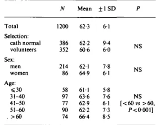
Table I Left ventricular ejection fraction at rest
Total Selection: cath normal volunteers Sex: men women Age: <30 31-40 41-50 51-60 . >60 N 1200 386 352 214 86 58 97 77 90 74 Mean 62-3 62-2 60-6 621 64-9 611 63-6 62-9 62-2 66-4 ±1SD 6 1 9-4 6 0 7-8 6 1 5-8 7-6 6 1 7-3 8-5 P w e N o XJC NS [ < 6 0 V J > 6 0 , /><0001] TO 6 0 50 40 A -LVEF RVEF ( A M 2 0 0 ) (/V = 365)
Figure 1 Normal values for left and right ventricular ejection fraction at rest. Bars indicate weighted mean values O O ± 1 weighted mean standard deviation (SDW), dots
represent mean values of different centres.
defined by catheterization findings (62-2 ±9-4%, A'=386) vs. normal volunteers with low prob-ability of disease (60-6 ± 6 0 % , JV=352). Further-more, there were no significant differences in normal LVEF at rest in sex and age subgroups, except for subjects older than 60 years whose resting LVEF was 66-4±8-5% vs. 62-6±6-9% in patients less than 60 years (/"< 0-001) (Table 1). Most values reported were based on gated blood pool studies (85%) acquired over 4-4±l-0min using an automated computer algorhythm (70%) with a variable LV region of interest (91% of answers). There was no significant difference between normal values obtained with first pass or gated equilibrium techniques.
LEFT VENTRICULAR EJECTION FRACTION DURING EXERCISE
During exercise (supine in 67%) which had to be symptom-limited (24 of 25 answers) or at least attained 85% maximal heart rate (18 of 24 answers), a good consensus was found concerning a normal change in left ventricular function: EF had to increase > 5 absolute EF percent over a normal resting value in 18 of 26 centres whereas 4 responders felt that a larger, 2 that a smaller increase and 2 that no fall below a normal resting value were normal criteria. Based on a study in 60 subjects with normal catheterization findings, one laboratory felt that due to the wide variation of EF responses to exercise, no 'normal' change could
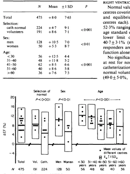
be defined'7' but the data of this laboratory was included, too. The mean change in LVEF during exercise of 475 subjects was + 8 0 % (range 3-15%) (Table 2, Fig. 2). Normal volunteers with low probability of coronary artery disease increased their LVEF significantly more than subjects de-fined as normals based on normal coronary angio-graphic findings ( + 8-6 vs. +4-7%; /><0001). Similarly, the exercise-induced rise in LVEF was
Table 2 exercise
Change in left ventricular ejection fraction during
Total N Mean ± 1 SD 475 +8-0 7-65 Selection: cath normal volunteers Sex: men women Age: < 3 0 3 1 ^ 0 41-50 51-60 > 6 0 224 191 128 50 56 48 62 40 36 + 4-7 + 8-6 + 10-5 + 5-3 + 12-5 + 11-8 + 8-5 + 8-6 + 7-6 9 1 7 1 7-0 8-7 4-4 5-2 6-6 5-8 7-3 <0-001 <0-01 < 0-001
significantly larger in men than in women (+10-5
vs. +5-3%; /><001). Finally, there was a gradual and significant decline of the rise in LVEF during exercise with age (P < 0001) (Table 2).
In 73% of responders who used the gated blood pool technique the data was acquired during a 2 min exercise period. There was good agreement that clearly submaximal exercise tests had to be interpreted cautiously, results being of question-able relevance. Only 14 of 26 responders felt that a LVEF of 75% at rest not changing during exercise was still a normal finding.
RIGHT VENTRICULAR EJECTION FRACTION AT REST
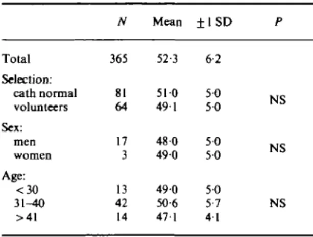
Normal values for RVEF were reported from 14 centres covering 365 subjects studied with first pass and equilibrium radionuclide techniques (in 7 centres each). Mean normal values at rest were 52-3% ranging between 47 and 59% with an aver-age standard deviation of ±6-2% (Table 3). The lower limit of normal was considered to be 40-7 ± 3 1% (range 35-45%). Twenty-two of 26 responders are using visual interpretation of RV function alone or in addition to calculation of EF.
No significant differences between RVEF values at rest for normal subjects as defined by normal catheterization findings ( 5 1 0 ± 5 0 % , # = 8 1 ) VS. normal volunteers with low probability of disease ( 4 9 0 ± 5 0 % , N=64) w e r e found. Again, there
20 16 12 8 - 4 Selection of normal Sex P<0-0\ Total N 475 Vol. Cath. 191 224 Men Women 128 50 . Mean values of different centres Q iwi I S O . <3O 31-40 41-50 51-60 >6O years years years years years
56 48 62 40 36
Figure 2 Normal change in LVEF from rest to symptom-limited exercise and its relation to selection of normal, sex and age subgroups. Bars indicate weighted mean values (x.) ± 1 weighted mean standard deviation (SDW), dots represent mean values of different centres [cath. — normal subjects as defined by normal coronary angiograms; vol. — normal subjects as defined by low probability of disease (volunteers)].
Normal values for ejection fraction 651
Table 3 Right ventricular ejection fraction at rest
Total N Mean ± 1 SD 365 52-3 6-2 Selection: cath normal volunteers Sex: men women Age: <30 31-40 >41 81 64 17 3 13 42 14 510 491 480 490 490 50-6 471 5 0 5 0 50 50 50 5-7 4 1 NS NS NS
Table 4 Variability of measurements
Interobserver: LVEF RVEF Intraobserver LVEF RVEF Over time: LVEF RVEF N ±1SD Range 558 155 548 155 164 59 2-6% 3-8% 2-3% 3-5% 3-6% 3-0% 1-4-50% 2-0-5-0% 1-0-3-0% 2-0-6-0% 1-6-10-0% 2-0-10-0%
were no significant differences in mean normal values assessed by first pass or equilibrium angio-cardiographic techniques (52-1% vs. 52-3%) but no intra-patient or intra-centre comparative data were available. The same is true for age and sex subgroups, but here the groups were too small to draw definite conclusions (Table 3).
RIGHT VENTRICULAR EJECTION FRACTION DURING EXERCISE
During exercise, RVEF is accepted to increase by at least 5 absolute EF percent over a normal resting value by most laboratories which quanti-tate RVEF routinely. In addition, RV function is generally assessed by visual interpretation of radionuclide data displayed in movie format (19 of 22 laboratories).
REPRODUCIBILITY OF MEASUREMENTS
Interobserver variability of LVEF for repeat analysis of the same data was small (range of l S D ± l - 4 % to ± 5 0 % , mean ±2-6%; JV = 558) as was intraobserver variability (range of 1 SD
± 1 0 % to ± 3 0 % , mean ±2-3%; J V = 5 4 8 ; Table
4). Variability of LVEF over time, i.e. in repeated acquisitions with an interval of 15 min to 14 days was good, too: ±3-6% (range of 1 SD ±1-6% to
± 100%; N= 164) resulting in a mean correlation coefficient for reproducibility of r = 0-95 (range r = 0-77 to r = 0-99) for EF values ranging from very low to high normal.
Inter- and intra-observer variability of RVEF was about 1 % larger than that for LVEF (Table 4), resulting in a reproducibility of these measure-ments with /--values between 0-80 and 0-98 (mean 0-92).
REGIONAL LEFT VENTRICULAR FUNCTION/WALL MOTION
Regional LV function or wall motion is assessed in all 28 centres by visual interpretation of cine-films in movie-format. Only in 13 centres is regional EF calculated regularly and three centres use numerical values for comparison of rest/ intervention studies only. There was only a fair agreement regarding the method used to calculate regional EF: a majority of those answering use radial sectors from a centre of gravity or mass. Due to the different analyses applied it was not possible to reach a consensus on normal values for regional EF. It is noteworthy, however, that in six of seven laboratories with detailed results, regional function was lowest in the septal area as compared to infero-apical and posterolateral regions.
Discussion
As part of an attempt to describe the state of the art of radionuclide angiocardiography today, the spread of normal values used in 28 leading centres in the field of nuclear cardiology was compiled and analysed in this study. The observed range of normal values may serve as a guideline for future applications of this technique and help in com-parisons of data obtained by these methods in different laboratories. Important factors influenc-ing normal values, especially durinfluenc-ing exercise, were defined such as age, sex, selection of a normal population as well as the study protocol used. Although it has to be stressed that calculated mean normal values originating from different centres may be open to debate from a strictly statistical point of view, it seems that knowledge of the
range of normal values observed world-wide is of considerable clinical importance. The large num-ber of EF values collected which to our knowledge are unprecedented by any published analysis of normal haemodynamic data outweigh some of the inherent problems of such a multicentre investigation based on a questionnaire.
Based on the present survey, a world-wide con-sensus on the range of normal values for LVEF and RVEF could be obtained. According to these results and in the absence of clinical signs of heart disease, LVEF 3*50% (mean 62-3 ± 6 1%) and RVEF =s40% (mean 52-3±6-2%) may be con-sidered normal at rest; during symptom-limited exercise, an increase of at least 5 absolute EF percent over a normal resting value describe a normal change for both ventricles. These findings are valid only in the absence of any cardioactive medication. With the methodology used today, these values can be obtained with low variability and high reproducibility. Certain factors which may influence the normal range, especially during exercise, such as definition of normal, age, sex and study protocol used have to be recognized.
FACTORS INFLUENCING LEFT VENTRICULAR FUNCTION
Subgroup analysis of LV (and RV) function at rest showed no significant differences regarding selection of normal subjects, gender and method-ology used, except for subjects older than 60 years who had a somewhat higher LVEF than those below age 60. It seems most likely that this differ-ence was due to the selection process which is probably most important in the older age group. The assurance of a basal state at rest with which exercise values can be compared appears to be important since the higher the EF at rest, the less its increase with exercise171. This point is underlined by the relatively large 'physiologic' variability of EF measurements over time described here and reported previously1281.
In contrast, during exercise significantly larger increases in LVEF measurements were found for men vs. women, for normal volunteers vs. subjects selected as 'normals' based on normal coronary angiographic findings and for younger v5. older subjects (Fig. 2). A decline in the LVEF response to exercise with increasing age has recently been reported by Port and coworkers who found no change or even a decrease in the EF during exercise in most subjects above age 60129'. In the present pooled data a more gradual decline was noted
and there was still a significant exercise-induced increase in LVEF in the age group >60 years. Possible mechanisms for this age-associated decrease in EF response to exercise include decreased contractility due to aging1301 or silent myocardial disease which is more frequent at higher age1311, increased afterload with higher age1321, lower exercise heart rate133"351 and lower maximal work load attained by older individ-uals'361. Unfortunately, the data base of the present investigation did not allow us to differentiate between these factors, but it has been demon-strated that LVEF is dependent on changing loading conditions of the heart137'. It should be noted, however, that the present findings parallel results of haemodynamic parameters such as maximal work capacity, oxygen consumption, cardiac output, stroke volume and left ventricular filling pressures during exercise in relation to a g elio, 13.38.39). The causes for the sex differences found in the present study remain speculative, too, but again, the differences parallel sex-related differences in maximal work capacity and haemodynamic variables reported112'40'411.
CHEST PAIN AND NORMAL CORONARY ANGIOGRAM The EF response to exercise in patients with chest pain but angiographically normal coronary arteries represents another important issue. This group may represent an as yet undefined new pathophysiologic entity rather than 'true' normals. Berger et al. observed LV dysfunction despite normal resting performance in a substantial num-ber of such patients with chest pain, an ischaemic-appearing exercise electrocardiogram and normal coronary arteries'42'. Maddahi et a/.*9' found objective signs of exercise-induced ischaemia more frequently in subjects with normal coronary arteriograms as compared with an alternative population with less than 1 % likelihood of coron-ary artery disease suggesting presence of disease despite normal coronary arteries. Such consider-ations may explain the significantly lower rise in EF during exercise found in the present study in subjects selected as normals based on a normal coronary angiogram vs. the higher increase in normal volunteers. This interpretation is sup-ported by a follow-up study of angiographically normal subjects which demonstrated that incipient heart disease may be present in subjects in whom coronary angiographic examination has removed a previous suspicion of coronary artery disease143'. The selection process has also been shown to cause
Normal values for ejection fraction 653
an apparent decline in specificity of exercise radio-nuclide angiocardiography in recently studied patients as compared with those studied when the technique was first introduced1441. Rozanski and coworkers'441 felt that differences in the selection and definition of 'normal' individuals during the two time periods could account for differences in specificity (referral bias), since recently studied patients had a markedly higher pretest probability of coronary artery disease as opposed to those studied earlier.
NORMAL RIGHT VENTRICULAR FUNCTION
There is less data on RV function at rest and during exercise. We noted a good agreement in these normal values despite the different methods used. Although first pass radionuclide angio-cardiography may be the preferred method to assess RVEF, the equilibrium technique seems to provide comparable normal values; the limits of this method might be more apparent in various disease states where the different cardiac structures may not be as easily separated as in normal sub-jects. A comparative validation of both techniques was, however, beyond the scope of this investi-gation. Reproducibility and variability of RVEF determination was somewhat worse than that for LVEF as assessed here and reported earlier143461, but still well within acceptable limits, most likely due to manually outlined RV regions-of-interest
vs. observer-independent edge-detection programs
used for LV regions'47'.
REGIONAL LEFT VENTRICULAR FUNCTION
No consensus could be reached for regional EF or shortening measurements due to the multitude of analyses used'48"51'. Therefore all responding centres still use a visual and subjective inter-pretation of wall motion disorders in addition to or instead of quantitative measurements. These differences in methodology between the various laboratories suggest that no one optimal solution has been described or accepted to date. It was interesting to note, however, that in 6 of 7 laboratories with detailed analyses, regional func-tion of the left ventricle was lowest in the antero-septal area as compared with the inferoapical and posterolateral regions, which may either be due to the left anterior oblique projection used for analysis or to the interaction with the right ventricle.
Conclusions
In conclusion, this investigation demonstrated
that radionuclide angiocardiography is a highly reproducible method for the assessment of LVEF and RVEF. A world-wide consensus on the range of normal values at rest and during exercise could be reached based on pooled data of 1200 normal subjects. These normal values may serve as general guidelines for future applications of these tech-niques, but factors which may influence the normal range as defined and discussed in this study should be recognized.
We appreciate the excellent statistical support given by J. E. Dowd, statistician, Epidemiological and Statistical Methodology, World Health Organization, Geneva, Switzerland. We also thank C. Faes for her secretarial assistance.
References
[1] Bodenheimer MM, Banka VS, Helfant RH. Nuclear cardiology I. Radionuclide angiographic assessment of left ventricular contraction: uses, limitations and future directions. Am J Cardiol 1980; 45: 661-73.
[2] Okada RD, Boucher CA, Strauss HW, Pohost GM. Exercise radionuclide imaging approaches to coronary artery disease. Am J Cardiol 1980; 46: 1188-204. [3] Berman DS, Mason DT (eds). Clinical nuclear
cardio-logy. New York: Grune and Stratton, 1981: 187-320. [4] Pfisterer M. Nukleannedizinische Herzdiagnostik.
Kliniktaschenbucher. Berlin: Springer-Verlag, 1980: 4-38.
[5] Borer JS, Kent KM, Bacharach SL et al. Sensitivity, specificity and predictive accuracy of radionuclide cineangiography during exercise in patients with coronary artery disease. Circulation 1979; 60: 572-80. [6] Pfisterer ME, Slutsky R, Schuler G et al. Profiles of
radionuclide left ventricular ejection fraction changes induced by supine bicycle exercise in normals and patients with coronary heart disease. Cathet Cardio-vascDiagn 1979; 5: 305-17.
[7] Gibbons RJ, Lee KL, Cobb F, Jones RH. Ejection frac-tion response to exercise in patients with chest pain and normal coronary arteriograms. Circulation 1981; 64: 952-7.
[8] Lichtlen PR. Hemodynamics of clinical ischemic heart disease. Ann Clin Res 1971; 3: 333-45.
[9] Maddahi J, Rozanski A, Becerra A et al. Patients with a calculated very low likelihood of coronary artery disease: an alternative population of cardiac normals. Circulation 1982; 66 (II): 62 (Abstr).
[10] Julius S, Amery A, Whitlock LS, Conway J. Influence of age on the hemodynamic response to exercise. Circulation 1967; 36: 222-30.
[11] Gerstenblith G, Lacatta EG, Weisfeldt M. Age changes in myocardial function and exercise response. Prog Cardiovasc Dis 1976; 19: 1-21.
[12] Buonanno C, Arbustini E, Rossi L et al. Left ventricular function in men and women. Another difference between sexes. Eur Heart J 1982; 3: 525-8. [13] Ehrsam RE, Perruchoud A, Oberholzer M, Burkart F,
Herzog H. Influence of age on pulmonary haemo-dynamics at rest and during supine exercise. Clin Sci
[14] Barnard RJ, McAlpin R, Cattus AA, Buckberg GD. Effect of training on myocardial oxygen supply/ demand balance. Circulation 1977; 56- 289-91. [15] Rerych SK, Shulz PM, Sabiston DC, Jones RH. Effects
of exercise training on left ventricular function in normal subjects: a longitudinal study by radionuclide angiography. Am J Cardiol 1980; 45 244-52.
[16] Hackett TP, Rosenbaum JF. Emotion, psychiatric disorders and the heart. In: Braunwald E, ed. Heart disease. Philadelphia: SB Saunders Co, 1980; 1923-41. [17] Battler A, Ross J Jr, Slutsky R, Pfisterer M, Ashburn
W, Froelicjier V. Improvement of exercise-induced left ventricular dysfunction with oral propranolol in patients with coronary heart disease. Am J Cardiol
1979; 44: 318-24.
[18] Pfisterer M, Glaus L, Burkart F. Comparative effects of nitroglycerine, nifedipine and metoprolol on regional left ventricular function in patients with one vessel coronary disease. Circulation 1983; 67: 291-301. [19] Slutsky, R. Response of the left ventricle to stress:
effects of exercise, atrial pacing, afterload stress and drugs. Am J Cardiol 1981; 47: 357-64.
[20] Jordan LJ, Borer JS, Zullo M et al. Exercise versus cold temperature stimulation during radionuclide cineangio-graphy: diagnostic accuracy in coronary artery disease. Am J Cardiol 1983; 51: 1091-7.
[21] Astrand PO, Cuddy TE, Saltin B, Steinberg J. Cardiac output during submaximal and maximal work. J Appl Physiol 1964; 19: 268-74.
[22] Sorrensen SG, Ritchie JL, Caudwell JH, Hamilton GW, Kennedy JW. Serial exercise radionuclide angiography. Validation of count-derived changes in cardiac output and quantitation of maximal exercise ventricular volume change after nitroglycerin and propranolol in normal men. Circulation 1980; 61: 600-9.
[23] Brady TJ, Thrall JH, Pitt B. The importance of adequate exercise in the detection of coronary heart disease by radionuclide ventriculography. J Nucl Med
1980; 21: 1125-30.
[24] Poliner LR, Dehmer GJ, Levis SE, Parkey RW, Blomqvist CG, Willerson JT. Left ventricular perform-ance in normal subjects: a comparison of the responses to exercise in the upright and supine positions. Circulation 1980; 62: 528-34.
[25] Freeman MR, Berman DS, Staniloff H et al. Com-parison of upright and supine bicycle exercise in the detection and evaluation of the extent of coronary artery disease by equilibrium radionuclide ventriculo-graphy. Am Heart J 1981; 102: 182-9.
[26] Kaul S, Boucher CA, Okada RD, Newell JB, Strauss HW, Pohost GM. Sources of variability in the radio-nuclide angiographic assessment of ejection fraction: a comparison of first-pass and gated equilibrium techniques. Am J Cardiol 1984; 83: 823-8.
[27] Cochran WG. The combination of estimates from different experiments. Biometrics 1954; 10: 101-29. [28] Wackers FJC, Berger HJ, Johnstone DE et al. Multiple
gated cardiac blood pool imaging for left ventricular ejection fraction: validation of the techniques and assessment of variability. Am J Cardiol 1979; 43: 1159-66.
[29] Port S, Cobb FR, Coleman E, Jones RH. Effect of age on the left ventricular ejection fraction response to exercise. N Engl J Med 1980; 303: 1133-6.
[30] Templeton GH, Platt MR, Willerson JT, Weisfeldt ML. Influence of aging on left ventricular hemo-dynamics and stiffness in beagles. Circ Res 1979; 44:
189-94.
[31] White NK, Edwards JE, Dry TJ. The relationship of the degTee of coronary atherosclerosis with age in man. Circulation 1950; 1.645-51.
[32] Yin FCP, Spurgeon HA, Greene HL, Lakatta EG, Weisfeldt ML. Age-associated decrease in heart response to isoproterenol in dogs. Mech Aging Dev 1979; 10: 17-25.
[33] Robinson S. Experimental studies of physical fitness in relation to age. Arbeitsphysiologie 1938; 10: 251-323. [34] Bruce RA, Hornsten RT. Exercise stress testing in
evaluation of patients with ischemic heart disease. Prog Cardiovasc Dis 1969; 11: 371-90.
[35] Pfisterer M, Burkart F. Test zur Bestimmung der korperlichen Leistungsfahigkeit drei Wochen nach Myokardinfarkt. Z Kardiol 1975; 64: 1143-53. [36] Raven PB, Mitchell JH. Effect of aging on the
cardio-vascular response to dynamic and static exercise. In: Weisfeldt ML, ed. The aging heart: its function and response to stress. New York: Raven Press, 1980; 28: 473-A.
[37] Mahler F, Ross J Jr, O'Rourke RA, Covell JW. Effect of changes in preload, afterload and inotropic state on ejection and isovolumic phase of measures of contrac-tility in the conscious dog. Am J Cardiol 1975; 35: 626-34.
[38] Astrand I, Astrand P-O, Hallback I, Kilbom A. Reduc-tion in maximal oxygen uptake with age. J Appl Physiol
1973; 35: 649-73.
[39] Robinson S, Dill DB, Ross JC, Robinson RD, Wagner JA, Tzankoff SP. Training and physiological aging in man. Fed Proc 1943; 32: 1628-56.
[40] Astrand I. Aerobic work capacity in men and women with special reference to age. Acta Physiol Scand 1960; 49(Suppl 169): 11-92.
[41] Burkart F, Pfisterer M. Bicycle ergometry during the first weeks after early mobilization in 600 patients with myocardial infarction. In: Early mobilization and exer-cise testing after myocardial infarction. Konig K, ed. Monheim: Pharma Schwarz, 1980: 57-64.
[42] Berger HJ, Sands MJ, Davies RA el al. Exercise left ventricular performance in patients with chest pain, ischemic-appearing exercise electrocardiograms and angiographically normal coronary angiograms. Ann Intern Med 1981; 94: 186-91.
[43] Erikssen J, Dale J, Rootwelt K, Myhre E. False suspicion of coronary heart disease: a 7 year follow-up study of 36 apparently healthy middle-aged men. Circulation 1983; 48: 490-7.
[44] Rozanski A, Diamond GA, Berman D, Forrester JS, Morris D, Swan HJC. The declining specificity of exer-cise radionuclide ventriculography. N Engl J Med 1983; 309: 518-22.
[45] Pfisterer M, Battler A, Swanson SM, Slutsky R, Frodicher V, Ashburn WL. Reproducibility of ejection fraction determinations by equilibrium radionuclide angiography in response to supine bicycle exercise. J Nucl Med 1979; 20:491-5.
[46] Upton MP, Rerych SK, Newman GE, Bounous EP, Jones RH. The reproducibility of radionuclide angio-graphic measurements of left ventricular function in normal subjects at rest and during exercise. Circulation 1980; 62: 126-32.
Normal values for ejection fraction 655
[47] Slutsky R, Pfisterer M, Verba J, Battler A, Ashburn W. Influence of different background and left ventricular assignments on ejection fraction in equilibrium radio-nuclide angiography. Radiology 1980; 135: 725-30. [48] Brady TJ, Thrall HJ, Keyes JW, Brymer JF, Walton JF,
Pitt B. Segmcntal wall-motion analysis in the right anterior oblique projection: comparison of exercise-equilibrium radionuclide ventriculography and exercise contrast ventriculography. J Nucl Med 1980; 21: 617-21.
[49] Maddox DE, Winne J, Uren R el al. Regional ejection fraction: a quantitative radionuclide index of regional left ventricular performance. Circulation 1979; 59-1001-9.
[50] Papapietro SE, Yestro MV, Logic JR el al. Method for quantitative analysis of regional left ventricular func-tion with first pass and gated blood pool scintigraphy. AmJCardiol 1981; 47: 618-25.
[51] Pfisterer M, Emmenegger H. Non-invasive quanti-fication of exercise-induced changes in regional left ventricular function in normals and patients with one vessel coronary artery disease using radionuclide ventriculography. Eur Heart J 1982; 3: 203-11.
Appendix 1
TASK FORCE ON NUCLEAR CARDIOLOGY
A. Battler, Tel-Hashomer, Israel H. Berger, New Haven, USA M. Bodenheimer, Philadelphia, USA J. Borer, New York, USA
M. Brochier, Tours, France M. Pfisterer, Basel, Switzerland B. Zaret, New Haven, USA (chairman) Z. Pisa, Geneva, Switzerland, WHO
P. Hugenholtz, Rotterdam, The Netherlands, ISFC H. Neufeld, Tel Hashomer, Israel, ISFC
E. Rapaport, San Francisco, USA, ISFC
Appendix 2
PARTICIPATING CENTRES—in parenthesis: number of subjects Europe
University of Liege, Belgium (10) University Hospital, Nancy, France (•) University Hospital, Tours, France (12) University Hospital, Goettingen, Germany (25) University Hospital, Heidelberg, Germany (30) University Hospital, Munich, Germany (30)
St Bartholomew's Hospital,.London, Great Britain (150) University Hospital, Basel, Switzerland (20)
University Hospital, Geneva, Switzerland (25) Thoraxcenter, Erasmus University, Rotterdam,
Netherlands (•) United States of America
University of Michigan, Ann Arbor, Michigan (50) National Institution of Health, Bethesda, Maryland and New York Hospital, New York (53)
•Harvard Medical School, Boston, Massachusetts (22) University of Vermont, Burlington, Vermont (42) Duke University, Durham, North Carolina (300) University of Texas, Houston, Texas (25)
UCLA, Los Angeles, California (16)
Mount Sinai Medical Center, Milwaukee, Wisconsin (*) Yale University, New Haven, Connecticut (45) Columbia University, New York, New York (21) VA Medical Center, Oklahoma City, Oklahoma (55) Presbyterian-University Medical Center, Philadelphia (24) University Hospital, San Diego, California (75)
UCSF, San Francisco, California (30) VA Hospital, Seattle, Washington (20) Other
Royal Prince Alfred Hospital, Sydney, Australia (30) Cardiovascular Center, Osaka, Japan (50)
Heart Institute, Tel Hashomer, Israel (40)
(•Answers only regarding methodology; no detailed results)
Appendix 3
Weighted mean values were calculated according to the formula: x _ = — y x, • w,, where w = >. w. and with
no = total no. of subjects,
k = no. of centres,
n, = no. of subjects in the i-th centre, x, = mean of centre i,
x . = weighted mean value,
CT( = standard deviation of centre 1,
ao = standard deviation pooled over all centres,
Weighted mean values for the standard deviation were calculated as:
fc / \ / —
where
K 1 - 1
Subgroup differences were calculated according to the formula:
_ difference in mean values
standard error of difference' where
standard error of difference = ( —2"* 1 **— I 1,2 = subgroups,