Purpose: Median nerve somatosensory evoked responses (MnSSER) alterations were compared to clinical tests (cold and pinprick) variations, in 20 ASA I adult patients following infraclavicular block obtained with 40 mL ropivacaine 0.5% to assess first, the difference of time course of the respective electrophysiological and clinical signs, and second, the objectiv-ity and the reproducibilobjectiv-ity of MnSSER changes.
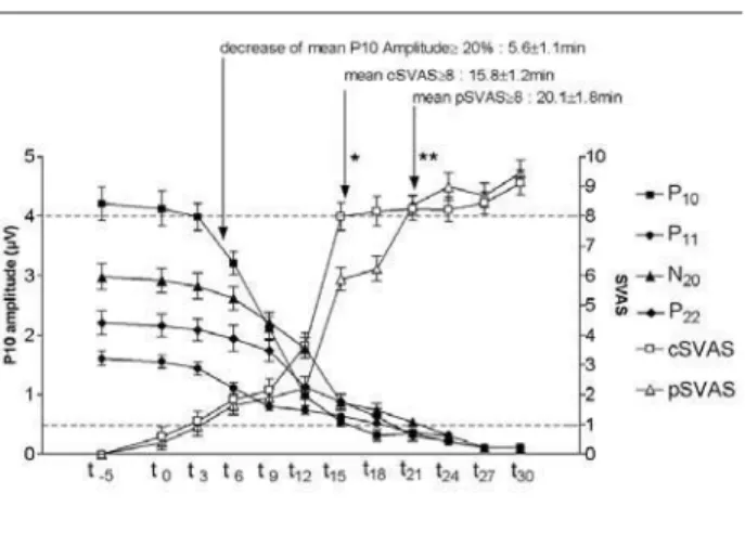
Clinical features: Four MnSSER derivations (Erb’s point; cuta-neous projection of peripheral end of brachial plexus; posterior neck at C6 level, frontal and controlateral parietal scalp) were monitored and recorded for retrospective analysis. Continuous data acquisition were started before ropivacaine injection (baseline) and maintained for 30 min thereafter. Every three minutes after ropivacaine injection, cold and pinprick tests were performed in the hand median nerve cutaneous supply zone and were assessed using a sensory visual score (varying from 0–10). Data were compared using analysis of variance. Although MnSSER values were stable during baseline period, after ropiva-caine administration, severe progressive amplitude depressions of selected MnSSER were detected in every patient. While clinical cold and pinprick tests became positive (score > 8) only 15.8 ± 1.2 min and 20.1 ± 1.8 min respectively after ropiva-caine administration, the mean time to observe the earliest MnSSER 20% amplitude decrease at Erb’s point derivation was reduced to 5.6 ± 1.1 min (P < 0.01).
Conclusion: Selected MnSSER amplitude reduction indicates objectively the onset of median nerve anesthesia following infraclavicular brachial plexus block before the appearance of clinical signs.
Objectif : Les modifications des potentiels évoqués somesthésiques du nerf médian (PESNM) induit par un block infraclaviculaire (40 mL de ropivacaine 0,5 %) on été comparés avec les tests cliniques usuels (tests au froid et à la piqûre) chez 20 patients adultes ASA I afin d’évaluer la différence d’évolution et de survenue des signes électrophysiologiques et cliniques, l’objectivité et la reproductibilité des modifications des PESNM et finalement le possible intérêt en pratique clinique.
Eléments cliniques : Les PESMN étaient enregistrés en continu au point d’Erb (projection cutanée de l’extrémité latérale du plexus brachial) et successivement au niveau cervical en regard de la six-ième vertèbre), frontal et pariétal controlatéral pour une comparai-son secondaire. L’enregistrement a commencé avant l’injection de ropivacaine et jusqu’à 30 min après. Toutes les trois minutes après la réalisation du bloc, les tests cliniques étaient réalisés au niveau de la main d’innervation du nerf médian en utilisant une échelle visuelle analogique (0–10). Une analyse de variance a été utilisée pour comparer les résultats. Si les PESNM étaient remarquable-ment stables avant la réalisation du bloc, après leur amplitude diminue précocement et de façon importante, chez tous les sujets. Alors que les tests au froid et à la piqûre peuvent être considérés comme positifs (> 8 sur l’échelle) seulement après 15,8 ± 1,2 min et 20,1 ± 1,8 min respectivement après l’injection de ropivacaïne, la diminution la plus précocement détectable de l’amplitude du potentiel (20 %) au point d’Erb, survenait en moyenne en 5,6 ± 1,1 min (P < 0,01).
Conclusion : La diminution de l’amplitude des PESNM indique objectivement la survenue du bloc du nerf médian après la réalisa-tion d’un bloc infraclaviculaire avant toute modificaréalisa-tion clinique.
CAN J ANESTH 2006 / 53: 1 / pp 67–72
Somatosensory evoked potentials as an objective
assessment of the sensory median nerve blockade
after infraclavicular block
[Les potentiels évoqués somesthésiques comme évaluation objective du bloc sensitif
du nerf médian après un bloc sous-claviculaire]
Pierre Pandin MD,* Jean Corentin Salengros MD,* Alain d’Hollander MDPhD,† Christiane Tchekap MD,*
Arlette Vandesteene MDPhD*
From the Department of Anesthesiology,* Erasmus Hospital, Free University of Brussels, Brussels, Belgium; and the Division of Anesthesiology,† Cantonal University Hospital, Geneva, Switzerland.
Address correspondence to: Dr. Pierre C. Pandin, Department of Anesthesiology and Intensive Care, Erasmus Hospital, Lennik Drive 808,
B-1070 Brussels, Belgium. Phone: +32-2-555-39-19; Fax: +32-2-555-43-63; E-mail: ppandin@ulb.ac.be
Assessed January 19, 2005
Accepted for publication June 23, 2005. Revision accepted July 19, 2005.
C
LINICAL assessment of the onset and extent of sensory block during regional anesthesia can be a challenging task. The problem of subjectivity in both percep-tion and expression of sensory block is unresolved, justifying the proposal of an “ideal multimodal test-ing sequence” based on recently compiled methods.1Swelling illusion2 can be an alternative, but
unfortu-nately is sometimes difficult to understand by some patients. Somatosensory evoked potential (SSEP) monitoring either for peripheral nerve block3–6 and iv
regional anesthesia7 or for epidural anesthesia8 has the
potential to provide an objective assessment of nerve block. Nevertheless, preliminary results cannot neces-sarily be extrapolated to clinical practice. Our study was thus designed: first, to describe the effect of ropi-vacaine, on the brachial plexus using the infraclavicu-lar approach, guided by median nerve somatosensory evoked responses (MnSSER) and second, to monitor clinically the sensory visual analogue score (SVAS) variations2 in response to cold and pinprick tests
applied in the hand’s median nerve cutaneous supply zone. We postulated that the median nerve blockade onset in the brachial plexus can be anticipated by quantitative variations of MnSSER recordings. Material and method
Patient selection
After giving written informed consent, nine males and 11 females non obese patients (18–42 yr old, ASA class I), were enrolled in this study approved by the Local Authors Institutional Human Investigation Committee (Erasmus Hospital in Brussels, Belgium). After alprazolam 0.5 mg po given one hour before anesthesia, no other sedative or analgesic drug was used before, during or after the block initiation until study completion.
Median nerve somatosensory evoked response recordings Before baseline and for 30 min after the ropivacaine administration, the median nerve was stimulated at the wrist (frequency 1 Hz and duration 0.2 msec) using the electrical stimulator of an Epoch 2000™ (Axon Systems Inc, San Francisco, CA, USA). The stimula-tion intensity was decreased by 2 mA when the evoked muscular twitches were observed (between 12 and 17 mA). Somatosensory evoked responses were recorded from sc needle electrodes at Erb’s point (cutaneous projection of the peripheral end of the brachial plexus 2–3 cm above the clavicle), designated as “Erb”; from the cervical spine (just above the cutaneous projection of the spinous process of the sixth cervical vertebrae) designated as “C6” - and at the scalp, at the median
prefrontal level (Fpz)- designated as “Fpz” and at the contralateral parietal level (P’4) - designated as “P’4”. A common reference electrode (positive pole) was positioned on the ipsilateral ear designated as “A”. All electrode impedances were automatically checked for values less than 5 kOhms. The recording frequency bandpass was 30 to 750 Hz and the sampling window 50 msec. Artifact rejection threshold was fixed at 25 µV. The 50 Hz notch filter was active.
Median nerve somatosensory evoked responses data acquisition methodology
Monitored MnSSER were respectively: P10, a peak present 10 ± 0.5 msec after stimulation, in A-Erb derivation; P11, at 11 ± 0.5 msec in the A-C6 deriva-tion; N20, at 20 ± 0.5 msec in the controlateral A-P’4 derivation and P22, at 22 ± 0.5 msec in the A-Fpz derivation. Before acquisition of baseline measure-ments, three groups of 500 averaged responses were required to assess a good visual reproducibility of evoked potential recordings. Triplicate MnSSER base-line measurement acquisitions were then performed approximatively five minutes prior to the ropivacaine injection. The average of the amplitude and latency values obtained (labelled “t-5”) were used as refer-ence for subsequent comparison. Immediately after the ropivacaine injection (labelled “t0”) and during the next 30 min, the averaging technique was applied continuously to obtain the MnSSER responses every 100 sweeps (MnSSER every 15–20 sec). The pre- and post-ropivacaine administration previously defined signals were automatically saved for retrospective analysis from each recording site. Besides manual retrospective analysis, the automatic tracker software of our monitoring system was able to detect, from baseline values, a 20% variation either in amplitude or in latency, widely accepted as the correct threshold for significant changes. The maximal electrophysiological effect, based on a non-automated analysis by a blinded observer, was defined as an evoked response amplitude of less than 0.5 µV present in three successive averag-ing periods.
Infraclavicular brachial plexus block
The infraclavicular block was performed following the Raj’s approach.9 After local anesthesia, a 10-cm long,
short bevel insulated needle (Stimuplex ATM, BBraun
Inc., Melsungen, Germany) connected to the nega-tive pole of a nerve stimulator (Stimuplex HNS11TM
BBraun Inc., Melsungen, Germany - 100 µsec pulse width, 1.5 mA initial current intensity at 1 Hz), was inserted. After median nerve location (muscular twitches corresponding to wrist, second and third
finger flexion and forearm pronation), with a minimal stimulation current intensity of 0.5 mA, 40 mL of ropivacaine 0.5% were injected.
Clinical assessment of sensory blockade
After ropivacaine administration, cold and pinprick tests were performed every three minutes to assess anesthesia on the palmar face of the base of the sec-ond and third fingers (median nerve cutaneous supply zone). The patient documented his/her perceptions according to a SVAS, similarly to the Benzon’s meth-od.3 The scale consisted of a 10-cm line, where the 0
cm mark stated “my cutaneous zone is not numbed at all”, while 10 cm corresponded to “my cutaneous zone is completely numbed”; scores less than 1 were matched as a “no block” whereas those higher than 8 as “a good and effective block”. The latter score was considered adequate for quatifying the clinical effectiveness of the block. Following cold or pinprick stimulation, we obtained cold SVAS (cSVAS) or pin-prick SVAS (pSVAS), respectively.
Data analysis
Mean values of amplitude and latency of each MnSSER (P10, P11, N20 and P22) at time t-5 where considered as baseline values. The MnSSER waves, registered before and immediately after each SVAS clinical
assess-ment performed every three minutes from t0 to t30, were extracted from the raw data and averaged. The parameters produced every three minutes after ropi-vacaine administration therefore consisted of mean MnSSER amplitudes and latencies, cSVAS and pSVAS values.
For statistical analysis, one way and repeated mea-sures ANOVA tests were used (Instat 3.05TM and
Prism 3.03 TM softwares - Graph Pad Software Inc,
San Diego, CA, USA).
The analysis permitted: 1) evaluation of individual stability and reproducibility of the baseline MnSSER by comparison of the respective amplitudes and laten-cies of baseline evoked potentials from the three initial averages of 500 sweeps to confirm the initial on-line visual inspection of the profiles of the different waves; 2) testing the existence or not of a possible bias induced by the physical effect of the 40 mL anesthesia solution injection, by comparing the mean amplitudes and latencies of each monitored MnSSER at baseline and t0 (just after the injection) and; 3) the time evolu-tion of amplitude and latency of each MnSSER and the two SVAS scores. The results are presented as mean ± SD. A P value < 0.05 was considered statisti-cally significant.
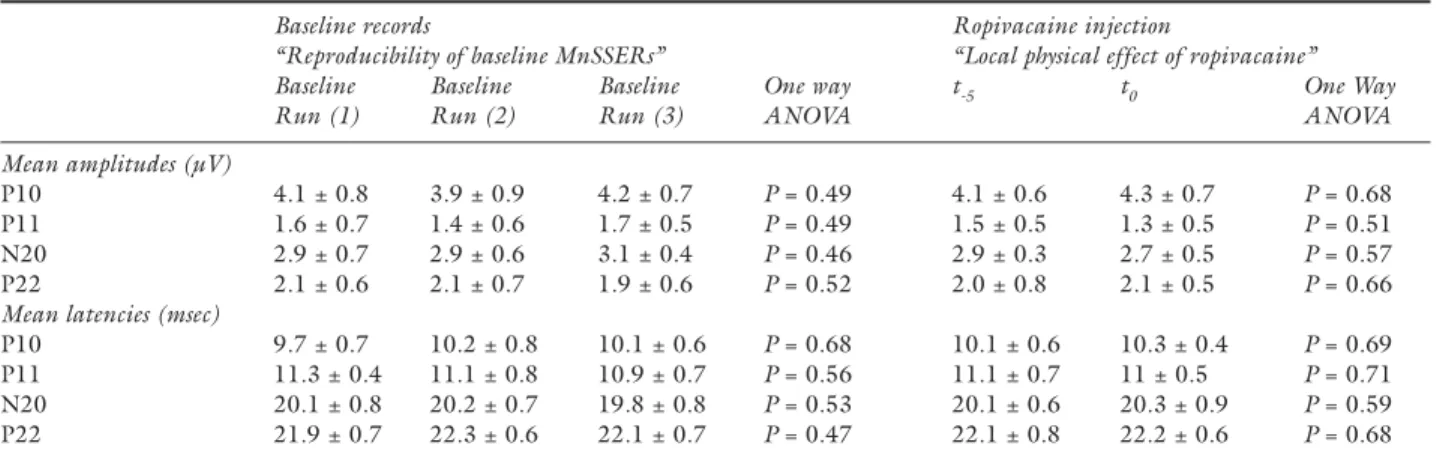
TABLE I Somatosensory evoked responses before and after ropivacaine
Baseline records Ropivacaine injection
“Reproducibility of baseline MnSSERs” “Local physical effect of ropivacaine” Baseline Baseline Baseline One way t-5 t0 One Way Run (1) Run (2) Run (3) ANOVA ANOVA Mean amplitudes (µV)
P10 4.1 ± 0.8 3.9 ± 0.9 4.2 ± 0.7 P = 0.49 4.1 ± 0.6 4.3 ± 0.7 P = 0.68
P11 1.6 ± 0.7 1.4 ± 0.6 1.7 ± 0.5 P = 0.49 1.5 ± 0.5 1.3 ± 0.5 P = 0.51
N20 2.9 ± 0.7 2.9 ± 0.6 3.1 ± 0.4 P = 0.46 2.9 ± 0.3 2.7 ± 0.5 P = 0.57
P22 2.1 ± 0.6 2.1 ± 0.7 1.9 ± 0.6 P = 0.52 2.0 ± 0.8 2.1 ± 0.5 P = 0.66 Mean latencies (msec)
P10 9.7 ± 0.7 10.2 ± 0.8 10.1 ± 0.6 P = 0.68 10.1 ± 0.6 10.3 ± 0.4 P = 0.69
P11 11.3 ± 0.4 11.1 ± 0.8 10.9 ± 0.7 P = 0.56 11.1 ± 0.7 11 ± 0.5 P = 0.71
N20 20.1 ± 0.8 20.2 ± 0.7 19.8 ± 0.8 P = 0.53 20.1 ± 0.6 20.3 ± 0.9 P = 0.59
P22 21.9 ± 0.7 22.3 ± 0.6 22.1 ± 0.7 P = 0.47 22.1 ± 0.8 22.2 ± 0.6 P = 0.68
Median nerve somatosensory evoked responses (MnSSERs) during baseline records (four columns on right including ANOVA compari-son) and before and after the ropivacaine injection (three columns on left including ANOVA comparicompari-son). Results are expressed as mean values ± standard deviation. For the baseline records, mean amplitude and latency values of each MnSSER have been calculated from the respective 20 individual values. The statisqtical comparison of these averages relates the stability and the reproducibility of these evoked potentials. For the ropivacaine injection physical effect, the mean amplitudes and latencies of the respective waves prior to the injection (t
-5) correspond to the average of the three successive baseline mean values (baseline run 1–3). They have been considered as baseline values
for further comparison. Just after the ropivacaine injection (t0), mean amplitudes and latencies have been calculated from the respective 20 individual values at t0. The statistical comparison between t-5 and t0 demonstrates the lack of local physical effect of the ropivacaine solu-tion on the MnSSER recordings.
Results
Regional anesthesia procedure
Infraclavicular block was successfully performed in all 20 patients by five different anesthesiologists. The neural blockade obtained was effective in the median nerve supply, as well as regions of the ulnar and the radial nerves, allowing the scheduled surgery. The mean infraclavicular block duration was 6.2 ± 0.6 min (time elapsed between local anesthesia and end of the ropivacaine injection). During the study period, the plexus block failed to occur (no median nerve block) in three patients from the 20 included, in spite of the correct median nerve stimulation. In these three patients, no changes were recorded. Our comparison between electrophysiological and clinical assessment of the nerve block was based only on successful blocks. Electrophysiological signal variations
The mean time to install the MnSSER equipment set and electrode insertion was 5.3 ± 0.4 min.
Values and reproducibility of baseline measurements Before ropivacaine administration, mean MnSSER amplitudes and latencies recorded in three consecutive runs were stable (one-way ANOVA; NS) and docu-mented normal baseline numerical values (Table I). Lack of immediate physical effects of 40 mL ropiva-caine administration
Comparison of mean amplitude and latency values of P10, P11, N20 and P22 before and just after anes-thetic solution injection did not demonstrate any dif-ference (Table I) whatever the MnSSER considered (one way ANOVA, NS).
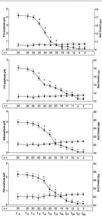
FIGURE 1 Changes of median nerve somatosensory
evoked responses (MnSSER; P10, P11, N20, P22) ampli-tudes (black squares) and latencies (black triangles) over time. The successive amplitude values of each MnSSER were compared to the respective initial one (baseline) using one way ANOVA (*P < 0.05). The number of patients with recordable amplitude signal (> 0.5 µV) is indicated in the rectangular area located at the bottom of each figure.
TABLE II Sensory visual analogue scores
Delays (min) One way ANOVA
P10 5.6 ± 1.1 -P11 5.8 ± 1.3 P = 0.60 N20 7.6 ± 1.3 P = 0.021 P22 8.4 ± 1.4 P = 0.026 cSVAS 15.8 ± 1.2 P = 0.0081 pSVAS 20.1 ± 1.8 P = 0.0093
SVAS = sensory visual analogue scale. Mean delays for the record-ing of a detectable and readable automatic 20% amplitude decrease for P10, P11, N20, P22 and the observation of equal or greater than 8 cSVAS and pSVAS (left column; results are presented as mean value ± SD). For statistical analysis (right column), the
dif-ferent delays observed were all compared to the delay observed for P10.
Median nerve somatosensory evoked responses amplitude and latency evolution after ropivacaine administration Figure 1 depicts the evolution of mean amplitude and latency of the MnSSER over the time from base-line (t-5) until 30 min after local anesthetic injection. Independently from the MnSSER, mean amplitudes have decreased progressively and significantly until they became very often undetectable. A significant reduction in peripheral MnSSER (P10 and P11) was observed faster than in central ones (N20 and P22 - Table II and Figure 1). Table II summarizes the time elapsed after ropivacaine administration and the stan-dard 20% amplitude decrease observed at P10, P11, N20 and P22. N20 and P22 alteration times were different compared to the P10 delay (P = 0.021 and P = 0.026, one way ANOVA, respectively).
Sensory visual analogic score and clinical block
Assessment of the mean time elapsed for confirming the clinical signs of a valuable median nerve block (cor-responding to a cSVAS or a pSVAS score ≥ 8) showed mean delays of 15.8 ± 1.2 min for cold and after 20.1 ± 1.8 min for pinprick tests (Table II and Figure 2). Comparison between MnSSER and SVAS
The differences between the delays documented with MnSSER recordings and the clinical criteria for establishment of median nerve block are presented in Table II (for comparison between P10 and cSVAS or a pSVAS). A difference of about ten minutes was observed between the P10 20% decrease time, and cSVAS (P = 0.0081, one way ANOVA); for pSVAS,
the mean supplementary waiting delay increased to 14 min (P = 0.0093, one way ANOVA).
Discussion
Somatosensory evoked potentials are effective to qual-ify and/or quantqual-ify the occurrence and time course of electrophysiological changes associated with neural block induced by local anesthetics.3–8 Nevertheless,
previous studies have not been able to identify direct clinical applications. Consequently, the goal of our study was to describe the MnSSER alterations indicat-ing a successful median nerve block of the brachial plexus by adapting Benzon’s3 methodology in surgical
patients. The median nerve was preferred because the ease of its location by the infraclavicular approach10
using a single injection technique, verifying optimal position of the needle tip near the brachial plexus.9
Consistent with other published data,3,7 no
sig-nificant changes in the latencies were observed, but depression of MnSSER amplitude in the diverse chan-nels appears as a good indice for detecting the onset and the progression of a chemically induced neural blockade. In the present context, (quantification with a possible anticipation of a potentially successful neural blockade), it seems evident that limited ampli-tude reduction (by 20%) is a good clinical indicator. Each time a 20% amplitude reduction is observed, the MnSSER will eventually disappear, indicating effective median nerve blockade. The 20% value was chosen because the default tracking method of our moni-toring system permitted an automatic detection of MnSSER alterations. However, for a human observer, the depression of amplitude and its time course can be more easily noticed and detected on the periph-eral MnSSER collected at Erb’s point, as their initial amplitudes (3–5 µV) are always higher than the ones recorded more centrally (< 2.5–3 µV). On the Erb’s point derivation, the detection occurred during the early phase (ranging between three to six minutes) of the ropivacaine induced block.
An unresolved issue is whether or not SSEP assessment of one brachial plexus nerve is sufficient to conclude that the plexus block will be complete. Based upon our experience, we think “no”. Although MnSSER can detect objectively the early occurrence of an effective brachial plexus block compared to clini-cal assessment, the correlation between median nerve location and plexus anatomy is variable. Accordingly, a more systematic SSEP investigation of median, ulnar and radial nerves during brachial plexus block tech-niques (not only infraclavicular, but also axillary, inter-scalene and subclavicular) may simplify the technique and clarify its usefulness in clinical practice.
FIGURE 2 Time evolution of the cSVAS (white square) and pSVAS (white triangle). Lower horizontal dot line matches for threshold of clinically absent nerve block (score < 1), while the upper one corresponds to the presence of an effective block (score > 8).
In conclusion, MnSSER monitoring in adult patients permits objective quantification of the onset of clinical median nerve blockade after 40 mL of 0.5% ropivacaine for brachial plexus blockade. The P10 amplitude decrease is always associated with a clini-cally successful block, and appears about ten minutes earlier than the clinical confirmation of median nerve block. This electrophysiologically-based, pain-free monitoring may be important in clinical anesthesia, and provides a method for studying more objectively the clinical action of local anesthetic solutions. References
1 Curatolo M, Petersen-Felix S, Arendt-Nielsen L. Sensory assessment of regional analgesia in humans: a review of methods and applications. Anesthesiology 2000; 93: 1517–30.
2 Paqueron X, Gentili ME, Willer JC, Coriat P, Riou B. Time sequence of sensory changes after upper extrem-ity block: swelling sensation is an early and accurate predictor of success. Anesthesiology 2004; 101: 162–8. 3 Benzon HT, Toleikis JR, Shanks C, Ramseur A, Sloan T. Somatosensory evoked potential quantification of ulnar nerve blockade. Anesth Analg 1986; 65: 843–8. 4 Richardson J, Jones J, Atkinson R. The effect of thoracic
paravertebral blockade on intercostal somatosensory evoked potentials. Anesth Analg 1998; 87: 373–6. 5 Murphy BA, Haavik Taylor H, Wilson SA, Knight JA,
Mathers KM, Schug S. Changes in median nerve soam-tosensory transmission and motor output following transient deafferentation of the radial nerve in humans. Clin Neurophysiol 2003; 114: 1477–88.
6 Weiss T, Miltner WH, Liepert J, Meissner W, Taub E. Rapid functional plasticity in the primary somatomotor cortex and perceptual changes after nerve block. Eur J Neurosci 2004; 20: 3413–23.
7 Lang E, Erdmann K, Gerbershagen HU. Median nerve blockade during diagnostic intravenous regional anesthesia as measured by somatosensory evoked potentials. Anesth Analg 1993; 76: 118–22.
8 Benzon HT, Toleikis JR, Dixit P, Goodman I, Hill JA. Onset, intensity of blockade and somatosensory evoked potential changes of the lumbosacral dermatomes after epidural anesthesia with alkalinized lidocaine. Anesth Analg 1993; 76: 328–32.
9 Raj PP, Montgomery SJ, Nettles D, Jenkins MT. Infraclavicular brachial plexus block - a new approach. Anesth Analg 1973; 52: 897–904.
10 Borene SC, Edwards JN, Boezaart AP. At the cords, the pinkie towards: interpreting infraclavicular motor responses to neurostimulation. Reg Anesth Pain Med 2004; 29: 125–9.