CASE REPORT
Interdisciplinary endoscopic assisted surgery of a patient
with a complete transorbital intracranial impalement
through the dominant hemisphere
Jan-Karl Burkhardt&David Holzmann&Lisa Strobl&
Christoph M. Woernle&Martina M. Bosch&
Spyros S. Kollias&Robert Reisch
Received: 5 December 2011 / Accepted: 28 December 2011 / Published online: 10 March 2012 # Springer-Verlag 2012
Introduction
Transorbital injuries are common and represent about 24% to 45% of all penetrating head injuries in adults and children [1]. However, only <1% of the cases show a skull- or brain-penetrating route with consecutive intracranial injuries [1,2]. The direction and orbital entry route depends on the velocity of the impalement and the trajectory of the foreign body. In
low-velocity injuries, the foreign body may be directed by the configuration of the orbital walls and enters most likely through the superior and inferior orbital fissures or the orbital canal [3]. In cases of higher-velocity impalements, the orbital wall may directly fracture following a perpendicular trajectory line. Here, we describe the first case in the literature of a complete impale-ment injury through the dominant hemisphere entering the inner orbital angle, fracturing the medial aspects of the skull base and leaving the skull contralaterally through the occipital bone, treated successfully in interdisciplinary cooperation via a combined transnasal–transcranial approach.
Case report
Patient history and clinical examination
A 4-year-old left-handed boy was transferred to our depart-ment after a traumatic impaledepart-ment injury through his left eye while playing with building bricks at home. Initially, he presented with a rapid progressive loss of consciousness at the University Children's Hospital, Zurich, with a wooden stick (30×1 cm) from a building set entering the medial angle of his left eye displacing the left globe laterally and inferiorly (Fig.1a–c). The assessment of the left pupil was difficult due to the surrounding swelling but showed normal afferent and efferent function of the pupil. The right eye showed a dilated pupil without reaction to light. The foreign body penetrated through the left superior eyelid.
Imaging
CT scan of the head confirmed the impalement with a transcranial penetrating route through the contralateral side,
J.-K. Burkhardt
:
L. Strobl:
C. M. Woernle:
R. Reisch Department of Neurosurgery,University Hospital, University of Zurich, Zurich, Switzerland
J.-K. Burkhardt
Department of Neurological Surgery,
Weill Cornell Medical College, New York Presbyterian Hospital, New York, NY, USA
D. Holzmann
Department of Otorhinolaryngology,
Head and Neck Surgery University Hospital, University of Zurich, Zurich, Switzerland
M. M. Bosch
Department of Ophthalmology,
University Hospital, University of Zurich, Zurich, Switzerland
S. S. Kollias
Department of Neuroradiology,
University Hospital, University of Zurich, Zurich, Switzerland
R. Reisch (*)
Center for Endoscopic and Minimally Invasive Neurosurgery, Clinic Hirslanden,
Witellikerstrasse 40, 8032 Zurich, Switzerland
e-mail: [email protected] Childs Nerv Syst (2012) 28:951–954 DOI 10.1007/s00381-011-1674-8
fracturing the occipital bone (Fig.1). The penetrating route led from the nasal half of the left upper eyelid fracturing the lamina papyracea, the posterior ethmoidal cells to the contra-lateral sphenoid sinus on the right. Here, the wooden foreign body continued penetrating the right cavernous sinus and was in close contact with the cavernous segment of the internal carotid artery. Further posteriorly, the middle cranial fossa was entered and left the right-sided occipital bone after piercing through the temporo-parieto-occipital brain tissue. No major intracranial bleeding was seen on the CT.
Operative course
After transferring the boy to the operating room, interdisciplin-ary endoscopic assisted emergency surgery (neurosurgery, ENT surgery, and ophthalmology) was performed. Since we were uncertain whether the internal carotid artery (ICA) was injured by the foreign body or could be injured by manipulating
the latter, we decided to expose the contact area of the foreign body with the ICA from below and above to guarantee a secure exposure and to be able to manage potential bleeding out of the ICA. First, a transnasal endoscopic approach was performed (D.H.) exposing the ICA in its proximal segment as well as the foreign body from below using a wide right sphenoethmoidec-tomy. To expose the cavernous segment of the ICA and the optic canal, the small sphenoid sinus was widely opened, and the foreign body was seen exactly in the right lateral opto-carotid recess (Fig.2). Thereafter, a pterional craniotomy was performed (R.R.), and the skull base was inspected through the transcranial way. The wooden stick was found entering the middle fossa through the lateral wall of the cavernous sinus. The stick was completely blocked within the skull base and could not be removed. As there was a high potential risk of rupturing adjacent vessels by rough removal of the stick, we decided to cut it into two pieces above the level of the anterior knee of the ICA using a high-speed drill (Fig.2). The anterior
Fig. 1 a–b Preoperative clinical images of the patient with the transorbital intracranial impalement. c–g Axial CT images show the penetrating route of the wooden foreign body (arrow). h CT 3D reconstruction of the skull shows the fractured occipital bone
piece was carefully removed first under continuous transnasal endoscopic monitoring and then by transcranial microscopic monitoring. The simultaneous visualization allowed safe proximal and distal control of the carotid artery; there was no severe bleeding; however, the right optic nerve showed a complete rupture. The right ICA appeared spastic, and only limited blood flow was detected by Doppler sonography. Therefore, after removal of the posterior part of the stick, the pterional craniotomy was enlarged for additional decompres-sion prior to closure. The upper eyelid wound was sutured after inspection by the ophthalmic surgeon (M.M.B.). Postop-erative CT scan showed no severe brain injury. However, CT-angiography revealed a slight displacement and stenosis of the ICA, and we supposed a traumatic fistula. A digital subtrac-tion angiography verified a carotid–cavernous fistula, and an
occlusion of the vessel was performed in light of sufficient collateral supply of the affected dominant hemisphere (S.S.K.) (Fig.3).
Postoperative course
After surgery, the patient was stable and transferred to the pediatric ICU. Intracranial pressure monitoring showed nor-mal values; after weaning and extubation, the patient showed a high-grade left-sided hemiparesis and global aphasia due to the injured dominant right hemisphere. The ophthalmic ex-amination revealed ophthalmoplegia with mydriasis and am-aurosis on the right side, whereas the left eye showed a normal reactive pupil, adequate motility, and an unremarkable fundus. Three months after surgery, the frontoparietal bone was
re-Fig. 3 a Postoperative DS-angiography in the sagittal plane verified a carotid– cavernous fistula. b Occlusion of the right internal carotid artery was performed after sufficient collateral supply of the right dominant hemisphere through the left arterial territory was confirmed
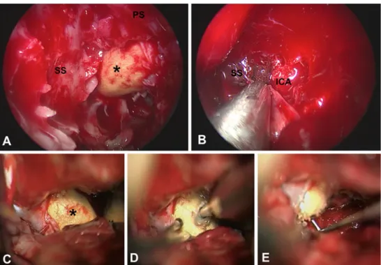
Fig. 2 a–b Transnasal intraoperative endoscopic view: a the wooden foreign body (asterisk) was exposed next to the lateral wall of the right sphenoid sinus (SS) and the planum sphenoidale (PS). b After removal of the wooden foreign body, the ICA was intact without signs of injury. c–d Transcranial intraoperative microscopic view before (c), during (d), and after (e) high-speed drilling of the wooden foreign body (asterisk) in two parts
implanted with an uneventful postoperative course. At 1-year follow-up and after neuro-rehabilitation, the boy showed a nearly complete regression of his hemiparesis with a slight remnant of his motor dysfunction and no impairment in un-derstanding and speech production. The right-sided ophthal-moplegia and mydriasis recovered completely; however, the right amaurosis remained unchanged (Fig.4).
Discussion
In this report, we present a case of a complete transcranial impalement through the patient's dominant hemisphere. Sharp or blunt transorbital impalement injuries are rarely described in children and occur most of the time during playing, as in this case [1,2,4–6]. Due to the thin anatomy of the orbita, where the bone is of least resistance, high-velocity injuries may lead to a continuation of the impalement intracranially.
In our case, the penetrating channel of the wooden foreign body involving the cavernous sinus potentially indicated in-jury of the carotid artery, thus necessitating safe proximal and distal control of a possible intraoperative fatal bleeding; this could be achieved through the combined transnasal–transcra-nial approach by exposing the wooden foreign body from medial and below and from lateral and above, respectively. With this combined approach, the ICA could be exactly dis-played, and a bleeding event would have been precisely controlled, had it occurred.
Endoscopic transnasal skull base surgery for young chil-dren is rarely described in the literature, especially if the cavernous sinus and the cavernous segment of the ICA need to be exposed [7]. We showed for the first time that our interdisciplinary transnasal–transcranial approach is a feasible and secure method to treat a traumatic impalement in a
4-year-old child. Since age-related differences in the anatomy of the skull base may limit endoscopic transnasal surgery in chil-dren, this technique should be performed by experienced endoscopic surgeons [8].
Here, we describe to our knowledge the first case of a complete transcranial penetration of a wooden foreign body, treated successfully via a combined transnasal–transcranial approach with a favorable clinical patient outcome after 1 year.
References
1. Turbin RE, Maxwell DN, Langer PD, Frohman LP, Hubbi B, Wolansky L, Mori M (2006) Patterns of transorbital intracranial injury: a review and comparison of occult and non-occult cases. Surv Ophthalmol 51:449–460
2. Dunn IF, Kim DH, Rubin PA, Blinder R, Gates J, Golby AJ (2009) Orbitocranial wooden foreign body: a pre-, intra-, and postoperative chronicle: case report. Neurosurgery 65:E383–E384, discussion E384 3. Balasubramanian C, Kaliaperumal C, Jadun CK, Dias PS (2009) Trans-orbital intracranial penetrating injury-an anatomical classification. Surg Neurol 71:238–240
4. Lee JS, Lee JE, Oum BS, Cha SH (1999) Orbitocranial injury caused by wood. Korean J Ophthalmol 13:128–132
5. Orszagh M, Zentner J, Pollak S (2009) Transorbital intracranial impalement injuries by wooden foreign bodies: clinical, radiological and forensic aspects. Forensic Sci Int 193:47–55
6. Park SH, Cho KH, Shin YS, Kim SH, Ahn YH, Cho KG, Yoon SH (2006) Penetrating craniofacial injuries in children with wooden and metal chopsticks. Pediatr Neurosurg 42:138–146
7. Kassam A, Thomas AJ, Snyderman C, Carrau R, Gardner P, Mintz A, Kanaan H, Horowitz M, Pollack IF (2007) Fully endoscopic expanded endonasal approach treating skull base lesions in pediatric patients. J Neurosurg 106:75–86
8. Tatreau JR, Patel MR, Shah RN, McKinney KA, Zanation AM (2010) Anatomical limitations for endoscopic endonasal skull base surgery in pediatric patients. Laryngoscope 120(Suppl 4):S229 Fig. 4 a and b Postoperative
clinical images of the patient show the favorable patient outcome with a regression of the left-sided hemiparesis and the right-sided ophthalmoplegia