Magnetic resonance imaging was used to establish the presence and nature of relationships between sulcal asymmetries and mid-sagittal callosal size in neurologically intact subjects, and to determine the influences of sex and handedness. Against a back-ground of long-standing disputes, effects of gender and handedness on callosal size, shape, and variability were additionally examined. Both positive and negative correlations between sulcal asymmetry and callosal size were observed, with effects influenced by sex and handedness. The direction of relationships, however, were depen-dent on the regional asymmetry measured and on whether real or absolute values were used to quantify sulcal asymmetries. Callosal measurements showed no significant effects of sex or handedness, although subtle differences in callosal shape were observed in anterior and posterior regions between males and females and surface variability was increased in males. Individual variations in callosal size appear to outrange any detectable divergences in size between groups. Relationships between sulcal asymmetries and callosal size, however, are influenced by both sex and handedness. Whether magnitudes of asymmetry are related to increases or decreases in callosal size appears dependent on the chosen indicators of asymmetry. It is an oversimplification, therefore, to assume a single relationship exists between cerebral asymmetries and callosal connections.
Introduction
Gender differences in callosal morphology have long been in dispute. Sexual dimorphisms reported from post-mortem and magnetic resonance imaging (MRI) studies include (i) larger total or forebrain volume-adjusted size of the corpus callosum (CC) in females (Holloway and DeLacoste, 1986; Johnson et al., 1994; Steinmetz et al., 1995); (ii) larger raw (Witelson, 1989; Steinmetz et al., 1992; Clarke and Zaidel, 1994) or proportional isthmus areas in females (isthmus relative to total CC) (Witelson, 1989); (iii) larger raw or proportional splenial areas in females (DeLacoste-Utamsing and Holloway, 1982; DeLacoste et al., 1986; Holloway and DeLacoste, 1986; Clarke et al., 1989; Davatzikos and Resnick, 1998); (iv) larger posterior callosal regions in males (Denenberg et al., 1991), and (v) shape differences indicating a more bulbous splenium in female compared to male subjects (DeLacoste-Utamsing and Holloway, 1982; Holloway and DeLacoste, 1986; Clarke et al., 1989; Allen et al., 1991). In addition, when comparing angles determined by lines connecting particular points of the CC and other brain regions, gender differences have been obser ved in the orien-tation of the CC (Oka et al., 1999). Although these findings suggest that sexual dimorphisms exist in callosal morphology, replications have not been consistent across laboratories, for a review see Bishop and Wahlstein (Bishop and Wahlstein, 1997).
Results are similarly mixed concerning relationships between callosal size and handedness. Post-mortem (Witelson, 1985, 1989; Witelson and Goldsmith, 1991) as well as in vivo studies (Denenberg et al., 1991; Habib et al., 1991) have shown smaller
callosal size in right compared with left handers and ambi-dextrous individuals. Gender by handedness interactions, as reported by Denenberg et al. (Denenberg et al., 1991) and Habib et al. (Habib et al., 1991) also appear present where larger callosal anterior midbody regions in right-handed females, left-handed and non-consistent right-left-handed males as well as larger posterior midbody regions in consistent right-handed females have been documented. Several studies, however, have failed to detect differences in callosal morphology in association with hand preference (Kertesz et al., 1987; Steinmetz et al., 1992, 1995). A number of explanations may account for discrepancies in results between laboratories including the composition of brains examined, and differences in the methods employed for measuring callosal subregions. The largest source of discrepancy, however, is likely attributable to the failure of most prior studies to take total brain weight or volume into account.
Cerebral lateralization, in addition to sex and handedness, may also inf luence callosal morphology. For example, if pro- cessing resources were shared between the hemispheres, this would lead to increased requirements for hemispheric com-munication, and thus a positive correlation between functional lateralization and callosal size may be obser ved. However, e.g. Hines and colleagues (Hines et al., 1992) observed a negative correlation between language lateralization and the size of the callosal splenium. This finding lends support to the hypothesis that when the processing takes place solely or dominantly in a single hemisphere, there may be less need for interhemispheric information exchange. This could be related to a decreased callosal size, and therefore a negative correlation may exist between functional lateralization and callosal size. In contrast, others view callosal size as an indicator for hemispheric isolation: the larger the CC, the greater the interhemispheric inhibition, and the more isolated the hemispheres in function (Clarke et al., 1993; Clarke and Zaidel, 1994; Hellige et al., 1998). Additionally, Cook (Cook, 1984a,b,c) proposed a mech- anism for interhemispheric communication in which the pattern of cortical activation in one hemisphere results in ipsilateral surround inhibition and contralateral homotopic inhibition. Therefore, if the CC is assumed to have an inhibitory function, and if we suppose a stronger inhibition in asymmetric brains, we might expect to find positive relationships between CC size and asymmetry measures.
Although functional lateralization has been hypothesized to be linked with the degree of structural asymmetry (Foundas et al., 1994, 1996; Jancke et al., 1994; Schlaug et al., 1995; Steinmetz, 1996; Amunts et al., 1996, 2000), surprisingly few human studies have examined the relationships between structural asymmetries and callosal morphology directly. Based on negative correlations revealed between the density of callosal projections and volumetric asymmetry in the cortex of the rat, Rosen and colleagues (Rosen et al., 1989, 1990) and Galaburda et
Relationships Between Sulcal
Asymmetries and Corpus Callosum Size:
Gender and Handedness Effects
E. Luders1,2, D.E. Rex2, K.L. Narr2, R.P. Woods2, L. Jancke3, P.M. Thompson2, J.C. Mazziotta2and A.W. Toga2
1Institute of Experimental and General Psychology, Otto-von-Guericke-University Magdeburg, Germany, 2Laboratory of Neuro Imaging, Department of Neurology, Center of Brain Mapping, UCLA School of Medicine, Los Angeles, USA and3Department of Neuropsychology, University of Zurich, Switzerland
al. (Galaburda et al., 1990) have proposed that an inverse relationship exists between the magnitude of anatomical cerebral asymmetry and the extent of commissural connections between the relevant cortical areas. Findings from some investigations of the human brain further suggest that such an inverse relationship exists. Specifically Aboitiz et al. (Aboitiz et al., 1992b,c) and Zaidel et al. (Zaidel et al., 1995) analyzed statistical associations between the degree of asymmetr y in perisylvian regions [Sylvian fissure (SF) curved length, planum temporale (PT) area and depth] and commissural connections (indexed by the regional number of callosal fibers or by regional callosal size) in post-mortem brains. In these investigations, a negative correlation was obser ved between perisylvian asym-metries and the total numbers of fibers in the isthmus of males, and in the anterior splenium of females (Aboitiz et al., 1992b). Negative correlations between SF and PT asymmetries and the size of the callosal isthmus were observed in males but not in females (Aboitiz et al., 1992c) and between PT asymmetry and the number of small diameter fibers in the isthmus of the CC, again only in male brains (Zaidel et al., 1995). Only one study, to our knowledge, has examined these relationships using in vivo imaging data, where links between midsagittal callosal area and overall cerebral asymmetry (measured from one matched sagittal brain slice in each hemisphere) were assessed (Dorion et al., 2000). Results showed a negative correlation between cerebral asymmetr y and total callosal size in male subjects. A lthough methods differ considerably, all of these studies suggest a sex-dependent decrease in callosal size and fiber number with increasing cerebral asymmetr y. However, not all of these investigations employed brain size corrections to control for differences in the magnitude of asymmetry between genders. Importantly, males are well known to possess larger brains and are thus expected to exhibit larger hemispheric differences. Furthermore, it is possible that post-mortem measurements may be compromised by a non-uniform shrinkage of the CC. Finally, measurements obtained from one brain slice in each hemisphere may be a less precise index of asymmetry and sensitive to the orientation of each brain volume.
In the present study, our first goal was to confirm the presence, and establish the nature of relationships between callosal size and well documented indices of sulcal asymmetries (Thompson et al., 2000; Blanton et al., 2001; Narr et al., 2001; Sowell et al., 2002a). Sulcal asymmetr y was defined as the interhemispheric difference in the surface location of a defined single point representing the anterior, posterior, superior or inferior end of a sulcus. Points rather than linear measures (although highly correlated) were used as asymmetry measures as based on previous analyses showing that points from the sulcal extremes are slightly better predictors of cerebral asym-metry than hemispheric differences in sulcal lengths or heights (Narr et al., 2001). For example, measures of SF posterior extrema are highly correlated with SF horizontal length, and measures of SF superior extrema are highly correlated with measures of SF height. Sulcal extreme points represent a proxy to the positions and extent of the tissue, encompassed by the respective sulci. For example, the posterior extent of the superior temporal sulcus ascending branch represents the angular gyrus. The termination of this sulcus is in the bend of the angular gyrus. It represents a single point that is sufficiently stable to ser ve as a proxy for the position of the angular gyrus. Thus, hemispheric differences in the location of sulcal extrema express the positional or size dependent asymmetr y of a particular brain region. The degree of structural asymmetry has been hypothesized to be linked with functional asymmetr y,
while both structural and functional asymmetry are proposed to be associated with the degree of interhemispheric communication or callosal connectivity. However, given that there is a disagreement in the existing literature concerning whether increased callosal connectivity is associated with more versus less lateralization and that existing empirical data are relatively sparse, our hypotheses were two-tailed.
Asymmetr y measures included sulcal measurements from frontal, temporal and parietal regions. Measures were specific-ally chosen from posterior brain regions established as asymmetric in normal individuals and as important for language processing. Finally, although less widely documented as asym-metric, we included sulcal measures surrounding anterior lan-guage regions from each hemisphere. Anatomical relationships were investigated between sulcal asymmetries and total CC area as well subareas of discrete callosal vertical partitions. To our knowledge, it is the first 3-D in vivo study examining associations between callosal sizes and cerebral asymmetr y using a refined set of sulcal extrema measures. Previous studies have used only a single SF and PT asymmetry measurement or an overall hemispheric asymmetr y measure as based on a single sagittal brain slice in each hemisphere.
As a second goal of this study we set out to examine whether relationships between sulcal asymmetries and callosal size were different in male and female subjects and between right- and left-handed individuals, given that gender, handedness, and their interactions appear to contribute to changes in callosal mor-phology. Furthermore, in light of above-mentioned controversies concerning the inf luences of sex and handedness on callosal morphology, we examined differences in callosal areas between groups defined by biological sex and handedness in a large and age-matched sample to help clarif y previous findings. Anatomical surface based mesh modeling methods (Thompson et al., 1996a,b, 1997; Narr et al., 2000, 2002) were further used to characterize average shape and detailed surface variability differences between groups.
Materials and Methods
Subjects
Subjects without any neurological, psychological or pathological impair-ment (n = 59) from the International Consortium for Brain Mapping (ICBM) database of normal adults were included for study. Groups were matched for biological sex, handedness and imaging site (McConnell Brain Imaging Center at the Montreal Neurological Institute (MNI) of McGill University, the Research Imaging Center (RIC) at the University of Texas Health Science Center at San Antonio, and the Division of Brain Mapping at the University of California, Los Angeles (UCLA) School of Medicine). Handedness was quantified with a Laterality Quotient (LQ) using the Edinburgh Handedness Inventory (EHI) (Oldfield, 1971). LQs that approach –100 represent left-dominant subjects and those that approach 100 represent right-dominant subjects. Only subjects with LQs that represent largely left-dominance and largely right-dominance were included (left handers mean LQ = 78.5, SD = 23.8; right handers mean LQ = 98, SD = 6.1). Cross-dominance was minimized to eliminate a potential confound in the study. UCLA’s ICBM database contained only four qualified handed females accounting for one less female left-handed subject from this site. Overall, there were 15 male right-left-handed subjects (mean age = 23.3 years, SD = 3.9), 15 male left-handed subjects (mean age = 25.3 years, SD = 6.2), 15 female right-handed subjects (mean age = 23.3 years, SD = 3.9), and 14 female left-handed subjects (mean age = 25.4 years, SD = 5.7). A ll subjects gave informed consent according to institutional guidelines.
MRI Acquisition
T1-weighted spoiled grass (SPGR) whole brain MRI scans were obtained for each subject (MNI: 1.5 T Siemens, 176 × 256 × 256 1 × 1 × 1 mm
voxels; RIC: 2 T Elscint, 190 × 256 × 256 1 × 1 × 1 mm voxels; UCLA: 3 T General Electric, 124 × 256 × 256 1.2 × 1 × 1 mm voxels).
Image Preprocessing
Sulcal Asymmetry Measures
To obtain sulcal asymmetry measures, image volumes passed through a number of preprocessing steps. First, image volumes were corrected for signal intensity inhomogeneities (Zijdenbos et al., 1994; Zijdenbos and Dawant, 1994; Sled et al., 1998), and transformed into ICBM 305 stereo-taxic space using an automatic nine-parameter linear transformation (Woods et al., 1998). Using signal intensity information a spherical mesh surface was created and continuously deformed to fit a threshold intensity value which best differentiates extra-cortical cerebrospinal f luid from underlying cortical gray matter (MacDonald et al., 1994). The resulting 3-D cortical surface models from each subject were used to manually outline primary cortical surface sulci and fissures in each hemisphere by two raters blind to group status (Thompson et al., 2000; Sowell et al., 2002a,b). Images volumes in coronal, axial and sagittal planes were available simultaneously to aid decisions when the brain anatomy was ambiguous. Detailed anatomic protocols for delineating cortical anatomy are available at http://w w w.loni.ucla.edu/NCRR/Protocols/ SulcalAnatomy.html and have been previously validated (Blanton et al., 2001; Narr et al., 2001; Sowell et al., 2002a). The reliability for the sulcal delineation procedure was established with an inter-rater error of <2 mm for all sulci (right hemisphere only) of six different randomly selected brains (Sowell et al., 2002a). A fter outlining the cortical surface sulci, we identified the surface locations of defined points representing sulcal openings and/or termination points from anterior, posterior, superior or inferior sulcal extrema [minima or maxima in z (horizontal) or y (vertical) dimensions]. Sulcal measures ref lect distances as measured from the anterior commissure, which was defined as the origin in each brain volume (0,0,0 in x,y,z coordinates).
For the present study, only sulcal parameters exhibiting highly significant hemispheric asymmetries in posterior temporo-parietal regions as identified in both normal and diseased populations (Narr et al., 2001) were used to assess associations with vertically partitioned
midsagittal areas of the CC. These measures were also chosen because they delimit posterior brain regions established as important for language processing. Measures included the posterior and superior extrema of the SF, the anterior extrema of the post central sulcus and the posterior extrema of the superior temporal sulcus ascending branch. Additionally, we examined relationships between callosal areas and sulcal parameters delimiting anterior language regions. These measures included the SF inferior extrema of the anterior horizontal ramus, the SF anterior extrema of the anterior vertical ramus and the sulcus triangularis inferior extrema (Fig. 1).
Callosal Measures
One rater, blind to gender and handedness, delineated the CC in the midsagittal sections of native brain volumes after correcting image volumes for signal intensity inhomogeneities (Zijdenbos et al., 1994; Zijdenbos and Dawant, 1994; Sled et al., 1998) using a 3-D visualization program (Woods, 2001). Midsagittal brain sections were defined by identifying the interhemispheric fissure in the coronal and sagittal planes and confirmed by the presence of the falx cerebri. For inter-rater reliability, the CC from six different randomly selected brains were contoured by two independent investigators. The intraclass correlation coefficient obtained for total CC area was r = 0.99. The midsagittal callosal renderings in native space were reoriented to maximize callosal length and divided into five vertical partitions representing the (i) splenium, (ii) isthmus, (iii) posterior midbody, (iv) anterior midbody and (v) anterior third (Fig. 2). This parcellation scheme was devised by Witelson (Witelson, 1989) based on electron microscopic study of callosal fibers in the macaque, and validated by Aboitz et al. (Aboitz et al., 1992a) in post-mortem analyses as well as by Clarke and Zaidel et al. (Zaidel et al., 1994) in in vivo analyses.
Midsagittal area measures were acquired in mm2for each segment as well as for the entire CC. In order to clarify findings concerning gender specific shape differences of splenial area (DeLacoste-Utamsing and Holloway, 1982; Holloway and DeLacoste, 1986; Clarke et al., 1989; A llen et al., 1991), the splenial extent in the y-dimension (length) was measured in mm.
Finally, to obtain maps of average callosal anatomy and detailed surface variability information, callosal surfaces were delineated in ICBM 305 stereotaxic space as detailed above, and surface-based anatomical
Figure 1. Three-dimensional model of the cerebral cortex showing sulcal landmarks and callosal area measures. Structures are visualized as follows: (1) splenium, (2) isthmus, (3) posterior midbody, (4) anterior midbody, (5) anterior third, (a) posterior extrema of the SF, (b) superior extrema of the SF, (c) SF inferior extrema of the anterior horizontal ramus, (d) SF anterior extrema of the anterior vertical ramus, (e) sulcus triangularis inferior extrema, (f) post central sulcus anterior extrema, and (g) posterior extrema of the superior temporal sulcus ascending branch. Although the SF posterior and superior extrema seem to represent the same extrema in Figure 1, the posterior ending of the SF is represented by an extrema in the vertical dimension, while the superior ending of the SF is represented by a different extrema in the horizontal dimension.
Figure 2. Delineation and partitioning of the midsagittal corpus callosum in T1-weighted MR images. The CC was traced in the most medial brain slices. The partitioning scheme adapted from Witelson (Witelson, 1989), Aboitiz and colleagues (Aboitiz et al., 1992a), and Clarke and Zaidel (Clarke and Zaidel, 1994) was employed to divide callosal areas into the splenium, representing the posterior fifth of callosal area, the isthmus representing two-fifteenths, the posterior midbody and anterior midbody, both representing one-sixth of callosal area; and the anterior third as shown.
mesh modeling methods were applied (Thompson et al., 1996a,b, 1997). For these procedures surface points making up each callosal outline were made spatially uniform in 3-D. Homologous callosal surface points were then matched between subjects within groups defined by biological sex and/or handedness in all three dimensions. The mean and root mean square distance between these sets of points were calculated to provide maps of average callosal shape and variability on a point-by-point basis across the entire surface in standard ICBM 305 space.
Brain Volume
Brain volume was estimated from the cortical surface models described above in raw scanner space. That is, the 3-D cortical surface models were mapped back onto each image volume and masks of brain tissue were obtained. A ny small errors in the masks were corrected manually. A ll extra-cerebral tissue was then removed from the image volumes based on the brain mask to provide measures of intracranial brain volume that included all tissue types (gray matter, white matter, and cerebrospinal f luid) but that excluded the cerebellum.
Statistical Analysis
Corpus Callosum Morphology and the Inf luence of Gender and Handedness
In order to control for the inf luence of total brain volume on midsagittal callosal (sub)areas and splenial length, the CC measures obtained in raw scanner space were residualized by the square of the cubed root (area measures) or by the cubed root (length) of each individual brain volume. For comparisons between groups defined by biological sex and handed-ness we used multivariate analyses of variance, which were followed by univariate analyses when appropriate. Morphometric variables were used as repeated measures in a two (Gender) by two (Handedness) design and included the residualized areas of (i) the total callosum, (ii) the anterior third, (iii) the anterior midbody, (iv) the posterior midbody, (v) the isthmus, (vi) the splenium and (vii) the splenial length as dependent measures. Although the primary objective of this study was to examine differences in callosal size after controlling for individual differences in brain volume, we also performed analyses without taking brain volume into account in order to compare our findings with others in the litera-ture. Using Bonferroni corrections for these tests was considered overly conservative given that MANOVAs already protect against inf lated Type I error (Tabachnick and Fidel, 1996). Finally, differences in total brain volume between groups defined by sex and handedness were examined using standard analyses of variance.
Relationships Between Sulcal Asymmetry and Callosal Area Measures The term ‘sulcal asymmetry’ refers to the interhemispheric difference of the location of defined points representing sulcal extrema. Asymmetry coefficients for sulcal extrema were calculated by subtracting right from left hemisphere sulcal measures (L–R). As described previously, sulcal measures were obtained after brain volumes had been transformed into average 305 stereotaxic space, thus already controlling for brain size contributions to increases in cortical asymmetry indices as may occur between genders. Given that brain size differences for sulcal parameters were controlled, it was therefore unnecessary to divide asymmetr y measures by the sum of left and right hemispheric values to obtain relative
asymmetr y measures. Pearson’s correlation coefficients were used to establish linkages between residual callosal areas and sulcal asymmetry measures.
In previous studies, both absolute and directional asymmetry measures have been used to examine relationships between hemispheric asymmetries and callosal size. To establish which of these approaches was more appropriate, we calculated the exact probability that distributions for asymmetry coefficients from each sulcal measure could have arisen by chance by modeling the data as a binomial distribution using equal probabilities that right or left measures were more extreme. For measures revealing significant deviations (P≤ 0.01) from this directionally unbiased binomial distribution, that is when measures indicated a significant leftward or rightward shift in sulcal asymmetry, we used real values (positive and negative) to examine if leftward or rightward asymmetries inf luence the direction of the correlations with callosal areas. For measures indicating no significant sulcal asymmetry shift in either direction, absolute values were used for correlations between callosal areas and asymmetr y. The results of these tests are presented in the Results section.
To determine the appropriate Bonferroni correction for having performing multiple correlations between sulcal asymmetry measures and callosal area measurements, we examined the relationships between different sulcal asymmetry indices (separately for absolute value and real value asymmetry indices as established above). Measures that were highly correlated were counted as a single measure for the purpose of Bonferroni correction. Correlations between the dependent measures are provided in the Results section. Significance was determined with a two-tailed probability value of alpha less than 0.05.
Results
Corpus Callosum Morphology and the Inf luence of Gender and Handedness
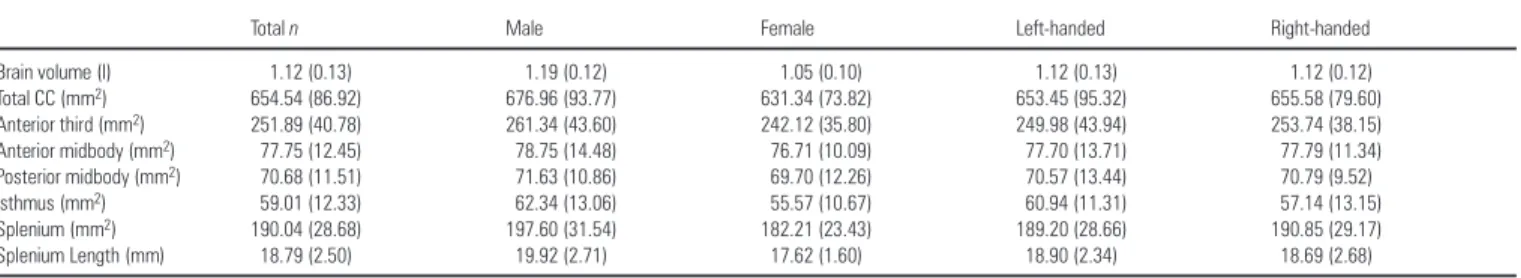
Table 1 shows means and standard deviations for total brain volume and callosal measures obtained in raw scanner space for the overall sample and for groups defined by biological sex and handedness.
Total brain volumes obtained from the present sample of young adults are somewhat smaller in comparison to other post-mortem and in vivo measures as found in the literature given that the cerebellum was excluded when estimating the brain volume. As expected, males possessed significantly larger brain volumes compared with females (P≤ 0.001; F = 29.34). Callosal (sub)regions revealed no significant differences between males and females both before and after brain size correction. Size and shape differences of callosal surface anatomy may be visualized between males and females in Figure 3 where surface averages are superimposed from each group in standard 305 stereotaxic space. As illustrated, differences between males and females appear situated predominantly in anterior and posterior callosal regions, and a subtle shape difference can be identified in the splenial region (although the gender difference of the splenial
Table 1
Means (and SD) of brain volume and areas of the corpus callosum
Total n Male Female Left-handed Right-handed
Brain volume (l) 1.12 (0.13) 1.19 (0.12) 1.05 (0.10) 1.12 (0.13) 1.12 (0.12) Total CC (mm2) 654.54 (86.92) 676.96 (93.77) 631.34 (73.82) 653.45 (95.32) 655.58 (79.60) Anterior third (mm2) 251.89 (40.78) 261.34 (43.60) 242.12 (35.80) 249.98 (43.94) 253.74 (38.15) Anterior midbody (mm2) 77.75 (12.45) 78.75 (14.48) 76.71 (10.09) 77.70 (13.71) 77.79 (11.34) Posterior midbody (mm2) 70.68 (11.51) 71.63 (10.86) 69.70 (12.26) 70.57 (13.44) 70.79 (9.52) Isthmus (mm2) 59.01 (12.33) 62.34 (13.06) 55.57 (10.67) 60.94 (11.31) 57.14 (13.15) Splenium (mm2) 190.04 (28.68) 197.60 (31.54) 182.21 (23.43) 189.20 (28.66) 190.85 (29.17) Splenium Length (mm) 18.79 (2.50) 19.92 (2.71) 17.62 (1.60) 18.90 (2.34) 18.69 (2.68)
length was not identified as significant in the omnibus analysis described above).
Total brain volume and callosal regions did not differ significantly between left- and right-handed subjects before and after brain size correction. Furthermore, no significant inter-actions between gender and handedness were observed for any of these measures. Superimposed surface maps of the CC averaged within right and left handers in 305 space are consistent with statistical analyses where surface averages are almost perfectly aligned, although small differences may exist in splenial regions (Fig. 3).
Corpus Callosum Surface Variability
The surface variability of the CC, shown in Figure 4, appears larger in males than in females for the overall sample and in groups defined by hand preference and sex. In contrast, similar variabilities exist in the CC when groups are divided by hand preference only. In callosal subregions, higher variabilities are seen in all groups in the anterior and posterior borders of the splenium and the anterior third of the CC. The smallest inter-individual differences in females are present in the inferior and superior borders of the isthmus and the posterior midbody as well as in the inferior surface of the CC anterior third. The least variability in males, left handers and right handers can be seen in the inferior surface of the CC anterior third.
Relationships Between Sulcal Asymmetry and Callosal Area Measures
Results from exact binomial modeling revealed significant (P≤ 0.01) leftward or rightward asymmetries for the following
sulcal measures: (i) the superior temporal sulcus ascending branch posterior extrema, (ii) post central sulcus anterior extrema, and (iii) SF posterior extrema. Relationships between the real values of these hemispheric asymmetry measures and callosal areas were thus assessed. All of these variables were significantly correlated (Table 2), and therefore considered as a single hypothesis test. Corrections for multiple comparisons were thus not applied.
Statistical tests assessing the relationships between callosal areas and these sulcal asymmetry measures revealed that when the asymmetr y was biased, increased asymmetries predicted Figure 3. Callosal surface averages mapped in groups defined by sex and handedness.
The average parametric midsagittal mesh models of the CC in the average ICBM 305 stereotaxic space are shown in different colors to illustrate differences between groups with females (n = 29) shown in red, males (n = 30) in blue, right-handed subjects (n = 30) in green, and left-handed subjects (n = 29) in pink. The anterior third is on the left and the splenium on the right.
Figure 4. Callosal surface variability mapped separately in groups defined by sex and handedness. Presented are the (a) overall female sample (n = 29), (b) overall male sample (n = 30), (c) left-handed females (n = 14), (d) left-handed males (n = 15), (e) right-handed females (n = 15), (f) right-handed males (n = 15), (g) overall left-handed sample (n = 29), (h) overall right-handed sample (n = 30). The color bar encodes the root mean square magnitude (in millimeters) of the displacement vectors required to map equivalent surface points from each individual to the group average. The anterior third is on the left and the splenium on the right.
Table 2
Correlations of real value asymmetry indices
STS PSA SFP
STS 1.000 – –
PSA 0.406** 1.000 –
SFP 0.488** 0.526** 1.000
**Significant correlations with a two-tailed probability value of 0.01 (STS = superior temporal sulcus ascending branch posterior extrema, PSA = post central sulcus anterior extrema, SFP = Sylvian fissure posterior extrema).
larger areas of the midsagittal CC. These relationships were significant for the SF posterior extrema and total callosal area, and for the SF posterior extrema and the CC anterior third (for the whole sample and in right handers). The inverse relation-ship, however, also appeared true for some highly asymmetric sulcal measures. That is, greater asymmetry bias and decreases in CC areas were revealed for the post central sulcus anterior extrema and the splenium in males and in left handers (Table 3, Fig. 5).
Measures indicating no significant sulcal asymmetry shift in any direction from exact binomial modeling were found for the (i) SF superior extrema, (ii) sulcus triangularis inferior extrema, (iii) SF anterior extrema of the anterior vertical ramus and (iv) SF inferior extrema of the anterior horizontal ramus. For these
sulcal measures relationships between the absolute values of hemispheric asymmetry and callosal areas were assessed. Again, Bonferroni corrections were not employed for these sulcal asymmetry measures given that all asymmetry indices were sig-nificantly correlated (Table 4).
In males, a significant negative correlation between SF superior extrema asymmetry and callosal isthmus was obser ved. There was also a significant negative correlation between the asymmetr y of the sulcus triangularis inferior extrema and splenium in left handers. In contrast, a positive correlation was found between the asymmetry index of the SF inferior extrema of the anterior horizontal ramus and the anterior midbody in females. Additionally, we obser ved positive correlations be-tween SF superior extrema asymmetry and anterior midbody in right handers, and splenium in females (Table 5).
Discussion
Gender Effects on Corpus Callosum Morphology
Significant differences were not obser ved in comparisons of callosal areas between groups defined by biological sex. These results are consistent with a number of studies that have failed to replicate some early findings of gender specific differences in callosal morphology. As argued by Bishop and Wahlstein (Bishop and Wahlstein, 1997) based on their review of 49 independent studies on the human CC from 1982 to 1994, there is insufficient evidence to support the presence of sex-related differences in the size or shape of the splenium, whether or not adjustments are made for overall brain size. Similarly, our results suggest that the effect of individual variation in callosal size is large enough to outrange any effect of callosal size differences between males and females.
The gender-specific maps of callosal surface anatomy (Fig. 3) demonstrate, however, after correcting for brain size by trans-forming image volumes into average stereotaxic 305 space, that
Table 3
Summary of significant correlations between real value sulcal asymmetry coefficients and callosal areas Region Total n (n = 59) Males (n = 30) Females (n = 29) Left-handed (n = 29) Right-handed (n = 30) Sylvian fissure posterior extrema (SFP)
Total area –0.288*,a – – – –0.399*,a
Anterior third –0.314*,a – – –0.444*,a
Post central sulcus anterior extrema (PSA)
Splenium – 0.361*,b – 0.370*,b –
*Significant correlations with a two-tailed probability value of 0.05.
aNegative r-values reflect associations between increased callosal areas and increased leftward
asymmetries.
bPositive r-values reflect associations between increased callosal areas and decreased leftward
asymmetries.
Figure 5. Relationships between real and absolute sulcal asymmetry measures and callosal size. Examples of correlations between absolute (a, b) and real (c, d) values of sulcal asymmetries (plotted on the x-axes in mm) and callosal regions (plotted on the y-axes in mm2). (a) Relationships between splenium and sulcus triangularis inferior extrema (STI) in left handers (LH). (b) Relationships between anterior midbody and Sylvian fissure inferior extrema of the anterior horizontal ramus (SFI) in females. (c) Relationships between anterior third and Sylvian fissure posterior extrema (SFP) in right handers (RH). (d) Relationships between splenium and post central sulcus anterior extrema (PSA) in males. The solid line marks the significant correlation, the dotted line the non-significant correlation.
Table 4
Correlations of absolute value asymmetry indices
SFS SFI STI SFA
SFS 1.000 – – –
SFI –0.305* 1.000 – –
STI –0.402** 0.650** 1.000 –
SFA 0.049 –0.308* –0.072 1.000
*Significant correlations with a two-tailed probability value of 0.05
**Significant correlations with a two-tailed probability value of 0.01 (SFS = Sylvian fissure superior extrema, SFI = Sylvian fissure inferior extrema of the anterior horizontal ramus, STI = sulcus triangularis inferior extrema, SFA = Sylvian fissure anterior extrema of the anterior vertical ramus).
Table 5
Summary of significant correlations between absolute value sulcal asymmetry coefficients and callosal areas Region Total n (n = 59) Males (n = 30) Females (n = 29) Left-handed (n = 29) Right-handed (n = 30) Sulcus triangularis inferior extrema (STI)
Splenium – – – –0.386* –
Sylvian fissure inferior extrema of the anterior horizontal ramus (SFI)
Anterior midbody – – 0.441* – –
Sylvian fissure superior extrema (SFS)
Anterior midbody – – – – 0.413*
Isthmus – –0.375* – – –
Splenium – – 0.368* – –
small differences between male and female CC are present and are predominantly situated in anterior and posterior callosal regions. These differences might be the result of a somewhat longer CC in females, although this hypothesis was not examined empirically. Maps of callosal surface anatomy may also suggest a slightly more bulbous splenial region in females that might agree with the reported, although controversial, sexual dimorphism of a more bulbous splenium in females (DeLacoste-Utamsing and Holloway, 1982; Holloway and DeLacoste, 1986; Clarke et al., 1989; Allen et al., 1991). This difference, however, was not determined as significant in our analysis of splenial length between groups defined by biological sex.
Handedness Effects on Corpus Callosum Morphology Statistical analyses comparing individuals based on hand prefer-ence did not yield any significant differprefer-ences for all callosal measures before or after brain size correction. These results are in contrast to some post-mortem findings by Witelson (Witelson, 1985, 1989) demonstrating that the CC of non-consistent right-handed males tend to be thicker than those of consistent right-handed males. A more recent study replicated these findings (Witelson and Goldsmith, 1991) and results from some in vivo investigations add further support to these findings (Denenberg et al., 1991; Habib et al., 1991). Notwithstanding, in line with our results, several other MRI studies have failed to detect differences in callosal morphology in relation to handedness (Kertesz et al., 1987; Steinmetz et al., 1992, 1995).
Maps of average callosal surface anatomy in right and left handers (Fig. 3) suggest that diversity exists in the shape of the splenium, but differences were not obser ved statistically. Never-theless, given that splenial shape differences have also been implicated previously between males and females, a more refined measure of splenial shape (in addition to splenial length) may be necessary to finally resolve the presence of both gender and handedness differences in splenial morphology.
Sulcal Asymmetries and Callosal Size
Our study demonstrates that both positive and negative associations exist between sulcal asymmetries in the human brain and callosal area measures. As demonstrated by Aboitiz et al. (Aboitiz et al., 1992a), an increased callosal area indicates an increase in the total number of fibers crossing the CC. Therefore, our findings support the hypothesis that increased asymmetry is associated with increased callosal connectivity, but also support the inverse relationship showing increased asymmetr y is associated with decreased callosal connectivity. Importantly, the direction of these relationships appear inf luenced by the respective local indicators of cerebral asymmetry suggesting that callosal fibers, whether through their primar y or secondar y connections with different cortical regions, subser ve different functions. These findings further suggest that relationships between cerebral asymmetry and callosal size are local in nature. The region-specific direction of associations between cerebral asymmetry and callosal size seems to be plausible given that the degree of structural asymmetry has been hypothesized to be linked with the degree of functional lateralization, and given that different brain functions subserved by different brain regions are processed through different callosal channels and in different ways (e.g. the non-dominant processing hemisphere might or might not receive information from the dominant processing hemisphere). Our data therefore show that it is an oversimplification to assume a general (inversed) relationship between cerebral asymmetry and callosal connectivity.
Previous anatomic studies support that different cortical
regions project through specific callosal channels (DeLacoste et al., 1985). Interestingly, in addition to significant correlations supporting the assumption that cortical areas are topo-graphically mapped in the CC, we also obser ved correlations between posterior callosal regions and anterior cortical asym-metries (e.g. between callosal splenium and sulcus triangularis inferior extrema) and, vice versa, between anterior callosal regions and posterior cortical asymmetries (e.g. between callosal anterior third and SF posterior extrema). Although these find-ings are not immediately understandable, they might lead to the assumption that the nature of callosal pathways is probably much more complex than pictured in present models of inter-hemispheric communication. Existing findings have already suggested that callosal connectivity is more complex than initially assumed. For example, it has been documented, that primar y and secondar y somatosensor y, auditor y, and visual information is transmitted via large diameter callosal fibers, while higher-order sensory and cognitive information is trans-mitted through small diameter fibers (Aboitiz et al., 1992a). A lthough connections across the corpus callosum have been shown to be topographically organized where the relay of sensor y, motor and cognitive information are transmitted from largely homologous regions in the two cerebral hemispheres, there are differential representations of large and small diameter fibers in different callosal channels (Aboitiz et al., 1992a). Anterior callosal regions (connecting frontal brain regions) may be more involved in the transmission of cognitive information, and some middle and posterior connections appear more involved in transmitting sensor y information (Clarke et al., 1998). The isthmus, however, has been demonstrated to connect myelinated fibers from posterior language regions and auditory association areas, and has shown links with the performance for the interhemispheric transfer of auditory stimuli as well as semantic–verbal information that may also ref lect processing capabilities in anterior language regions (Clarke and Zaidel, 1994).
The present results further demonstrate that the direction of the correlations between sulcal asymmetr y and callosal measurements are sensitive to variations in the methods (e.g. whether real or absolute asymmetry values are examined, or whether brain size corrections are employed). Finally, individual differences including sex and handedness are shown to be important when determining the direction, magnitude and significance of correlations between sulcal asymmetry measures and midsagittal callosal size.
Negative Relationships and Absolute Asymmetry Measures Negative correlations suggest that increased asymmetries in frontal and temporo-parietal regions are associated with decreased callosal size. In our study, negative relationships between the absolute asymmetry values of sulcus triangularis inferior extrema and the splenium in left handers, as well as between SF superior extrema and isthmus in males were demonstrated. These results agree with the in vivo findings of Dorion et al. (Dorion et al., 2000), the post-mortem-results of Aboitiz et al. (Aboitiz et al., 1992b,c) and Zaidel et al. (Zaidel et al., 1995) in the human brain, as well as with the findings of Rosen et al. examining asymmetries in rat cortices (Rosen et al., 1989, 1990).
A number of additional post-mortem (Witelson, 1985, 1989; Witelson and Goldsmith, 1991) and in vivo studies (Denenberg et al., 1991; Habib et al., 1991, Hines et al., 1992) assessing relationships between callosal size and hand preference also suggest that right-handed individuals, who are presumably more
lateralized, have smaller callosal size than non-consistent right-handed individuals. If we agree with the hypothesis that anatomical asymmetr y is the substrate of the functional lateralization and callosal size ref lects callosal connectivity, these findings suggest, that increased hemispheric asymmetry occurs with decreased callosal connectivity as already proposed by Rosen et al. (Rosen et al., 1989, 1990) and Galaburda et al. (Galaburda et al., 1990). This assumption is consistent with the findings of negative correlations between sulcal asymmetr y measures and CC areas obser ved in our study as mentioned above. Negative correlations between callosal size and cerebral asymmetr y might be due to a decreased interhemispheric information exchange with increasing cerebral asymmetr y. Based on the assumption that the degree of structural asymmetry is related to the degree of functional lateralization, increased hemispheric independence/dominance may result in less inter-hemispheric communication.
Positive Relationships and Absolute Asymmetry Measures Importantly, in addition to findings supporting inverse relation-ships between the magnitude of sulcal asymmetry measures and callosal size, we found evidence to support that the opposite relationship also applies for some sulcal asymmetry measures. Positive correlations were obser ved between absolute asym-metry values of the SF inferior extrema of the anterior horizontal ramus and the anterior midbody in female subjects, and between SF superior extrema and anterior midbody in right handers, and the splenium in females (Table 5). These findings might suggest, in contrast to some previous reports, that increases in the magnitude of cerebral asymmetries are associated with more, rather than less callosal connectivity. Positive correlations between callosal size and cerebral asymmetry might be due to an increased need of interhemispheric communication with in-creasing cerebral asymmetry in order to relay results from the dominant processing hemisphere to the non-dominant hemi-sphere. On the other hand, it might also be assumed, that an increase in callosal size is related to an increase of inter-hemispheric inhibition. Thus, positive correlations between callosal size and cerebral asymmetr y may arise from a de-creased interhemispheric information exchange with increasing asymmetry. This assumption agrees with former theories under-standing callosal size as an indicator for hemispheric isolation (Clarke et al., 1993; Clarke and Zaidel, 1994; Hellige et al., 1998), or indicative for ‘homotopic inhibition’ as described by Cook (1984a,b,c).
Real Asymmetry Measures
Results revealed that if the asymmetr y of the SF posterior extrema is shifted leftward (typical shift), then the brains showing the greatest shift exhibit larger overall areas of the CC and of the CC anterior third. In contrast, brains with the most extreme asymmetry of the post central sulcus anterior extrema in the typical shifted direction (leftward), tend to have the smallest spleniums. Therefore these results suggest that the direction of cerebral asymmetries (typical vs atypical) inf lu-ences the relationships between sulcal asymmetries and callosal size.
Effects of Sex and Handedness on Relationships Between Sulcal Asymmetries and Callosal Size
In this study, a positive relationship between the asymmetr y index of the SF inferior extrema of the anterior horizontal ramus and the CC anterior midbody, and between SF superior extrema and splenium was observed in females. In contrast, a significant
negative relationship was obser ved between the post central sulcus anterior extrema and the splenium in males if the asymmetr y was biased in the typical direction (leftward). In addition, we found a significant negative correlation between the asymmetry index of the SF superior extrema and isthmus in males. The latter finding agrees with those of Aboitiz et al. (Aboitiz et al., 1992b,c) and Zaidel et al. (Zaidel et al., 1995). Using post-mortem brains, investigators obser ved a negative correlation between perisylvian asymmetries and isthmal area or the total numbers of fibers in the isthmus, in males. Our findings together with those of Dorion et al. (Dorion et al., 2000), Aboitiz et al. (Aboitiz et al., 1992b,c) and Zaidel (Zaidel, 1995) support that sexually dimorphic processes in the brain might inf luence the relationships between cerebral asymmetries and callosal connectivity (Zaidel et al., 1995; Wisniewski, 1998). Never-theless, it may be too speculative to relate our results in detail to interpretations of gender-specific brain organization with regard to our findings of different correlations between sulcal and callosal measures in men and women. However, several studies have documented that male brains may be more lateralized in function (Shay witz et al., 1995; Hiscock et al., 2001) or asymmetric in structure (Jancke et al., 1994; Kulynych et al., 1994; Amunts et al., 2000) than female brains. Furthermore, on average, men appear to possess better spatial and mathematical abilities while women posses better verbal abilities (Kimura, 1999). Sex dependent relationships with respect to the SF inferior extrema, which borders anterior language regions (Broca’s), and the anterior midbody of the corpus callosum, connecting primarily frontal brain regions, may thus ref lect differences in the requirements for interhemispheric com-munication between males and females. It is possible that females, if less functionally asymmetric, may rely on increased interhemispheric communication and thus exhibit stronger relationships between structural asymmetries and callosal size in anterior callosal regions transmitting language related infor-mation.
Our finding of correlations in female subjects, results not observed previously, might ref lect differences in methods with regard to including brain size corrections. Brain size contributes to increases in cortical asymmetry, where the magnitudes of left–right differences are greater in larger brains. Differences in brain size between males and females may therefore have implications for gender differences in cerebral asymmetries. Similarly, relationships exist between callosal size and brain size (Rauch and Jinkins, 1994; Jancke et al., 1997). Brain size corrections are therefore clearly necessary to accurately estab-lish the direction and magnitude of relationships between structural asymmetr y measures and CC size. In addition, relationships between cerebral asymmetries and callosal variables appear dependent on the asymmetry measures used for assessment (area, volume, sulcal extreme, slope, length, height, etc.). This assumption may partially explain why previous studies found only negative relationships between callosal size and hemispheric asymmetr y measures while we found both positive and negative relationships to exist when using seven different sulcal asymmetr y measures from brain regions established as important for language processing. Other possible explanations for discrepancies in results regarding the direction of these relationships may be attributable to measuring fiber density as oppose to callosal area. As visualized in Figure 5, an additional gain of information is revealed by using real left minus right asymmetry measures in contrast to absolute value asymmetry indices, as statements regarding whether leftward or
rightward asymmetry has an inf luence on the direction of the correlation may be interpreted.
Hand preference in addition to biological sex appeared to inf luence the direction and magnitude of relationships between sulcal asymmetry measures and callosal areas. Positive relation-ships between the SF superior extrema and anterior midbody in right handers were exhibited, as well as between the SF posterior extrema, total callosal area, and the CC anterior third in right handers if the asymmetry was biased in the typical direc-tion (leftward). In contrast, we observed a negative correladirec-tion between the sulcus triangularis inferior extrema and splenium in left handers. An additional negative relationship was detected between the post central sulcus anterior extrema and the splenium in left handers if the asymmetr y was biased in the typical direction (leftward). Significant associations between asymmetr y measures and anterior callosal regions (anterior midbody, anterior third) were therefore more prominent in right handers while significant correlations with posterior callosal regions (splenium) were apparent in left handers. These results may suggest a dimorphic organization in the brains of left and right handers inf luencing regionally specific relationships between surface asymmetries and callosal connectivity.
Notes
This work was supported by Deutsche Forschungsgemeinschaft (DFG) JA 737/8-1, National Library of Medicine Grant LM/M H05639, NCRR Grant RR05056, and National Institute of Neurological Disorders and Stroke (NINDS) Grant NS38253, Human Brain Project Grant to the International Consortium for Brain Mapping (P20-MHDA52176), funded by National Institute of Mental Health (NIMH), National Institute on Drug Abuse, National Cancer Institute, and NINDS Grant P20 MH/DA52176, by P41 Resource Grant RR13642 from the NCRR. For generous support, the authors also wish to thank the Brain Mapping Medical Research Organization, the Pierson-Lovelace Foundation, the A hmanson Founda-tion, the Tampkin FoundaFounda-tion, the Jennifer Jones Simon FoundaFounda-tion, and the Robson Family.
Address correspondence to Dr Arthur W. Toga, Laboratory of Neuro Imaging, Department of Neurology, UCLA School of Medicine, 710 Westwood Plaza, 4238 Reed, Los Angeles, CA 90095-1769, USA. Email: toga@loni. ucla.edu.
References
Aboitiz F, Scheibel A B, Fisher RS, Zaidel E (1992a) Fiber composition of the human corpus callosum. Brain Res 598:143–153.
Aboitiz F, Scheibel A B, Fisher RS, Zaidel E (1992b) Individual differences in brain asymmetries and fiber composition in the human corpus callosum. Brain Res 598:154–161.
Aboitiz F, Scheibel A B, Zaidel E (1992c) Morphometr y of the Sylvian fissure and the corpus callosum, with emphasis on sex differences. Brain 115:1521–1541.
A llen LS, Richey MF, Chai YM, Gorski R A (1991) Sex differences in the corpus callosum of the living human being. J Neurosci 11:933–942. A munts K, Schlaug G, Schleicher A, Steinmetz H, Dabringhaus A,
Roland PE, Zilles K (1996) Asymmetry in the human motor cortex and handedness. Neuroimage 4:216–222.
A munts K, Jancke L, Mohlberg H, Steinmetz H, Zilles K (2000) Interhemispheric asymmetry of the human motor cortex related to handedness and gender. Neuropsychologia 38:304–312.
Bishop KM, Wahlsten D (1997) Sex differences in the human corpus callosum: myth or reality? Neurosci Biobehav Rev 21:581–601. Blanton RE, Levitt JG, Thompson PM, Narr KL, Capetillo-Cunliffe L, Nobel
A, Singerman JD, McCracken JT, Toga AW (2001) Mapping cortical asymmetry and complexity patterns in normal children. Psychiatry Res 107:29–43.
Clarke JM, Zaidel E (1994) Anatomical–behavioral relationships: corpus callosum morphometry and hemispheric specialization. Behav Brain Res 64:185–202.
Clarke JM, Luf kin RB, Zaidel E (1993) Corpus callosum morphometry and
dichotic listening performance: individual differences in functional interhemispheric inhibition? Neuropsychologia 31:547–557. Clarke JM, McCann CM, Zaidel E (1998) The corpus callosum and
language: anatomical--behavioral relationships. In: Right hemisphere language comprehension: perspectives from cognitive neuroscience (Beeman M, Chiarello C, eds), pp. 27–50. Mahwah, NJ: Lawrence Erlbaum.
Clarke S, Kraftsik R, Van der LH, Innocenti GM (1989) Forms and measures of adult and developing human corpus callosum: is there sexual dimorphism? J Comp Neurol 280:213–230.
Cook ND (1984a) Callosal inhibition: the key to the brain code. Behav Sci 29:98–110.
Cook ND (1984b) Homotopic callosal inhibition. Brain Lang 23:116–125. Cook ND (1984c) The transmission of information in natural systems. J
Theor Biol 108:349–367.
Davatzikos C, Resnick SM (1998) Sex differences in anatomic measures of interhemispheric connectivity: correlations with cognition in women but not men. Cereb Cortex 8:635–640.
DeLacoste-Utamsing C, Holloway RL (1982) Sexual dimorphism in the human corpus callosum. Science 216:1431–1432.
DeLacoste MC, Kirkpatrick JB, Ross ED (1985) Topography of the human corpus callosum. J Neuropathol Exp Neurol 44:578–591.
DeLacoste MC, Holloway RL, Woodward DJ (1986) Sex differences in the fetal human corpus callosum. Hum Neurobiol 5:93–96.
Denenberg VH, Kertesz A, Cowell PE (1991) A factor analysis of the human’s corpus callosum. Brain Res 548:126–132.
Dorion A A, Chantome M, Hasboun D, Zouaoui A, Marsault C, Capron C, Duyme M (2000) Hemispheric asymmetr y and corpus callosum morphometry: a magnetic resonance imaging study. Neurosci Res 36:9–13.
Foundas AL, Leonard CM, Gilmore R, Fennell E, Heilman KM (1994) Planum temporale asymmetr y and language dominance. Neuro-psychologia 32:1225–1231.
Foundas AL, Leonard CM, Gilmore RL, Fennell EB, Heilman KM (1996) Pars triangularis asymmetry and language dominance. Proc Natl Acad Sci USA 93:719–722.
Galaburda A M, Rosen GD, Sherman GF (1990) Individual variability in cortical organization: its relationship to brain laterality and implications to function. Neuropsychologia 28:529–546.
Habib M, Gayraud D, Oliva A, Regis J, Salamon G, Khalil R (1991) Effects of handedness and sex on the morphology of the corpus callosum: a study with brain magnetic resonance imaging. Brain Cogn 16:41–61. Hellige JB, Taylor KB, Lesmes L, Peterson S (1998) Relationships between
brain morphology and behavioral measures of hemispheric asym-metry and interhemispheric interaction. Brain Cogn 36:158–192. Hines M, Chiu L, McAdams LA, Bentler PM, Lipcamon J (1992) Cognition
and the corpus callosum: verbal f luency, visuospatial ability, and language lateralization related to midsagittal surface areas of callosal subregions. Behav Neurosci 106:3–14.
Hiscock M, Perachio N, Inch R (2001) Is there a sex difference in human laterality? IV. An exhaustive survey of dual-task interference studies from six neuropsychology journals. J Clin Exp Neuropsychol 23:137–148.
Holloway RL, DeLacoste MC (1986) Sexual dimorphism in the human corpus callosum: an extension and replication study. Hum Neurobiol 5:87–91.
Jancke L, Schlaug G, Huang Y, Steinmetz H (1994) Asymmetry of the planum parietale. Neuroreport 5:1161–1163.
Jancke L, Staiger JF, Schlaug G, Huang Y, Steinmetz H (1997) The relation-ship between corpus callosum size and forebrain volume. Cereb Cortex 7:48–56.
Johnson SC, Farnworth T, Pinkston JB, Bigler ED, Blatter DD (1994) Corpus callosum surface area across the human adult life span: effect of age and gender. Brain Res Bull 35:373–377.
Kertesz A, Polk M, Howell J, Black SE (1987) Cerebral dominance, sex, and callosal size in MRI. Neurology 37:1385–1388.
Kimura, D (1999) Sex and cognition. Boston, M A: MIT Press.
Kulynych JJ, V ladar K, Jones DW, Weinberger DR (1994) Gender differences in the normal lateralization of the supratemporal cortex: MRI surface-rendering morphometr y of Heschl’s gyrus and the planum temporale. Cereb Cortex 4:107–118.
MacDonald D, Avis D, Evans AC (1994) Multiple surface identification and matching in magnetic resonance imaging. Proc Int Soc Opt Eng 2359:160–169.
(2000) Mapping morphology of the corpus callosum in schizophrenia. Cereb Cortex 10:40–49.
Narr K, Thompson P, Sharma T, Moussai J, Zoumalan C, Rayman J, Toga A (2001) Three-dimensional mapping of gyral shape and cortical surface asymmetries in schizophrenia: gender effects. A m J Psychiatr y 158:244–255.
Narr KL, Cannon TD, Woods RP, Thompson PM, Kim S, Asunction D, van Erp TG, Poutanen VP, Huttunen M, Lonnqvist J, Standerksjold-Nordenstam CG, Kaprio J, Mazziotta JC, Toga AW (2002) Genetic contributions to altered callosal morphology in schizophrenia. J Neurosci 22:3720–3729.
Oka S, Miyamoto O, Janjua NA, Honjo-Fujiwara N, Ohkawa M, Nagao S, Kondo H, Minami T, Toyoshima T, Itano T (1999) Re-evaluation of sexual dimorphism in human corpus callosum. Neuroreport 10:937–940.
Oldfield RC (1971) The assessment and analysis of handedness: the Edinburgh inventory. Neuropsychologia 9:97–113.
Rauch R A, Jinkins JR (1994) Analysis of cross-sectional area measure-ments of the corpus callosum adjusted for brain size in male and female subjects from childhood to adulthood. Behav Brain Res 64:65–78.
Rosen GD, Sherman GF, and Galaburda A M (1989) Interhemispheric connections differ between symmetrical and asymmetrical brain regions. Neuroscience 33:525–533.
Rosen GD, Galaburda A M, and Sherman GF (1990) The ontogeny of anatomical asymmetry: constraints derived from basic mechanisms. In: Neurobiology of higher cognitive function (Scheibel A B, Wechsler AF, eds), pp. 215–238. New York: Guilford Press.
Schlaug G, Jancke L, Huang Y, Steinmetz H (1995) In vivo evidence of structural brain asymmetry in musicians. Science 267:699–701. Shay witz BA, Shay witz SE, Pugh KR, Constable RT, Skudlarski P,
Fulbright RK, Bronen R A, Fletcher JM, Shankweiler DP, Katz L (1995) Sex differences in the functional organization of the brain for language. Nature 373:607–609.
Sled JG, Zijdenbos A P, Evans AC (1998) A nonparametric method for automatic correction of intensity nonuniformity in MRI data. IEEE Trans Med Imaging 17:87–97.
Sowell ER, Thompson PM, Rex D, Kornsand D, Tessner KD, Jernigan TL, Toga AW (2002a) Mapping sulcal pattern asymmetry and local cortical surface gray matter distribution in vivo: maturation in perisylvian cortices. Cereb Cortex 12:17–26.
Sowell ER, Trauner DA, Gamst A, Jernigan TL (2002b) Development of cortical and subcortical brain structures in childhood and adolescence: a structural MRI study. Dev Med Child Neurol 44:4–16. Steinmetz H (1996) Structure, functional and cerebral asymmetr y: in
vivo morphometry of the planum temporale. Neurosci Biobehav Rev 20:587–591.
Steinmetz H, Jancke L, Kleinschmidt A, Schlaug G, Volkmann J, Huang Y (1992) Sex but no hand difference in the isthmus of the corpus callosum. Neurology 42:749–752.
Steinmetz H, Staiger JF, Schlaug G, Huang Y, Jancke L (1995) Corpus callosum and brain volume in women and men. Neuroreport 6:1002–1004.
Tabachnick BG, Fidell LS (1996) Using multivariate statistics, 3rd edn. New York: HarperCollins College Publishers.
Thompson PM, Schwartz C, Lin RT, Khan A A, Toga AW (1996a) Three-dimensional statistical analysis of sulcal variability in the human brain. J Neurosci 16:4261–4274.
Thompson PM, Schwartz C, Toga AW (1996b) High-resolution random mesh algorithms for creating a probabilistic 3D surface atlas of the human brain. Neuroimage 3:19–34.
Thompson PM, MacDonald D, Mega MS, Holmes CJ, Evans AC, Toga AW (1997) Detection and mapping of abnormal brain structure with a probabilistic atlas of cortical surfaces. J Comput Assist Tomogr 21:567–581.
Thompson PM, Woods RP, Mega MS, Toga AW (2000) Mathematical/ computational challenges in creating deformable and probabilistic atlases of the human brain. Hum Brain Mapp 9:81–92.
Wisniewski A B (1998) Sexually-dimorphic patterns of cortical asym-metry, and the role for sex steroid hormones in determining cortical patterns of lateralization. Psychoneuroendocrinology 23:519–547. Witelson SF (1985) The brain connection: the corpus callosum is larger in
left-handers. Science 229:665–668.
Witelson SF (1989) Hand and sex differences in the isthmus and genu of the human corpus callosum. A postmortem morphological study. Brain 112:799–835.
Witelson SF, Goldsmith CH (1991) The relationship of hand preference to anatomy of the corpus callosum in men. Brain Res 545:175–182. Woods RP (2001) Tracer: java based 3D-tracing program (http://
bishopw.loni.ucla.edu/Tracer/Tracer.html). UCLA Brain Mapping Center, Department of Neurology, University of California at Los Angeles School of Medicine, Los Angeles, CA.
Woods RP, Grafton ST, Watson JD, Sicotte NL, Mazziotta JC (1998) Automated image registration: II. Intersubject validation of linear and nonlinear models. J Comput Assist Tomogr 22:153–165.
Zaidel E, Aboitiz F, Clarke J (1995) Sexual dimorphism in inter-hemispheric relations: anatomical–behavioral convergence. Biol Res 28:27–43.
Zijdenbos A P, Dawant BM (1994) Brain segmentation and white matter lesion detection in MR images. Crit Rev Biomed Eng 22:401–465. Zijdenbos A P, Dawant BM, Margolin R A (1994) Automatic detection of
intracranial contours in MR images. Comput Med Imaging Graph 18:11–23.