Review
Nastassja Lewinski*, Halshka Graczyk and Michael Riediker
Human inhalation exposure to iron oxide particles
Abstract: In the past decade, many studies have been
conducted to determine the health effects induced by exposure to engineered nanomaterials (NMs). Specifi-cally for exposure via inhalation, numerous in vitro and animal in vivo inhalation toxicity studies on several types of NMs have been published. However, these results are not easily extrapolated to judge the effects of inhaling NMs in humans, and few published studies on the human response to inhalation of NMs exist. Given the emergence of more industries utilizing iron oxide nanoparticles as well as more nanomedicine applications of superpara-magnetic iron oxide nanoparticles (SPIONs), this review presents an overview of the inhalation studies that have been conducted in humans on iron oxides. Both occupa-tional exposure studies on complex iron oxide dusts and fumes, as well as human clinical studies on aerosolized, micron-size iron oxide particles are discussed. Iron oxide particles have not been described to elicit acute inhalation response nor promote lung disease after chronic expo-sure. The few human clinical studies comparing inhala-tion of fine and ultrafine metal oxide particles report no acute changes in the health parameters measured. Taken together existing evidence suggests that controlled human exposure to iron oxide nanoparticles, such as SPIONs, could be conducted safely.
Keywords: human; inhalation; iron oxide; nanoparticle;
occupational health.
*Corresponding author: Nastassja Lewinski, Institute for Work and Health, University of Lausanne and Geneva, Rte de la Corniche 2, 1066 Epalinges-Lausanne, Switzerland, Phone: +41-(0)21-314-03-19, Fax: +41-(0)21-314-74-20, E-mail: [email protected] Halshka Graczyk and Michael Riediker: Institute for Work and Health, University of Lausanne and Geneva, 1066 Epalinges-Lausanne, Switzerland
Introduction
Nanotechnology is one of the few material technologies that researchers have proactively examined for human health effects in parallel with its development. However, given the complexity of many engineered nanomaterials
(NMs), which are often multi-component structures versus pure materials, researchers have faced challenges in measuring toxicokinetic parameters and interpreting data to determine mechanisms of action. Although much knowledge on the toxicity of NMs has been gained in the past decade, the nanotoxicology research community remains hesitant to answer the public’s question: Are NMs toxic or not? As researchers continue to study this question, human exposures to NMs are occurring through consumer products and occupational exposure. Workers manufacturing and handling NMs will likely be the first subpopulation to exhibit any potential chronic effects due to often daily exposures at the workplace. Inhalation is of significant concern since inhaled particulates are known to induce various respiratory conditions. In addition, com-pared to dermal and oral exposures, inhalation is more likely to result in a systemic effect [1–3]. Due to their small size, NMs can deposit in the lower, gas exchange region of the lungs. Therefore, exposure to high concentrations of airborne NMs could lead to physiologic effects. Particle deposition and biokinetics of NMs in the lungs have been reviewed in depth in recent articles [4, 5].
Although numerous in vitro and animal in vivo inhalation toxicity studies on several types of NMs have been published, these results are not easily extrapolated to judge the effects in humans. Currently, there are few published studies on the human response to inhalation of NMs. Many safety and ethical concerns restrict the possibility of conducting controlled exposure studies in humans. In addition, how should it be determined which NMs should advance to human testing? At the preclinical level, researchers are presented with a daunting number of NMs to test due to varying chemical composition, size, shape, surface characteristics, and dispersion. This only names a few of the major parameters that can be manipu-lated, and the number will increase as our ability to control matter at the nanoscale continues to become more sophis-ticated. To date, nanotoxicology research has focused on NMs with high production levels categorized by chemical composition. In addition, the Organization for Economic Cooperation and Development (OECD) identified fourteen priority NMs for evaluation [6]. These are carbon black,
C60, single and multi-walled carbon nanotubes, Ag, Au, Fe,
Of these, pharmaceutical formulations of Ag, Au, and Fe nanoparticles plus dendrimers have undergone human clinical trials for intravenous (IV) administration [7].
Only iron nanoparticles, specifically superpara-magnetic iron oxide nanoparticles (SPIONs), have been approved by both the US Food and Drug Administration and the European Medicines Agency for IV medical use. To the authors’ knowledge, no controlled human inhala-tion exposure study has been conducted using SPIONs. However, micron-sized bare and radiolabeled iron oxide particles have been used as routine tracer aerosols for magnetopneumography, lung function, and particle clearance measurements. In addition, occupational expo-sure studies on complex iron oxide dusts and fumes pro-duced during industrial processes have been conducted. The potential hazards associated with inhalation of par-ticulates, including nanoparticles, have been discussed extensively in the literature, and the consequences of exposure include the onset of lung disease and systemic effects due to particle translocation [8–10]. This review presents an overview of the inhalation studies that have been conducted in humans on iron oxides particles, with some discussion of in vivo animal inhalation studies using iron nanoparticles, to guide future studies on human inhalation of iron oxide nanoparticles.
Occupational inhalation exposure
to iron oxide particles
Reports on the human health effects due to inhalation of iron oxides date back to 1867 with Zenker suggesting a link between lung fibrosis and iron oxide exposure [11]. The X-ray shadows often observed in iron oxide exposed workers were suggested by Collis in 1923 not to be signs of fibrosis but visualization of retained iron oxide parti-cles in the lungs [12]. More recently, a case study corrobo-rates this retention theory by demonstrating a significant recovery of particles from the lungs as well as a reduction in computerized tomography (CT) contrast in the lungs of a welder after undergoing bronchial alveolar lavage [13]. These reports suggest that inhalation of iron oxide, despite particle retention in the lungs, results in little to no gross adverse health effects. However, an increased incidence of lung disease is associated with workers in occupations involving exposure to iron oxide dusts or fumes. The indus-tries of most concern for human exposure to inhaled iron oxide particles include four distinct and historically rele-vant groups: iron welders, iron foundry workers, iron and steel manufacturers, and iron ore miners. Epidemiologi-cal evidence on exposed cohorts from these four groups
indicates higher risk of lung fibrosis, siderosis, and silico-sis. In addition, iron oxide exposure is suspected to lead to an increased risk of lung cancer for workers in these industries [14, 15]. However, these studies contain several methodological drawbacks, due in part to their retrospec-tive nature, that do not directly correlate iron oxide expo-sure with the observed health effects, perpetuating the uncertainty of a causal relationship.
In response, industrial hygienists included iron oxides when crafting the first list of Threshold Limit Values (TLV), the exposure level that is deemed acceptable over a working lifetime [16]. The limit has changed over time
from the original 1949 TLV of 15 mg/m3 (total particulates)
to 10 mg/m3 (total particulates) in 1964 to 5 mg/m3
(respir-able fraction) in 2006 [17]. While the International Agency for Research on Cancer (IARC) classifies iron and steel founding exposures as Group 1 substances, which are considered carcinogenic to humans, hematite and ferric oxides are listed as Group 3 substances, which are consid-ered not classifiable as a carcinogen to humans [18]. Few human epidemiological studies specifically investigate the risk of cancer in relation to iron oxide exposure. Occu-pational cohort study results are difficult to interpret due to potential confounding with multiple exposures, namely to other potential and proven carcinogens such as PAHs, silica, and formaldehyde [19–21]. Additionally, failure to report on the use of personal protective equipment (PPE), namely respiratory protective devices, makes it impossible to accurately discern differences between workplace con-centration of iron oxide and personal exposure, specifi-cally, the inhaled dose. The following studies summarized in Table 1 on workers in steel factories show inconsistent results and have shared a similar concomitant exposure problem. Bourgkard et al. investigated a cohort of 16,742 males and 959 females employed for at least 1 year in a French carbon steel-producing factory between 1959 and 1997 [14]. Overall, no correlation was determined between iron oxide exposure and mortality from lung cancer rela-tive risk adjusted with asbestos, silica, and polycyclic aro-matic hydrocarbon (PAH) exposures (RR = 0.80; 95% CI, 0.55–1.17). However, this retrospective study suffers from an incomplete exposure assessment for iron oxides. Char-acterization of iron oxide exposure was mostly qualitative with the mineralogy and particle size not reported. The exposure assessment was primarily based on a job expo-sure matrix and historical air monitoring meaexpo-surements performed in the factory. Only total dust concentrations,
30% of which were above 10 mg/m3, were collected from
workplace air measurements. The authors noted that the percentage of iron in total dust ranged from 10% to 50%, and reported quartiles of total iron ranging from 0.18, 0.32
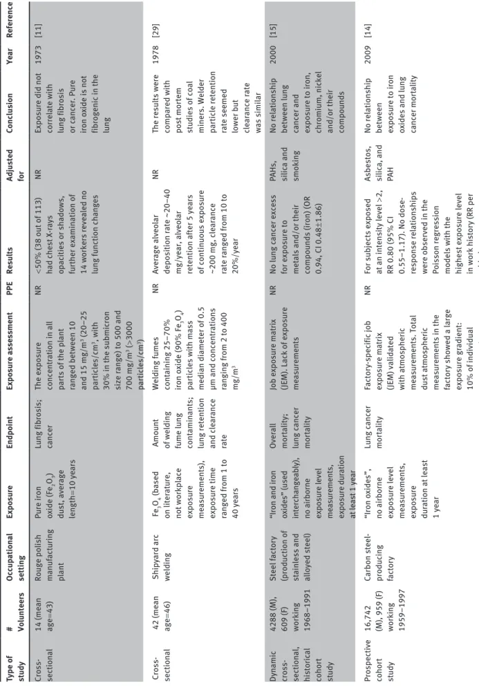
Tab le 1 List of ep idemio logic st udies invo lving hu m an occ up ation al inh al ation of iron o xides. Type of study # Volu nteer s Occ up ationa l setting Expo su re Endpo int Expo su re a sse ss ment PPE Resul ts Ad ju sted for Conc lu sion Ye ar Reference Cross- section al 14 (me an age = 43) Ro uge po lish m anuf act ur ing pla nt Pu re iron ox ide (Fe 2 O3 ) dust , aver age leng th = 10 ye ars Lu ng fibrosis; cancer The e xposu re concent ration in a ll par ts of the p lant ranged between 10 and 15 mg/m 3 (20–25 par tic les/c m 3, w ith 30% in the submic ron size r ange) to 500 and 700 mg/m 3 ( > 3000 par tic les/c m 3) NR < 50% (38 o ut of 113) had chest X -ra ys op ac ities or sh ado ws, fu rther e xamin ation of 14 workers reve aled no lu ng f unction ch anges NR Exposu re did not correl ate w ith lu ng fibrosis or c ancer . P ure iron o xide is not fibrogenic in the lu ng 1973 [11] C ross-section al 42 (me an age = 46) Ship yard arc welding Fe3 O4 (b ased on liter at ure, not workp lace exposu re me asu rements), exposu re time ranged from 1 to 40 ye ars Amo unt of welding fu me lu ng cont amin ants; lu ng retention and cle ar ance rate Welding f umes cont aining 25–70% iron o xide (90% Fe 3 O4 ) par tic les w ith m ass medi an di ameter of 0.5 µm and concent rations ranging f rom 2 to 400 mg/m 3 NR Aver age a lveo lar deposition r ate ~20–40 mg/ye ar , a lveo lar retention af ter 5 ye ars of continuo us e xposu re ~200 mg, c le ar ance rate r anged from 10 to 20%/ye ar NR The resu lts were comp ared w ith post mor tem st udies of co al
miners. Welder partic
le retention
rate seemed lower b
ut cle ar ance r ate was simi lar 1978 [29] Dy namic cross- section al , histor ic al cohor t st udy 4288 (M), 609 (F) work ing 1968–1991 Steel factor y (production of stain less and al lo yed steel) “Iron and iron ox ides” (used interch ange ab ly), no airborne exposu re level me asu rements, exposu re du ration at le ast 1 ye ar Ov era ll mor ta lity; lu ng c ancer mor ta lity Job e xposu re m at rix (JEM). Lack of e xposu re me asu rements NR No lu ng c ancer e xcess for e xposu re to met
als and/or their
compo
unds (iron) (OR
0.94, CI 0.48 ± 1.86) PAHs, silic a and smok ing No rel ationship between lu ng
cancer and exposu
re to iron,
chromiu
m, nickel
and/or their compo
unds 2000 [15] Prospective cohor t st udy 16,742 (M), 959 (F) work ing 1959–1997
Carbon steel- produc
ing factor y “Iron o xides”, no airborne exposu re level me asu rements, exposu re du ration at le ast 1 ye ar Lu ng c ancer mor ta lity Factor y-spec ific job exposu re m at rix (JEM) va lid ated with at mospher ic me asu rements. Tot al dust at mospher ic me asu rements in the factor y sho wed a l arge exposu re gr adient: 10% of individua l me asu rements NR For sub jects e xposed at an intensity level > 2,
RR 0.80 (95% CI 0.55–1.17). No dose- response rel
ationships were observed in the Po isson regression models w ith the highest e xposu re level in work histor y (RR per added Asbestos, silic a, and PA H No rel ationship between exposu re to iron ox ides and lu ng cancer mor ta lity 2009 [14]
Type of study # Volu nteer s Occ up ationa l setting Expo su re Endpo int Expo su re a sse ss ment PPE Resul ts Ad ju sted for Conc lu sion Ye ar Reference and 30% of are a me asu rements were > 10 mg/m 3 level 0.98, 95% CI 0.87 to 1.10) Workp lace exposu re assessment N/A Seven f ac ilities (sm al l, mediu m, l arge m anuf act urers, and end users of n anosc ale par tic les) Exposu re
assessment surveys were conducted at < 0.1
μm di ameter met al ox ides, inc luding iron o xides Exposu re Ha lf and fu ll shif t samp ling b ased on direct re ading, m ass based are a, and person al aeroso l samp ling. M ajor ity of the p ar tic les were agglomer ated , w ith the predomin ant p ar tic le size r ange 0.1–1 μm NR Iron o xide is e xpressed as tit aniu m equiva lent for comp ar ison pu
rposes, thus not
providing an act ua l m ass for p ure iron ox ide p ar tic les NR The gre atest potenti al for exposu re
to workers occurred
du
ring
the h
and
ling
process, levels were wel
l belo w est ab lished limits 2011 [35] NR, not repor ted; M, m ale; F , fem ale. (T ab le 1 Continued)
4.24 mg/m3 for a 50% total iron content. However, these
levels are estimated from sporadic measurements leaving uncertainty in the actual dust composition. Moulin et al. conducted a dynamic, cross-sectional, cohort study of 4288 male and 609 female workers employed for at least 1 year in a French steel factory between 1968 and 1991 [15]. Overall, the authors did not find any excess risk of lung cancer in relation to exposure to iron oxides (Odds Ratio adjusted for asbestos, PAHs, silica and smoking < 0.50). However, the authors note that the job exposure matrix showed simultaneous exposures to some chemicals and dusts may have occurred, thus making it difficult to dis-tinguish the individual effects of pure iron oxide to the risk of lung cancer mortality. Also, similar to the Bourg-kard study, the exposure assessment of iron oxide fails to accurately assess true worker exposure. Due to the lack of exposure measurements, speciation of the metals at the workplace was not considered, indicating that iron oxide exposure estimates may have been inaccurate. In addition to inadequate exposure assessment, many studies such as the ones previously described have focused on occu-pational tasks that result in iron oxide exposure and their relationship to carcinogenicity instead of directly linking health effects to iron oxide exposure [22, 23].
Iron welders utilize an industrial process that releases small, solid particles into the air creating a plume, known as welding fume. The contents of these fumes are complex and depend on the components of the base metal, coat-ings, filler materials, and temperatures used in the welding process [24, 25]. Iron represents the predominant component of welding fumes, containing 80–95 wt% iron, and this relates to the fact that most welding fumes are generated from mild steel or carbon steel materials [26]. In regards to specific welding processes, iron and steel arc welding, including gas metal arc welding (GMAW) and shielded metal arc welding (SMAW), are known to produce iron containing fumes [25, 26]. A characterization of welding fumes conducted by Jenkins determined the
presence of magnetite (Fe3O4) in the GMAW process and
MnFe2O4 in the SMAW process [25]. A more recent welding
fume characterization study assessed the components of arc welding fume and found three crystalline phases of
iron: Fe0, FeO and Fe
3O4 [27]. It is important to note that
characterization of welding fumes has confirmed the pres-ence of nanosized iron oxide particles, providing likely evidence for occupational exposure to inhaled particles of this size [25, 28]. Kalliomaki et al. studied three welder cohorts (2 years, 5 years, 13 years continuous exposure) who were exposed to welding fumes containing 25–70%
iron oxide (90% Fe3O4) particles with mass median
400 mg/m3 [29]. The methods used to characterize the
dust content and the use of personal protective equip-ment (PPE) by the workers were not reported. From these workers, they report that a constant lung contamination was reached in < 9 years with a balance between particle retention and clearance reached between 5 and 9 years of continuous exposure. The average amount retained for 5–30 years exposure was determined to be approximately 200 mg by converting measurements collected using a SQUID magnetometer. Therefore particle clearance was calculated to be approximately 23% of the deposited dose per year. Interestingly, more recent occupational studies of welders overlook iron oxide, focusing on other work-place hazards such as asbestos, hexavalent chromium, and manganese [30]. The lung effects including carcino-genicity of welding fumes have been reviewed in depth and current evidence points to co-exposure with known carcinogens (i.e., asbestos, Cr, Mg, Ni) as a possible expla-nation for elevated cancer risk [26, 31].
It is evident that the lack of accurate exposure assess-ment and the inability to differentiate complex, occupa-tional exposure scenarios, which may or may not involve use of PPE, makes the relationship between pure iron oxide exposure and related health effects difficult to ascertain from epidemiological studies. While semi-quantitative exposure estimates (such as job exposure matrixes) are often used in occupational cohort studies when exposure measurements are not always documented, this lack of quantitative expo-sure levels to iron oxide weakens any potential study con-clusions [32]. Few occupational exposures involve pure iron oxide dusts or fumes. Teculescu and Albu reported the pul-monary function of male workers exposed to pure iron oxide
(Fe2O3) dust in a plant manufacturing rouge polish [11]. The
exposure concentration in all parts of the plant were above 10
mg/m3, with the lowest at 10–15 mg/m3 or 20–25 particles/cm3
with 30% of particles in the submicron size range measured.
The highest concentration reported was 500 and 700 mg/m3
or more than 3000 particles/cm3. The methods used to
characterize the dust content and the use of PPE by the workers were not reported. Less than half of the examined workers (38 out of 113) had opacities or shadows in their chest X-rays, and further examination of 14 workers with an average exposure duration of 10 years revealed no lung function changes to suggest lung fibrosis. A more recent review of occupational lung diseases corroborates their con-clusion stating that siderosis, or iron oxide accumulation in lung macrophages, does not lead to fibrosis or impair-ment in lung function and adds that the X-ray abnormalities observed are reversible [33].
It is important to note that occupational exposures to iron oxide particles are not limited to metal workers,
miners, and iron oxide manufacturers. Iron oxides have become increasingly important as a pigment due to their pure hue, consistent properties, and tinting strength. Single-component forms are mainly produced with red
(hematite, Fe2O3, 70% Fe), yellow (limonite/goethite,
FeO(OH), 63% Fe), orange (lepidocrocite, γ-FeO(OH), 63%
Fe) and black (magnetite, Fe3O4, 72% Fe) colors. Its use is
highest in the construction and coatings industries, with uses also in ceramics, paint, ink, rubber, plastics, and cos-metics [34]. There are many other applications including: (a) additives in fertilizers; (b) catalysts; (c) fluid tracers; (d) magnetic materials; (e) water purification adsorbers; and (f) biomedical imaging and therapeutic agents. There-fore, a new group of workers potentially exposed to iron oxide particles include producers and users of nanoscale iron oxide for medical, scientific, or industrial purposes. However, the novel applications of iron oxide nanoparti-cles have not yet given rise to epidemiological studies of these uniquely exposed occupational groups. The limited number of workers directly exposed to NMs in such occu-pational settings further hinders such research [35].
A recent study from Curwin and Bertke presents expo-sure data for various metal oxides (including iron oxide) in seven companies that produce or utilize nanoscale metal oxides [35]. Half and full shift sampling based on direct reading and mass based area and personal aerosol sampling was employed to measure metal oxide exposure. Overall, the authors found that medium-sized facilities had higher particle number and particle surface area concentrations in the air, followed by small facili-ties for all particle sizes measured. Production processes had the highest particle number concentrations, par-ticularly for the smaller particles when compared with handling processes. However, the authors note that the greatest potential for exposure to all workers in the study occurred during the handling process. The majority of the particles were agglomerated, with the predominant par-ticle size being between 100 and 1000 nm (measured by TEM). The authors concluded that exposure levels were well below established and proposed limits in the US. Unfortunately because the predominant metal analyzed was titanium dioxide, other metal oxides, including iron oxide, are expressed as titanium equivalents for com-parison purposes, thus not providing an actual mass for pure iron oxide particles. This study pointed out that the number of employees specifically involved in producing and handling the metal oxide nanoparticles in each facil-ity was minimal, with usually only one or two employees involved, highlighting the difficulties of modern day epi-demiological studies of workers exposed to iron oxide nan-oparticles. Overall, it should be noted that characterizing
nanoparticle exposure in the workplace is challenging given the lack of standard methods for assessing exposure scenarios. Despite this fact, this study provided salient information on occupational exposures to metal oxide nanoparticles and highlighted the importance of accurate characterization methods in the workplace.
Controlled human volunteer
inhalation studies
Epidemiological studies involve assessing the health effects from chronic exposure to aerosol mixtures. While more representative of real world scenarios, they are limited with regard to identification of (a) biomark-ers of exposure, (b) dose-response relationships, and (c) individual substances responsible for measured effects. Therefore, controlled human volunteer studies, which comprise clinical studies, can fill this knowledge gap by contributing human exposure data where many exposure para meters are defined. Surprisingly to the authors’ knowledge, there are no standardized methods of conducting controlled human inhalation exposures. While there are discussions in the literature about the benefit of controlled human inhalation studies, no stand-ard such as those published by OECD (403, 412, 413) and the International Organization for Standardization (ISO) (10801, 10808) for conducting controlled animal inhala-tion studies exist for human clinical studies [36, 37].
Since the 1950s, iron oxide (Fe2O3 or Fe3O4) particles
have been studied in human volunteer inhalation experi-ments. The iron oxide particles served as a tracer aerosol utilizing either the particles’ inherent magnetic
proper-ties or radiolabeling (59Fe, 198Au, 111In, or 99mTc) for
detec-tion and measurement. The primary aim of these earlier studies was to understand human lung physiology as well as particle deposition, clearance rate, and clear-ance mechanism(s) in the lungs of healthy volunteers and patients with lung disease. The human experimental data generated from these studies was used to develop a model of the human respiratory tract which is discussed exten-sively in the International Commission on Radiological Protection (ICRP) Publication 66 [38]. For the purposes of this review, these experiments were examined for toxico-logical endpoints. Overall, none of the reviewed studies reported acute toxicity or adverse effects due to inhalation of iron oxide aerosols. The reviewed studies spanned over 30 years and included over 475 volunteers. All of these studies employed micron-sized iron oxide particles with physical diameters ranging from 1 to 6 µm and about half
used Fe2O3 and half Fe3O4. Exposure durations ranged
from < 1 min to 30 min with multiple exposures conducted in some cases. A summary of the human inhalation studies reviewed is presented in Table 2.
Besides inherent toxicity, a substance can also elicit adverse effects if it is persistent or bioaccumulative. The epidemiological studies presented in the previous section suggest iron oxides exhibit both of these qualities since X-ray shadows resulting from iron oxide retained in the lungs were reported for exposed workers. Interestingly, the ICRP clearance model for particles is based in large part on measurements of iron oxide particle clearance in human volunteers [38]. Two phases of clearance, a fast phase on the order of days representing mucociliary clear-ance in the tracheobronchial region and a slow phase on the order of years representing macrophage clearance in
the alveolar region were defined. Studying 59Fe labeled
iron oxide dust, Albert and Arnett found that particle clearance rate was dependent on size [39]. When the same dose of 100 μCi was inhaled, clearance of ~47% was meas-ured after 2.4 h for particles with diameters of 1.4–2.3 µm while ~87% of larger particles with diameters of 3.5–4.3 µm cleared after 2 h. Note that in this study and for about half of all studies reviewed, the methods used to characterize the aerosol content were not reported. This size depend-ent clearance was further investigated by Stahlhofen et al. using particles with aerodynamic equivalent diameters of 1, 2, 3, and 6 µm [40]. Their results, which gave the frac-tion of particles quickly cleared as ~75% for 6 µm parti-cles compared to ~40% for 1–2 µm partiparti-cles, corroborate the observations of Albert and Arnett. A later study by
Stahlhofen et al. examined Fe3O4 particles with an average
aerodynamic equivalent diameter of 1.3 µm and reported a slow phase clearance half-life of ~110 days [41]. More than 1 year post exposure, particle retention in the lungs was also detected, approximately 15% of the initial measured signal, without any associated health effects reported.
Not only has iron oxide been administered by aerosol inhalation, healthy human volunteers have also under-gone intrapulmonary instillation, which involves instill-ing a solution of particles directly into the lungs. Lay
et al. investigated clearance of instilled Fe2O3 particles
with average physical diameter of 2.5 µm by conduct-ing bronchioalveolar lavage (BAL) to harvest alveolar macrophages and to determine the number of particles recovered [42, 43]. Clearance after instillation was also found to be similar to inhalation, with a measured fast phase clearance half-life of 0.5 days and a calculated slow phase clearance half-life of 110.1 days. Uneven distribu-tion of particles in alveolar macrophages, some contain-ing 0–1 particle and some loaded with > 70 particles, was
Tab le 2 List of iron o xide hu m an inh al ation st udies. NP Type Su rface label Method Di ameter , µ m Nu mber conc ., par tic le s/c m 3 M as s conc ., mg/m 3 Expo su re conditions # Vo lu nteer s As sa ys per formed Ye ar Reference Fe2 O3 198Au Sp inning disc atomizer ( Technic al m achine co .) 2.04 (MMD b y mic roscope with gr atic ule), GSD = 1.08 2.5 (me asu red b y Timbrel l aeroso l spect rometer) 0.033 5 min, 15 bre aths/ min 19 (16 M, 3 F , me an age = 29 ± 9) Pu lmon ar y f unction test , γ camer a an alysis 1971 [46] Fe2 O3 99m Tc Sp inning disc aeroso l gener ator 1.7–4.7 (MMD b y mic roscope w ith fil ar mic rometer) 1–13 0.1 1–1.5 min 7 (age r ange = 23–41) γ co unting, sp iromet ry 1976 [94] Fe3 O4 Bare Daut reb ande gener ator 1.1 (MMAD b y unrepor ted method), GS D = 1.41 2.9 ×10 4 60 30–45 min, mo uth on ly inh al ation exposu re 41 [15 he al th y, 14 smokers, 8 CF (ages < 35); 4 emph ysem a (age range = 58–71)] M agneto-pneu mogr aph y 1988 [95] Fe2 O3 198Au, 99m Tc Sp inning disc aeroso l gener ator 3.5, 6 (b y unrepor ted method) NR NR 2 min ( 198Au t agged Fe2 O3 , before exposu re), 2 min ( 99m Tc -Fe2 O3 , af ter exposu re) 10 M (me an age = 32 ± 9) Resp irator y mech anics (forced e xp irator y vo lu me, forced vit al cap ac ity , mid-m ax im al exp irator y flo w r ate, pe ak e xp irator y flo w rate), sc inti llation detector 1989 [96] Fe2 O3 99m Tc Sp inning top aeroso l gener ator 6 (MMAD b y light mic roscope an alysis) GS D < 1.14 NR NR 10 min, 30 bre aths/ min, 2 times 12 (7 M, 5 F , me an age = 29) γ c amer a an alysis 1990 [97] Fe2 O3 198Au, 111In Sp inning top aeroso l gener ator 1, 2, 3, 6 (AED) NR NR 50 c m 3 bo lus, 250 c m 3/s 1 M Body co unter ( γ ra y an alysis) 1990 [40] Fe2 O3 99m Tc Sp inning top aeroso l gener ator 5 (MMAD b y unrepor ted method), GS D = 1.25 NR (30 µC i) NR (30 µC i) 5 min, 30 bre aths/ min, 3 times 10 (5 M, 5 F , me an age = 33) γ c amer a an alysis 1992 [47] Fe3 O4 Bare Sp inning top aeroso l gener ator 1.3 (b y sediment ation cel l) 10 10 (10 8 p ar tic les deposited) 50 (0.5 mg deposited) 30 min (30 bre aths), 250 c m 3/s, 1 L tit al vo lu me 1 M agneto-pneu mogr aph y 1992 [41] Fe2 O3 99m Tc Sp inning top aeroso l gener ator , Par i bo lus deliver y system 3.5 (MMAD b y unrepor ted method) NR (10–15 µC i) NR (10–15 µCi) 15–20 min, 10–20 boluses of 40 mL, mo uth on ly inh al ation e xposu re 16 (10 M, 6 F , age range = 20–43) γ c amer a an alysis, atomic de ad sp ace, effective air sp ace dimensions 1998 [98]
NP Type Su rface label Method Di ameter , µ m Nu mber conc ., par tic le s/c m 3 M as s conc ., mg/m 3 Expo su re conditions # Vo lu nteer s As sa ys per formed Ye ar Reference Fe2 O3 Bare Sp inning disc aeroso l gener ator 2.6 (CMD b y unrepor ted method), GS D = 1.3 (3 × 10 7 p ar tic les/ mL insti lled – co unted b y hemocytometer) (8.3 mg insti lled) Insti llation 30 (24 M, 6 F , me an age = 25.5 ± 4.3) BAL, mic roscop y 1998 [43] Fe2 O3 99m Tc Sp inning top aeroso l gener ator , Par i bo lus deliver y system 3.5 (MMAD b y unrepor ted method) NR (10–15 µC i) NR (10–15 µCi) 15–20 min, 10–20 boluses of 40 mL, mo uth on ly inh al ation e xposu re 11 (age r ange = 20– 43) γ c amer a an alysis, atomic de ad sp ace, effective air sp ace dimensions 1999 [99] Fe2 O3 Bare Sp inning disc aeroso l gener ator 2.6 (CMD b y S EM), GSD = 1.3 (3 × 10 7 p ar tic les/ mL insti lled) (8.3 mg insti lled) Int rap ulmon ar y insti llation 34 (27 M, 7 F , me an age = 25.8 ± 4.3) Bronchoscop y, B AL 1999 [42] Fe2 O3 99m Tc NR 5 (MMAD b y unrepor ted method), GS D < 1.2 NR (20–30 µC i) NR (20–30 µCi) 15 bre aths/min 22 (11 M, me an age = 26 ± 6; 3 M CF , 7 F CF , me an age = 31 ± 8) γ c amer a an alysis, region al deposition, region al venti lation 2001 [50] Fe2 O3 Bare Neid le & B ar ab sy nthesis, u ltr asonic neb ulizer (DeV ilb is Ul tra-Neb 99) 1.5 (MMAD b y Anderson c asc ade imp actor), GS D = 2.1 2.4 ×10 3 (1.8– 3.1 ×10 3) 12.7 ± 0.5 mg/m 3 (9.4–16.3 mg/m 3)
30 min, 2 times, mouth on
ly inh al ation 16 (8 M, 8 F , age range = 18–34) Pu lmon ar y c le ar ance ha lftime, p ulmon ar y fu nction test , resp irator y ep itheli al perme ab ility 2001 [45] Fe3 O4 Bare Sp inning top aeroso l gener ator 2.9 (AED b y sediment ation cel l), 1.35 (GMD b y EM), GS D < 1.1 1.6–1.8 ×10 3 60–67 40 min (40 bre aths), 250 c m 3/s, 1 L tid al vo lu me 39 He al th y (19 w ith age < 39, 20 w ith me an age = 53 ± 7), 15 C OB (9 M, 6 F , me an age = 60 ± 8), 12 IPF (5 M, 7 F , me an age = 49 ± 15 ), 15 SAR (5 M, 10 F , me an age = 48 ± 14) Pleth ysmogr aph y, sp iromet ry , lu ng fu nction test , m agneto-pneu mogr aph y (30 min, 2 d , 1 week & 1,5,9 month) 2001 [53] Fe3 O4 Bare Sp inning top aeroso l gener ator 2.9 (AED b y sediment ation cel l), 1.35 (GMD b y EM), GS D < 1.1 NR NR 2 bre aths 17 He al th y (me an age = 54 ± 7), 12 IPF (me an age = 49 ± 15) M agneto-pneu mogr aph y 2001 [55] Fe3 O4 99m Tc Sp inning top aeroso l gener ator 2, 3, 4 (AED b y convection f ree sediment ation cel l) NR NR 1000–2000 c m 3, 200 c m 3/s, mo uth on ly inh al ation 10 (7 M, 3 F , me an age = 54) Pu lmon ar y f unction test , γ camer a an alysis 2003 [51] Fe3 O4 Bare Sp inning top aeroso l gener ator 4.2 (AED b y sediment ation cel l), 1.9 (GMD b y EM) NR NR 100 c m 3 bo lus w/ bre ath ho ld , 250 c m 3/s, 13 (me an age = 37 ± 11) Body p leth ysmogr aph y and sp iromet ry , lu ng fu nction test , m agneto-pneu mogr aph y 2004 [100] (T ab le 2 Continued)
NP Type Su rface label Method Di ameter , µ m Nu mber conc ., par tic le s/c m 3 M as s conc ., mg/m 3 Expo su re conditions # Vo lu nteer s As sa ys per formed Ye ar Reference Fe3 O4 Bare Sp inning top aeroso l gener ator 2.9 (AED b y sediment ation cel l), 1.35 (GMD b y EM), GS D < 1.1 1.6–1.8 ×10 3 60–67
40 min (30–40 breaths), 250 3cm
/s, 1 L tid al vo lu me 17 He al th y (12 M, 5 F), 18 C OB (10 M, 8 F), 12 IPF (5 M, 7 F), 15 SAR (5 M, 10 F) Body p leth ysmogr aph y and sp iromet ry , lu ng fu nction test , m agneto-pneu mogr aph y 2006 [52] Fe3 O4 Bare Sp inning top aeroso l gener ator 4.2 (AED b y sediment ation cel l), 1.9 (GMD b y EM) NR NR 100 c m 3 bo lus 7 (PC D, me an age = 35 ± 12) Pu lmon ar y f unction test , ph ysio logic al de ad sp ace, m agneto-pneu mogr aph y 2006 [54] Fe3 O4 99m Tc Sp inning top aeroso l gener ator 3.52 ± 0.17 (AED b y sediment ation cel l), 1.59 ± 0.80 (GMD by E M ) NR NR 7–9 bre aths, 250 cm 3/s 14 He al th y (13 M, 1 F, me an age = 36 ± 10), 10 BHR (5 M, 5 F , me an age = 51 ± 13), 23 C OPD (17 M, 6 F , me an age = 63 ± 8) Pu lmon ar y f unction test , Fo wler de ad sp ace, hist amine ch al lenge, γ ra y spect romet ry 2008 [56] NR, not repor ted; M, m ale; F , fem ale. aVa lue c alc ul ated using aerody namic di ameter and a reference va lue p ar tic le density of 3 g/c m 3 [38]. (T ab le 2 Continued)
suggested to indicate intracellular overload and release of particles which are rephagocytized by other macrophages. It is important to note that instillation is known to result in different lung deposition of particles compared to
inhala-tion [44]. At the instilled concentrainhala-tion (3 × 10⁸ particles or
3.2 particles per alveolar macrophage), an acute inflamma-tory response was observed one day post-instillation with reactive oxygen species generation leading to measurable lipid peroxidation and cell injury [42]. In a follow-up
inha-lation study with healthy volunteers exposed to ~12 mg/m3
of Fe2O3 particles with aerodynamic diameter of 1.5 µm, no
signs of inflammation or altered pulmonary function were detecting using non-invasive techniques [45]. In addi-tion, using another tracer aerosol, technetium labeled
diethylene triamine pentaacetic acid (99mTc-DTPA),
clear-ance half-lives were similar for air and iron oxide exposed volunteers, approximately 50–200 min post-inhalation. These results suggest that short-term iron oxide particle inhalation does not alter normal lung function.
No adverse effects were reported in studies using iron oxide particles to measure particle deposition and clear-ance differences between non-smokers and smokers as
well as patients with lung disease. Both radiolabeled Fe2O3
and Fe3O4 particles as well as magnetite (Fe3O4) particles
served as the tracer aerosol. Deposition patterns for 198Au
labeled Fe2O3 particles with an average physical
diam-eter of 2 µm were found to be similar between smokers and non-smokers by Lourenco et al.; however, clearance was significantly slower in the first hour after exposure in smokers [46]. This was not the case for asymptomatic smokers as Bennett et al. reported their mucociliary
clear-ance of 99mTc labeled Fe
2O3 as comparable to healthy
vol-unteers over time [47]. Although fast phase clearance was slower in smokers vs. non-smokers, Cohen et al. found the impairment of clearance was even more pronounced around 1 year post-exposure with smokers retaining ~50% of the administered particles while non-smokers retaining ~10% [48]. In the Cohen study and predominantly in more recent studies, retained particles were measured by mag-netopneumography which uses the inherent magnetic
property of Fe3O4 for detection, making it better suited for
monitoring over longer time periods compared to radio-active methods. Magnetometry, which includes magneto-pneumography, and human studies using this method were recently reviewed by Aizawa and Kudo [49]. Mag-netic relaxation was found to be delayed following differ-ent types of chemical exposures.
In addition to smokers, the lung function of cystic fibrosis (CF), chronic obstructive pulmonary disease (COPD), emphysema, bronchial hyper-responsiveness (BHR), sarcoidosis (SAR), idiopathic pulmonary fibrosis
(IPF), chronic obstructive bronchitis (COB), and primary cilia dyskinesia (PCD) patients were also studied with iron oxide particles [50–56]. The primary objective of the studies by Brown, Meyer and Scheuch was to determine the influence of controlled breathing on particle deposi-tion in order to optimize the delivery of therapeutic aero-sols in lung disease patients. In comparison, the studies by Moller et al. focused on alveolar clearance kinetics and differences in slow phase clearance between healthy and diseased patients. Interestingly, iron oxides were still chosen as the tracer aerosol in these more recent studies despite evidence of long-term particle retention. We suspect that the risks associated with lung retention of iron oxide particles do not outweigh their utility in lung function studies.
All of the before mentioned studies on iron oxide par-ticles were conducted using parpar-ticles with average diame-ters in the micron size range. The authors are not aware of any published human inhalation studies using iron oxide nanoparticles. However, several controlled human inha-lation studies have exposed volunteers to other ultrafine particles [57–66]. In addition, many human inhalation
studies have been conducted using Technegas or 99mTc
labeled ultrafine carbon particles; however, this literature is beyond the scope of this review. There are two published human inhalation studies comparing the effects of nano-sized and submicron nano-sized metal oxide particles. Kusch-ner et al. conducted a study comparing the physiological response to inhaling ultrafine and fine magnesium oxide particles in healthy and former smoker, male and female volunteers [61]. Six volunteers were exposed for 15–45 min to MgO generated by a furnace system at a median
con-centration of 133 mg/m3, which consisted primarily of
par-ticles < 1.8 µm in aerodynamic diameter, determined by micro-orifice uniform deposit impactor analysis. No sig-nificant differences in pulmonary function, hematology and bronchoscopy/bronchoalveolar lavage were meas-ured after 18–20 h post-exposure.
Beckett et al. conducted a study comparing the physi-ological response to inhaling ultrafine and fine zinc oxide particles in healthy male and female volunteers [59]. Twelve volunteers were exposed for 2 h to ZnO generated by an electric arc discharge system brought to a concentration of
500 µg/m3, which consisted of either 4.6 × 10⁷ particles for
the ultrafine (~40 nm in aerodynamic diameter) particles
or 1.9 × 10⁵ particles for the fine (~300 nm in aerodynamic
diameter) particles. Particle concentration and size were determined by a condensation particle counter and an electrostatic classifier respectively. Several effects of metal fume fever were monitored, but no significant changes in these parameters were measured. The concentration was
ultimately deemed below the level where acute systemic effects occur. In earlier papers, the same research group and another reported observing symptoms of metal fume
fever in healthy volunteers exposed to 4.5, 5 and 33 mg/m3
ZnO dust, which are concentrations above the 2 mg/m3
TLV for ZnO [67–71]. In these studies, the ZnO particles had average diameters of 300 nm (Fine and Gordon) and 170 nm (Kuschner), the latter having primary particle sizes of 8–40 nm. These studies suggest that controlled human exposure to some metal oxide nanoparticles can be con-ducted safely and that current threshold limit values can serve as reference concentrations for study design.
Animal inhalation studies on iron
oxide nanoparticles
Although to date no human inhalation clinical studies have been conducted using iron oxide nanoparticles, there are beagle dog and several rodent inhalation studies reported in the literature. Of over 30 papers reviewed, 43% of the studies report exposure via instillation while 57% of the studies report generating an aerosol for exposure via inhalation. Focusing on the inhalation studies, iron
oxide particle concentrations ranged from 2 × 10³–2 × 10⁹
particles/cm3 or 0.04–640 mg/m3. Most of the authors
(90%) reported concentration in terms of mass, and for reference, particle concentrations were calculated based
on the reference density of 3 g/cm3 in Table 3. Particle sizes
ranged from the nanoscale (0.01 µm) to comparable parti-cle sizes found in the previously discussed human studies (1.5 µm). No acute toxicity was reported in the studies that tested micron sized particles [72–74]. While oxida-tive stress and inflammation were reported in some nano-particle inhalation studies, these experiments involved multiple exposures or very high (orders of magnitude above the current TLV) exposure concentrations [75–77]. Beyond acute toxicity, a comprehensive carcinogenicity evaluation of several types of iron oxides was conducted in rats by Steinhoff et al. [78]. The shortest dimension of the seven iron oxide particles tested ranged from 0.03 to 1 µm. An instilled dose of 1530 mg/kg resulted in tumor induction in 1–2% of exposed rats; however, the tumors were attributed to non-specific stress effects rather than specific carcinogenic effects of the other iron oxide formu-lations. Therefore based on these findings, iron oxides are not considered to be carcinogenic.
Although a few studies reported oxidative stress and inflammation responses to iron oxide nanoparticle inha-lation, the effects occurred without associated acute
Tab le 3 List of iron o xide anim al inh al ation st udies. NP T ype Su rface char acter Method Di ameter , µ m Nu mber conc ., par tic le s/c m 3 M as s conc ., mg/m 3 Ro ute of admin. Spec ie s # Anima ls Expo su re conditions Ass ays per formed Ye ar Reference Fe2 O3 Bare Dust feed ap ar at us < 1 (88%) 1500–2000, 2500–3000, 5000 2.3–7.8 a Inh al ation Rats 12 (NR) 3 h/d , 140 d Radiogr aph y, histo log y 1947 [101] Fe2 O3 59Fe D30 jet asp irator with 10 ψ air pressu re 0.090 (CMD by EM), GSD = 1.8 2.6 × 10 7 a 30 Nose on ly aeroso l exposu re Be agle dogs 6 (F) 1 h nose on ly exposu re w/ post -e xposu re eva luation up to 6 month Sc inti llation, Te xas wel l co unter 1962 [102] Fe2 O3 59Fe Lauterb ach gener ator f rom so lution w ith 30 ψ pressu rized air 0.068 (CMD by EM), GS D 1.62 0.6–1.8 × 10 9 a 300–900 Nose on ly aeroso l exposu re Rochester rats 45 (M) 45 min nose only e xposu re w/ post -exposu re eva luation up to 30 d Te xas wel l co unter , histo log y 1966 [103] Iron o xide Bare 0.3–3 mg Fe deposited 1 d post -exposu re, shor t term e xposu re t1/2 = 1 d , t1/2 = 33 d (r at), 70 d (m an) 0.3 (MAD b y EM), GS D = 1.8 1.6 × 10 7 a 700 Inh al ation Alb ino r ats NR (M) 16, 30, 235 min inhal ation w/ post -e xposu re eva luation up to 10 d NA A 1974 [104] Fe2 O3 Bare Iron pent ac arbon yl comb ustion 0.1 (d) 0.1–3 (l) (S EM) 1.1–1.3 × 10 8 a 170–200 Who le body exposu re CD-1 mice NR 3 h, up to 14 months Histo log y b y light and elect ron mic roscop y 1975 [105] Fe2 O3 Bare Iron pent ac arbon yl comb ustion 0.005 (pr im ar y par tic le), 0.15 (MMAD), GSD = 2.2 5.7 × 10 7 a 300 Inh al ation CD-1 mice 10 (M) 3 h, w/ post exposu re eva luation up to 7 d Histo log y by light and elect ron mic roscop y 1979 [106] Fe3 O4 59Fe Aeroso l gener ation method referenced (c an′t access) 1.5 (GS D 1.8, by u nkno wn method) 2.9 × 10 3 a 15.4 ± 4.5 Nose on ly aeroso l exposu re Fischer 344 rats 320 (M) 2 h nose on ly exposu re NaI detector 1984 [74] γ- Fe 2 O3 Bare Aeroso l di luted st raight from comb ustion gener ator 0.155 ( TEM), 0.73 (MMAD by concent ric aeroso l spect rometer , 1.26 GS D) 3.3–4.9 × 10 5 200–300 Nose on ly aeroso l exposu re New Zeal and white rabb its 50 (M) 0.5–1 h nose only e xposu re M agnetomet ry 1984 [107]
NP T ype Su rface char acter Method Di ameter , µ m Nu mber conc ., par tic le s/c m 3 M as s conc ., mg/m 3 Ro ute of admin. Spec ie s # Anima ls Expo su re conditions Ass ays per formed Ye ar Reference γ- Fe 2 O3 Bare Lamin ar diff usion flame system 0.072 (0.020– 0.140, b y TEM) 0.97– 1.5 × 10 5 a 0.057, 0.090 Who le body exposu re Spr ague-Da wley rats 24 (M) 6 h/d for 3 d BAL, LDH, TNB, CDNB, IL -1b , TNF-a, ferr itin 2003 [77] γ- Fe 2 O3 Bare Co llison neb ulizer 0.025 ± 0.002 (TEM); 0.2 (GS D 1.3, SM PS ) 3–6 × 10 5 a 3.55, 7.62 Who le body exposu re C57Bl/6 mice 63 (M) 4 h/d , 5 d/week
for 2 weeks w/ post
-e xposu re eva luation up to 3 weeks BAL, LDH, histop atho log y, LCP-M S 2008 [75] Fe3 O4 (FeREX ) De xt ran LC St ar jet neb ulizers oper ated with Devi lb iss Pu lmoAide compressors 0.01 (manuf act .), 5.6 ± 0.8 [MMAD b y
Aerosizer (TSI) aeroso
l par tic le spect rometer , GS D 1.3 ± 0.03] 510 a 140 Nose on ly aeroso l exposu re BALB/c mice 12 (F) 2 h MRI 2008 [81] CoFe 2 O4 RITC -TE OS-PVP
TSI single jet atomizer
0.009 ( TEM, core); 0.049 (GS D 1.8, b y SMP S), 0.051 (GS D 1.7, b y SM PS ) 0.8–1.6 × 10 6 0.16–0.32 Nose on ly aeroso l exposu re Slc:ICR mice 30 (M/F) 4 h/d , 5 d/week for 4 weeks SMP S, confoc al , MRI 2008 [79] Fe3 O4 (Ferro xide Bl ack 88P) Bare W right Dust Feeder 1.5 (MMAD, GSD 2.1 by casc ade imp actor) 0.19–1.8 × 10 4 a 10.1 ± 1.44, 19.7 ± 3.27, 45.61 ± 6.77, and 95.84 ± 17.6 Nose on ly aeroso l exposu re W ist ar rats st rain Bor:WIS W (SPF-Cpb) 72 (M, 6 per gro up) 6 h/d , 5 d/week
for 4 weeks w/ post
-e xposu re eva luation up to 6 month BAL, lu ng weight , A AS 2009 [108] CoFe 2 O4 RITC -TE OS-PVP
TSI single jet atomizer
0.009 ( TEM, core); 0.049 (GS D 1.8, b y SMP S), 0.051 (GS D 1.7, b y SM PS ) 0.8–1.6 × 10 6 0.16–0.32 Nose on ly aeroso l exposu re Slc:ICR mice 30 (M/F) 4 h/d , 5 d/week for 4 weeks Hem ato log y, histo log y, weight & beh avior ch ange 2009 [80] Fe2 O3 Bare Diff usion fl ame system 0.072 (0.045– 0.110, b y TEM) 4.2 × 10 5 a 0.25 Who le body exposu re Spr ague-Da wley rats 32 (NR) 6 h/d for 3 d BAL, LDH, GSH, FRAP , ferr itin, IL-1 b, TNF-a 2010 [109] (T ab le 3 Continued)
NP T ype Su rface char acter Method Di ameter , µ m Nu mber conc ., par tic le s/c m 3 M as s conc ., mg/m 3 Ro ute of admin. Spec ie s # Anima ls Expo su re conditions Ass ays per formed Ye ar Reference Fe3 O4 Oleic ac id Ul trasonic atomizer (nomin al frequency 1.7 MHz, pu rch ased on line from M ain land-M ar t.com) 0.022 ± 0.002, 0.1 ± 0.013, 0.198 ± 0.031 (V MD b y DLS); 0.61 ± 1.8– 1.07 ± 2.03 (MMAD ± GS D by casc ade imp actor) 2 × 10 6 a 384 ± 30 Inh al ation CD-1 mice 9 (F) 5 min HPLC 2010 [82] Fe3 O4 (Ferro xide Bl ack 88P) Bare W right Dust feeder 1.4 (MMAD, GSD = 2 by casc ade imp actor) 0.7–2.3 × 10 4 a 30, 100 Nose on ly aeroso l exposu re W ist ar r ats 136 (M, 68 per gro up) 6 h/d , 5 d/week
for 4 weeks w/ post
-e xposu re eva luation up to 3 month BAL, A AS, TB ARS, qPCR
for heme oxigen
ase-1 & ferr itin mRNA , 8-o xoguanidine IHC, resp irator y fu nction testing 2011 [73] Fe3 O4 (Ferro xide Bl ack 88P) Bare W right Dust feeder 1.3–1.5 (MMAD, GS D 1.9–2.2, by casc ade imp actor) 0.2–2.3 × 10 4 a 10, 20, 50, 100 Nose on ly aeroso l exposu re W ist ar rats st rain HsdCpb:WU 75 (M/F , 5 per gro up) 6 h/d , 5 d/week for 13 weeks BAL, A AS, hem ato log y, ur in alysis, org an weights, histop atho log y 2011 [72] Fe2 O3 Bare Fl ame spr ay py ro lysis 0.05 (FMP S), GSD = 1.6 2–3 × 10 5 0.04–0.06 a Who le body exposu re Spr ague-Da wley rats NR (M) 5 h
In vivo chemi- luminescence
2012 [110] Fe3 O4 Bare W right Dust feeder 0.015–0.02 (manuf act .), 0.048 ± 0.007 (S EM), 0.651 (DLS), 2.25 (SMP S), GSD = 2.56 3.6 × 10 4 a 640 Nose on ly aeroso l exposu re W ist ar r ats 72 (M/F) 4 h Hem ato log y, BAL, IL -1b , TNF-a, IL -6, TB AR, GSH, SOD, c at al ase 2012 [76] aVa lue c alc ul ated using aerody namic di ameter and a reference va lue p ar tic le density of 3 g/c m 3 [38]. (T ab le 3 Continued)
toxicity, morbidity, or mortality. Pettibone et al. exposed
mice 4 h per day for 2 weeks to γ-Fe2O3 nanoparticle
con-centrations as high as 7.6 mg/m3 and found increased cell
counts in BAL fluid, which returned to baseline 3 weeks post exposure, with no acute toxicity or signs of pathology [75]. In a study by Zhou et al., rats exposed 6 h per day
for 3 days to 90 µg/m3 of γ-Fe
2O3 nanoparticles presented
with mild respiratory effects measured by BAL (i.e., induc-tion of ferritin, increased lavage protein, elevated oxida-tive stress and inflammatory markers) but no significant cytotoxicity [77]. Also testing rats, Srinivas et al. reported elevated oxidative stress and inflammation markers after
a single 4 h exposure to 640 mg/m3 Fe
3O4 nanoparticles
but no morbidity, mortality or changes in blood bio-chemistry, despite using a concentration that is over 100 times higher than the current TLV for iron oxides [76]. The observed oxidative stress and inflammation could be due to free iron released from the particles. Although iron oxides are relatively insoluble in aqueous conditions,
Beck-Speier et al. reported that Fe2O3 particles can dissolve
in the acidic lysosomal environment after phagocytosis by alveolar macrophages [27]. However, they also report that the intracellular free iron may suppress particle induced inflammation since the level of inflammatory marker IL-6 was not significantly elevated.
In addition to particle dissolution, impurity of the stock solution can also contribute free iron. Lay et al. measured an acute inflammatory response in human
vol-unteers instilled with Fe2O3 particles synthesized in their
laboratory [42]. Additional testing in rats led the authors to attribute the observed inflammatory response to free iron present in the laboratory made particles compared to
commercially available Fe2O3 particles from Alfa
Chemi-cals or Sigma ChemiChemi-cals. One should note that in both human and animal inhalation studies an analysis of the purity of the iron oxide particles is rarely reported. This becomes more important when testing complex NMs since multiple synthesis steps increase the number of possi-ble impurities. Only four animal studies were found that tested the inhalation of surface modified iron oxide nano-particles [79–82]. The coatings included dextran, oleic acid, and fluorescent-labeled silica. No acute toxicity or pulmonary effects were reported for mice exposed to aero-sols containing these surface modified iron oxide nano-particles for durations ranging from 5 min to 4 weeks. For the fluorescent-labeled iron oxide nanoparticles which were inhaled 4 h per day, 5 days per week for 4 weeks, sys-temic effects were reported with particles found not only in the lungs but also in the liver, spleen, brain and testes [79]. In addition, decreased body weight, increased white blood cell counts and extramedullary hematopoiesis were
observed [80]. As the latter two conditions suggest an immune response, this also raises questions on the purity of the test particles. Since many regulatory authorities require that materials tested in controlled human inhala-tion studies be produced under good manufacturing prac-tice (GMP) conditions, NMs that advance to human testing will be quality controlled, with impurities identified and within acceptable levels.
Respiratory medicine applications
of SPIONs
While no inhalation clinical studies have been conducted using iron oxide nanoparticles, SPIONs have undergone extensive preclinical and clinical studies, which has resulted in their regulatory approval for medical IV and oral administration [83–86]. These include SPION formu-lations in two size ranges (USPIO: < 50 nm and SPIO: > 50 nm) and with several surface modifications (aminosilane, citrate, dextran, polyethylene glycol-starch, polyglucose sorbitol carboxymethyl ether, siloxane, and sulphonated styrene–divinylbenzene copolymer) [87]. The indications include magnetic resonance imaging contrast agent for liver and gastrointestinal cancers (dextran and silicone coated SPIONs), and treatment of iron deficiency anemia (modified dextran coated SPIONs).
However, SPIONs have yet to be administered by inha-lation in humans. One of the most promising applications of SPION aerosols is targeted imaging and treatment of lung disease. Dames et al. showed theoretically by com-puter-aided simulation, and for the first time experimen-tally in mice, that targeted delivery of SPIONs in the lungs can be achieved with a directed magnetic gradient field [88]. A SPION aerosol was generated using an ultrasonic nebulizer, which outputs droplets of 2.5–4 µm in diam-eter. The SPIONs were delivered to mice via intratracheal intubation and an eight-fold increase in SPION lung depo-sition was found in the presence of a magnetic field, meas-ured by magnetorelaxometry and qualitatively confirmed by histology. More recently, this group demonstrated experimentally in mice that an increase in SPION deposi-tion can be achieved using more realistic exposure routes of nose-only and whole body inhalation [89]. The aerosol was generated using both ultrasonic and jet nebulizers, which output micron sized droplets. A two-fold increase in SPION deposition in the presence of a magnet was meas-ured by magnetorelaxometry and pDNA quantification.
Although Dames et al. determined dry powder formu-lations of SPIONs will not undergo magnetic direction,
Upadhyay et al. formulated drug and SPION containing lipid microparticles for dry powder inhaler based lung delivery [90]. Cascade impactor measurements showed that 30% of the inhaler-generated aerosol consisted of particles with a diameter < 2.5 µm, which would deposit deeper into the lungs. Magnetic mobility testing resulted in 100% recovery at a magnet distance of 5 mm and ~5% recovery at a magnet distance of 2 cm. Since a 0.2 T magnet was employed, higher recovery at longer distances may be achieved with a stronger magnet. For reference, a 1.3 T magnet was used in sheep for SPION mediated drug deliv-ery for treating inflammatory joint disease [91]. However, a major issue with magnetic direction is that the strength of the magnetic field decreases with distance to the fourth power compared to optical methods where light radiation decreases with the distance squared. Therefore, despite encouraging results in rodents, clinical use of SPIONs in human respiratory medicine is still in the horizon. Since no human clinical trials testing inhalation of SPIONs have been conducted, the determinants of pulmonary deposi-tion and kinetics of SPIONs after inhaladeposi-tion can only be extrapolated from in vitro and animal in vivo studies.
Discussion
Both human epidemiological and clinical studies contrib-ute to our understanding of the physiological effects of inhaling particles, and neither type of study alone gives a clear picture on the relationship between exposure and health effects. While occupational cohort studies provide data on real workplace conditions, they are often limited due to incomplete exposure assessment, exposures to complex mixtures, and potential confounding from mul-tiple exposures. In clinical studies, investigators have control over the exposure conditions; however, these studies are limited to assessing short-term effects. When examined together, the two study types provide comple-mentary information that present a better understanding of the health effects from exposure.
With their extensive use in industry as well as a multi-tude of emerging applications, this review has focused on iron oxide particles since iron oxide nanoparticles, such as SPIONs, are a strong candidate for controlled human inhalation studies. No adverse effects were reported in all of the reviewed clinical studies using iron oxide tracer aerosols. Although most were designed to determine dep-osition and clearance of particles in the lungs versus toxi-cological endpoints, the few that did assess biomarkers of exposure did not report acute effects from inhalation
[42, 43, 45]. Acute effects are also not associated with workplace exposure to dusts and fumes primarily com-posed of iron oxide. While increased risk of developing lung disease has been correlated with iron oxide expo-sure, co-exposure to other known carcinogens present in the dusts and fumes as well as smoking confound the relationship. Those that examined workers exposed to, at
times, very high concentrations of pure Fe2O3 dust did not
indicate acute toxicity but rather asymptomatic particle retention in the lungs [11].
Taking into account the findings from human inhala-tion studies on micron sized iron oxide particles and other ultrafine particles as well as animal inhalation studies on iron oxide nanoparticles, do we have enough data to extrapolate the consequences of iron oxide nanoparticle inhalation in humans? While past studies allow research-ers to formulate more precise hypotheses, these new hypotheses still need to be confirmed experimentally. For example, based on animal studies demonstrating
persis-tent inflammation after TiO2 nanoparticle exposure, many
countries have adopted lower occupational exposure
limits (OEL) for ultrafine TiO2 compared to fine TiO2
parti-cles. However, a recent study on a TiO2 nanoparticle
pro-duction plant reported exposure concentrations up to 30
mg/m3, which is significantly higher than European Union
OELs for inert dust [92, 93]. Elevated oxidative stress bio-markers were measured in exposed workers, although it is unclear whether this was due to the high exposure con-centration or specifically nanoparticle inhalation. Despite
40 years between the 1970s health effects study on Fe2O3
pigment factory workers and the 2010s study on TiO2
nan-oparticle production plant workers, assessments of work-place air still reveal instances where conditions are above established OELs and unfortunately PPE use continues to be unreported [11, 93]. This example illustrates that weight of evidence analysis for respiratory effects may not always be strong enough to support regulatory enforcement.
While human clinical studies should be maintained at a minimum, they will remain a requisite in some risk assessments until accepted and validated model systems for human inhalation and resulting effects exist. Direct correlation studies comparing human in vivo and human in vitro measurements could pave the way for developing modern in vitro techniques to potentially replace acute inhalation testing. However, approval of these human studies faces similar chal-lenges. In order to gain the most knowledge from con-trolled human inhalation experiments, consistency in reporting the physiochemical characterization of NMs along with the exposure parameters is essential. Inad-vertently, the type of study heavily influences which
parameters are measured and reported, resulting in some publications missing the aerosol particle concen-tration. The lack of a standard method of conducting controlled human inhalation exposures further sup-ports the need for consistently detailed documentation of the experimental conditions.
Nanotechnology is raising new questions and pro-voking additional oversight in regards to human health research. Although controlled human exposure studies play an important role alongside epidemiological, animal in vivo, and in vitro studies when investigating human health effects, the criteria to justify human testing of NMs remains unclear. The rapid pace of development, matched with various uncertainties produce additional hurdles to overcome. It is essential to recognize that nanotoxicology
researchers are testing a wide range of NMs, from simple and passive NMs to complex and active/interactive NMs. Many of these more sophisticated nanostructures are not ready and may never reach the stage of human testing. Nevertheless, the wide scope of nanotechnology should not block the onset of testing some NMs in controlled human inhalation studies.
Acknowledgements: The authors would like to thank the
Whitaker International Program for funding the postdoc-toral grant to N.L. and the Swiss National Science Founda-tion for funding the doctoral studies of H.G.
Received March 17, 2013; accepted July 25, 2013
References
1. Hoet PH, Bruske-Hohlfeld I, Salata OV. Nanoparticles – known and unknown health risks. J Nanobiotechnology 2004;2:12. 2. Hoyt VW, Mason E. Nanotechnology: emerging health issues. J
Chem Health Safety 2008;15:10–5.
3. Oberdorster G, Oberdorster E, Oberdorster J. Nanotoxicology: an emerging discipline evolving from studies of ultrafine particles. Environ Health Perspect 2005;113:823–39. 4. Kreyling WG, Semmler-Behnke M, Takenaka S, Möller W.
Differences in the biokinetics of inhaled nano- versus micrometer-sized particles. Acc Chem Res 2013;46:714–22. 5. Yang W, Peters JI, Williams RO, 3rd. Inhaled nanoparticles – a
current review. Int J Pharm 2008;35:239–47.
6. OECD. List of manufactured nanomaterials and list of endpoints for phase one of the sponsorship programme for the testing of manufactured nanomaterials: revision. Organisation for Economic Co-operation and Development, 2010.
7. Schütz C, Juillerat-Jeanneret L, Mueller H, Lynch I, Riediker M. Therapeutic nanoparticles in clinics and under clinical evaluation. Nanomedicine 2013;8:1–19.
8. Geiser M, Rothen-Rutishauser B, Kapp N, Schurch S, Kreyling W, Schulz H, et al. Ultrafine particles cross cellular membranes by nonphagocytic mechanisms in lungs and in cultured cells. Environ Health Perspect 2005;113:1555–60.
9. Kendall M, Holgate S. Health impact and toxicological effects of nanomaterials in the lung. Respirology 2012;17:743–58. 10. Roller M. Carcinogenicity of inhaled nanoparticles. Inhal Toxicol
2009;21(s1):144–57.
11. Teculescu D, Albu A. Pulmonary function in workers inhaling iron oxide dust. Internationales Archiv für Arbeitsmedizin 1973;31:163–70.
12. Collis EL. An inquiry into the mortality of coal- and metalliferous-miners in england and wales. Proc R Soc Med 1923;16(Sect Epidemiol State Med):85–101.
13. Yamada G, Igarashi T, Sonoda H, Morita S, Suzuki K, Yoshida Y, et al. Use of bronchopulmonary lavage for eliminating inhaled fume particles from a patient with arc welder’s lung. Intern Med 1998;37:962–4.
14. Bourgkard E, Wild P, Courcot B, Diss M, Ettlinger J, Goutet P, et al. Lung cancer mortality and iron oxide exposure in a French steel-producing factory. Occup Environ Med 2009; 66:175–81.
15. Moulin JJ, Clavel T, Roy D, Dananché B, Marquis N, Févotte J, et al. Risk of lung cancer in workers producing stainless steel and metallic alloys. Int Arch Occup Environ Health 2000;73: 171–80.
16. Stokinger HE. A review of world literature finds iron oxides noncarcinogenic. Am Ind Hyg Assoc J 1984;45:127–33. 17. ACGIH. Iron oxide. 7th ed. Cincinnati, OH: American Conference
of Governmental Industrial Hygienists, 2006.
18. IARC, IARC monographs on the evaluation of the carcinogenic risk of chemicals to humans, Overal evaluations of carcino-genicity – Haematite and Ferric Oxide. Geneva, Switzerland: International Agency for Research on Cancer, 1987.
19. Adzersen KH, Becker N, Steindorf K, Frentzel-Beyme R. Cancer mortality in a cohort of male German iron foundry workers. Am J Ind Med 2003;43:295–305.
20. Andjelkovich DA, Janszen DB, Brown MH, Richardson RB, Miller FJ. Mortality of iron foundry workers: IV. Analysis of a subcohort exposed to formaldehyde. J Occup Environ Med 1995;37:826–37.
21. Hansen E. A cohort mortality study of foundry workers. Am J Ind Med 1997;32:223–33.
22. Blot WJ, Brown LM, Pottern LM, Stone BJ, Fraumeni JF Jr. Lung cancer among long-term steel workers. Am J Epidemiol 1983;117:706–16.
23. Xu Z, Brown LM, Pan GW, Liu TF, Gao GS, Stone BJ, et al. Cancer risk among iron and steel workers in Anshan, China, part II: case-control studies of lung and stomach cancer. Am J Ind Med 1996;30:7–15.
24. Brand P, Lenz K, Reisgen U, Kraus T. Number size distribution of fine and ultrafine fume particles from various welding processes. Ann Occup Hyg 2013;57:305–13.
25. Jenkins NT, Eagar TW. Chemical analysis of welding fume particles. Weld Res 2005:87–93.