REVIEW PAPER
Plastid lipid droplets at the crossroads of prenylquinone
metabolism
Lucia Eugeni Piller1, Marion Abraham1,*, Peter Do¨rmann2, Felix Kessler1,†and Ce´line Besagni1
1
Laboratoire de Physiologie Ve´ge´tale, Universite´ de Neuchaˆtel, 2000 Neuchaˆtel, Switzerland 2
Institute of Molecular Physiology and Biotechnology of Plants (IMBIO), University of Bonn, D-53115 Bonn, Germany * Present address: Dongseo University, Division of Health Science, Jurye-2Dong, Sasang-gu, 617-764 Busan, South Korea
y
To whom correspondence should be addressed. E-mail: [email protected]
Received 14 December 2011; Revised 10 January 2012; Accepted 10 January 2012
Abstract
Lipid droplets called plastoglobules (PGs) exist in most plant tissues and plastid types. In chloroplasts, the polar lipid monolayer surrounding these low-density lipoprotein particles is continuous with the outer lipid leaflet of the thylakoid membrane. Often small clusters of two or three PGs, only one of them directly connected to thylakoids, are present. Structural proteins (known as plastid-lipid associated proteins/fibrillins or plastoglobulins) together with lipid metabolic enzymes coat the PGs. The hydrophobic core of PGs contains a range of neutral lipids including the prenylquinones [tocopherols (vitamin E), phylloquinone (vitamin K1), and plastoquinone (PQ-9)]. In this review the function of PGs and their associated enzymes in prenylquinone metabolism will be discussed.
Key words: Chloroplast prenylquinone metabolism, PG lipid droplets, plastochromanol, plastoquinone, tocopherol, phylloquinone.
Introduction
Plastoglobuli (PGs) were discovered ;40 years ago as osmiophilic globules in electron microscopy of plant tissues (Greenwood et al., 1963;Leggettbailey and Whyborn, 1963). They are present in all tissues and plastid types such as chloroplasts, chromoplasts, and leucoplasts. Easily iso-lated by flotation density centrifugation, PGs were charac-terized as low-density globules containing lipids and small amounts of protein (Greenwood et al., 1963;Leggettbailey and Whyborn, 1963; Lichtenthaler and Peveling, 1966;
Lichtenthaler, 1968;Kessler and Vidi, 2007).
The first PG protein to be discovered was named fibrillin because it was identified in the carotenoid fibrils of red pepper chromoplasts. Technically, the fibrils are elongated lipid droplets (Deruere et al., 1994). Later, fibrillins were also discovered in association with PGs in leaf tissue and termed plastid-lipid associated proteins (PAPs) or plastoglobulins (Deruere et al., 1994; Pozueta-Romero et al., 1997; Kessler et al., 1999).
Until recently, PGs were largely viewed as passive lipid storage droplets, their size and composition varying as
a function of the developmental stage or the type of plastids. However, recent proteome studies of the PGs isolated from Arabidopsis chloroplasts and red pepper chromoplasts (Vidi et al., 2006; Ytterberg et al., 2006) revealed the presence not only of an entire family of plastoglobulin proteins but also of enzymes. Many of these are predicted or known to participate in lipid metabolic pathways.
Electron tomographic experiments demonstrated that the PG constitutes a distinct structural and functional subcom-partment of the thylakoids. This is underscored by the fact that its hydrophobic core is surrounded by a polar lipid monolayer contiguous with the thylakoid outer lipid leaflet (Austin et al., 2006). PG dimensions range from 30 nm to 5 lm (Lichtenthaler, 1968;Thomson and Platt, 1973;Austin et al., 2006). Several studies demonstrated that under biotic and abiotic stress conditions, the size and number of the lipid droplets increase. Moreover, the PGs may connect, resulting in grape-like clusters (Austin et al., 2006). It has been suggested that PG clusters form by a two-step mechanism: ª The Author [2012]. Published by Oxford University Press [on behalf of the Society for Experimental Biology]. All rights reserved.
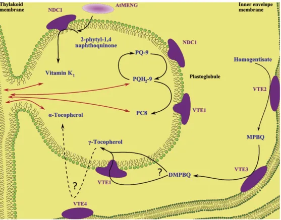
a primary ‘blistering’ event at the outer thylakoid lipid leaflet followed by a secondary blistering event at the surface of an existing PG. The resulting connections between PGs and thylakoids as well as those between PGs themselves provide the basis for a bidirectional metabolite conduit between PGs and the thylakoid membrane (Fig. 1).
PGs in various plant species differ with regard to size and number (Lichtenthaler, 2007). In older leaves of herbaceous plants such as spinach, the number of PGs with a small diameter (0.1–0.2 lm) increases to several hundred per chloroplast (Lichtenthaler, 1969). In older, sun-exposed leaves of beech and oak, PGs are less numerous but significantly enlarged (Lichtenthaler, 1968). Moreover, in several-year-old Ficus leaves PGs may reach diameters of 0.3–3.0 lm (Lichtenthaler and Weinert, 1970).
Studies of the PG core identified members of the neutral lipid class including prenylquinones, triacylglycerols (TAGs), carotenoid, and others. The prenylquinones, plastoquinol-9 (PQH2-9) and tocopherol (vitamin E), are among the major constituents of PGs (Lichtenthaler and Peveling, 1966;Tevini and Steinmuller, 1985; Austin et al., 2006; Vidi et al., 2006) whereas phylloquinone (vitamin K1) is present in minor amounts (Lohmann et al., 2006) (Fig. 1). However, no full lipidome of PGs has been determined so far.
While traces of protein in PGs were observed long ago (Leggettbailey and Whyborn, 1963), it is now known that lipid droplets are coated with specific proteins. Two independent studies reported on the PG proteome, which consists of a total of about three dozen proteins. These belong
to three categories: PAPs/fibrillins, chloroplast metabolic enzymes, and unclassified proteins (Vidi et al., 2006; Ytter-berg et al., 2006).
The first group contains a total of eight of the 13 member Arabidopsis plastoglobulin/PAP/fibrillin family (Vidi et al., 2006; Ytterberg et al., 2006). Fractionation experiments demonstrated the enrichment and physical association of family members, PGL34 (At3g58010) and PGL35 (At4g04020), with PGs (Vidi et al., 2006, 2007). Based on their role in organizing red pepper carotenoid fibrils, the PAPs/fibrillins are hypothesized to fulfil a structural role in PGs too (Deruere et al., 1994). It is interesting to note that the cyanobacterial Synechocystis sp. genome also contains two PAP/fibrillin homologues. Mutant analysis demon-strated that they serve, by an unknown process, to protect the organism from photooxidative damage (Cunningham et al., 2010).
The expression of plastoglobulins is regulated in response to abiotic and biotic stress as well as hormone treatment (Brehelin and Kessler, 2008). Cold treatment induced the expression of a plastoglobulin in rice leaves (Lee et al., 2007). AtPGL30.4 (At3g23400) was identified as a phos-phorylated protein in the defence response to Pseudomonas syringae pv. tomato DC3000 (Jones et al., 2006). In a comparative proteome study, four members of the Arabidopsis plastoglobulin family were found to accumulate under high light stress (Giacomelli et al., 2006). Abscisic acid also induced the expression of several plastoglobulins, AtPGL35 in particular (Gillet et al., 1998; Yang et al.,
Fig. 1. Function of PG-localized enzymes in prenylquinone metabolism. Prenylquinones are shown in blue, enzymes in violet. Bidirectional trafficking between the PG and the thylakoid membrane is represented by red arrows. PQ-9, plastoquinone; PQH2-9, plastoquinol; PC8: plastochromanol-8; MPBQ, 2-methyl-6-phytyl-1,4-benzoquinol; DMPBQ, 2,3-dimethyl-5-phytyl-1,4-benzoquinol.
2006). Recently, it has been shown that plastoglobulins accumulate in response to light/cold stress-related jasmo-nate biosynthesis (Youssef et al., 2010).
The second group of proteins identified in the Arabidopsis PG proteome consists of known metabolic enzymes. This category includes the tocopherol cyclase VTE1 (vitamin E defective, At4g32770) involved in vitamin E synthesis, the carotenoid cleavage dioxygenase CCD4 (At4g19170) proba-bly implicated in carotenoid metabolism, the three isoforms of fructose bisphosphate aldolase of the Calvin cycle (At2g21330, At4g38970, and At2g01140), and the allene oxide synthase (AOS) in jasmonate biosynthesis (At5g42650) (Kazan and Manners, 2011).
The third group of PG proteins consists of unclassified proteins. Some of them are predicted to be involved in lipid metabolism. For instance, ELT1 and 2 (esterase/lipase/ thioesterase, At1g5440 and At3g26840) may be involved in thylakoid lipid metabolism, while ABC1 (activity of bc1 complex)-like kinases (At1g79600, At4g31390, At5g05200, and At5g71810) are predicted regulators of prenylquinone metabolism (Ytterberg et al., 2006). Thus, the available data strongly suggest that PGs, by the presence of enzymes, intervene in diverse aspects of thylakoid lipid metabolism.
Response of PGs to stress
Under oxidative stress-inducing conditions such as drought, high saline concentration, nitrogen deprivation, high light, viral infection, chilling, and ozone (Nordby and Yelenosky, 1985; Locy et al., 1996; Rey et al., 2000; Oksanen et al., 2001;Gaude et al., 2007;Lichtenthaler, 2007) as well as at different developmental stages (senescence and fruit de-velopment) (Kaup et al., 2002), PGs increase in size and number. This occurs in parallel to the disassembly of thylakoid membranes. Also, the lipid composition of PGs will change dramatically due to the accumulation of fatty acid phytyl esters (FAPEs) from thylakoid catabolism (Gaude et al., 2007;Brehelin and Kessler, 2008).
In the thylakoid membrane, reactive oxygen species (ROS) accumulate when the absorption of light by chloro-phyll exceeds the capacity for energy utilization by the photosynthetic apparatus (Pospisil, 2011). Photosystem I (PSI) and PSII are the major sites of free radical O2– generation. Plant responses against oxidative stress implicate different biochemical pathways including the enhanced synthesis of prenylquinones (Gruszka et al., 2008). Antioxidant action has been attributed to phylloqui-none and plastoquinol, although they are primarily known as electron carriers at PSI and PSII, respectively. Tocoph-erol does not play a role as an electron carrier but is a key antioxidant lipid during high light stress (Munne-Bosch, 2005).
While prenylquinones partly accumulate in PGs, their true site of action is probably the thylakoid membrane where they scavenge ROS and protect the photosystems. Of the proteins in the PG proteome, the tocopherol cyclase VTE1 (Vidi et al., 2006) and the NADPH quinone dehydrogenase
C1, NDC1 (Eugeni Piller et al., 2011), are known players in prenylquinone metabolism and implicate PGs as a metabolic compartment.
Implication of PGs in storage and
biosynthesis of tocopherols
Tocopherols belong to the amphipathic group of tocochro-manols (vitamin E) that also includes tocotrienols (Falk and Munne-Bosch, 2010). The two types of tocochromanols differ in the degree of saturation of their prenyl side chains. Synthesized only in photosynthetic organisms (plants, green algae, and cyanobacteria), tocopherols are composed of a polar region derived from tyrosine and a hydrophobic polyprenyl side chain from the isoprenoid pathway (Fig. 2) (Valentin and Qi, 2005).
The group of tocopherols consists of four different forms, a-, b-, c-, and d-, which differ by the number and position of methyl groups on the chromanol ring (Mene-Saffrane and DellaPenna, 2010).
The presence of tocopherols is universal in higher plants, albeit with differential tissue distribution of the various forms: a-tocopherol is predominant in leaves whereas in other organs, such as seeds, flowers, and roots, c-tocopherol is the principle form (Horvath et al., 2006). In plastids, tocopherol inserts in both the envelope and thylakoid membranes whereby the polar chromanol group faces the hydrophilic surface (Dormann, 2007). Around one-third of the total plastid tocopherol is contained in the Arabidopsis PG core (Vidi et al., 2006) (Fig. 1). Under oxidative stress-inducing conditions, such as high light, the production of tocopherols increases to protect membrane lipids from photooxidation and PSII from photoinactivation (DeLong and Steffen, 1997; Havaux et al., 2005). In older leaves under high light stress, the level of a-tocopherol increases >4-fold (Szymanska and Kruk, 2010). The elimination of tocopherols drastically reduces the tolerance of photosyn-thetic organisms to high light stress (Maeda et al., 2005). The a-tocopherol accumulation correlates with an increase in size and number of PGs in older leaves (Vidi et al., 2006;
Brehelin et al., 2007).
Three of the reactions of the tocopherol biosynthesis pathway, mediated by VTE2, VTE3 (Cheng et al., 2003), and VTE4, have been located at the chloroplast inner envelope (Soll et al., 1985). However, surprisingly, the tocopherol cyclase VTE1 (Porfirova et al., 2002) was identified in the PG proteome (Vidi et al., 2006; Ytterberg et al., 2006). Its localization in PGs was confirmed by physical fractionation, immunoelectron microscopy, and expression of a fluorescent fusion protein (Vidi et al., 2006). A serial immunoelectron tomography study revealed the penetration of VTE1 accros of the lipid PG monolayer. This may enable VTE1 to access substrates inside the PGs and carry out the cyclase reaction (Austin et al., 2006).
VTE1 catalyses the conversion of 2,3-dimethyl-5-phytyl-1,4-benzoquinol (DMPBQ) to c-tocopherol and is required for the formation of the chromanol ring of all tocopherols
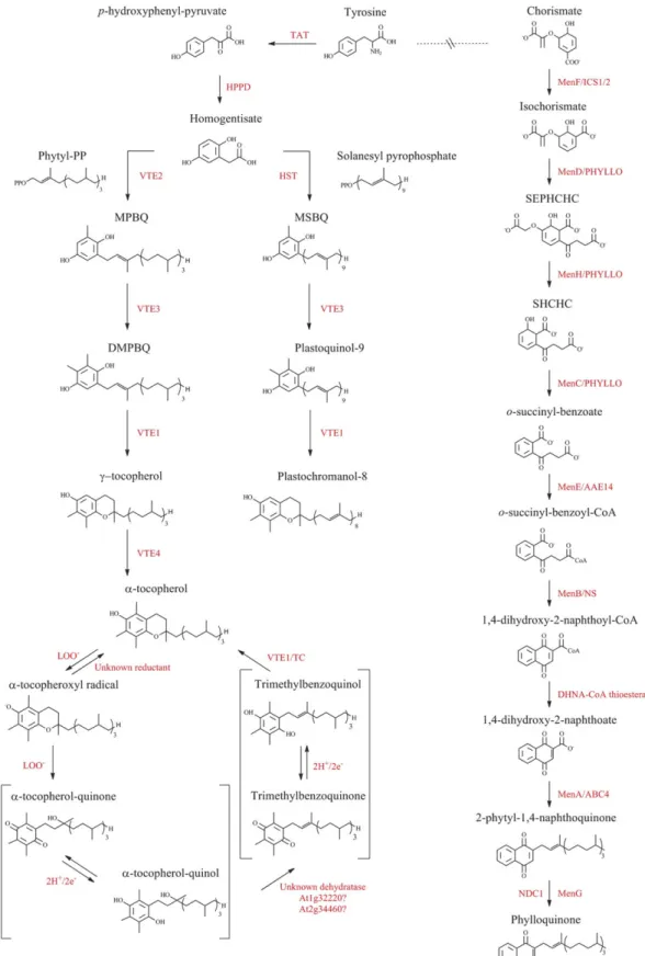
Fig. 2. The biosynthetic pathways of prenylquinones in Arabidopsis. Summary of tocopherol, plastoquinol, plastochromanol, and phylloquinone pathways in Arabidopsis. The enzyme abbreviations are shown in red. TAT, tyrosine aminotransferase; HPPD,
p-hydroxyphenyl-pyruvate dioxygenase; HST, homogentisic acid solanesyl transferase; VTE, enzymes of vitamin E synthesis; LOO–, lipid peroxy radical; TC, tocopherol cyclase; Men, menaquinone synthesis; ICS 1/2, isochorismate synthase 1 and 2; AAE14, acyl-CoA activating enzyme isoform 14; NS, naphthoate synthase; DHNA-CoA thioesterase, 1,4-dihydroxy-2-naphthoyl-CoA thioesterase; ECHId, enoyl-CoA hydratase/isomerase; PP, pyrophosphate; MPBQ, 2-methyl-6-phytyl-1,4-benzoquinone; DMPBQ, 2,3-dimethyl-6-phytyl-1,4-benzoquinone; MSBQ, 2-methyl-6-solanesyl-1,4-benzoquinol; PQH2, plastoquinol; SEPHCHC, 2-succinyl-5-enolpyruvyl-6-hydroxy-3-cyclohexene-1-carboxylate; SHCHC, 2-succinyl-6-hydroxy-2,4-cyclohexadiene-1-carboxylate.
(Soll et al., 1985) (Fig. 2). If VTE1 were uniquely present in PGs, DMPBQ would have to be moved from the inner envelope membrane to PGs where the cyclase would convert it to c-tocopherol. In support of this hypothesis, DMPBQ was indeed highly enriched in PGs of the vte1 mutant (Fig. 3).
The last step of a-tocopherol synthesis is carried out by the c-tocopherol methyl transferase, VTE4, located at the chloroplast envelope (Zbierzak et al., 2010) (Fig. 1). Again, if VTE1 were exclusively located at PGs, c-tocopherol would have to be transported back to the inner envelope membrane to complete the synthesis of a-tocopherol (Zbierzak et al., 2010). Alternatively, it has been proposed that sufficient VTE1 for vitamin E synthesis may still be present at the envelope membranes and that at PGs VTE1 serves other metabolic purposes such as the recycling of tocopherol oxidation products (DellaPenna and Kobayashi, 2008).
Role of PGs in the tocopherol redox cycle
Tocopherol oxidation products form in response to high light stress (DellaPenna and Kobayashi, 2008). By in vitro chemical treatment, 23 different oxidation products can be generated from a- and c-tocopherol, but only two of these were detected in vivo: wild-type plants accumulated a-tocopherol-quinol (a-TQH2) under high light conditions (DellaPenna and Kobayashi, 2008) and vte4 mutant plants(containing only c-tocopherol) accumulated c-TQH2. To determine whether a-TQH2 was degraded or recycled to a-tocopherol, isolated wild-type and vte1 chloroplasts were incubated with 14C-labelled a-TQH2 (DellaPenna
and Kobayashi, 2008; Mene-Saffrane and DellaPenna, 2010). In wild-type chloroplasts, the incubation led to a-tocopherol accumulation, whereas in vte1 mutants a sub-strate of tocopherol cyclase, trimethylphytylbenzoquinone (TMPBQ), was detected (DellaPenna and Kobayashi, 2008). This is clear evidence that tocopherol oxidation products are recycled in higher plants. The a-tocopherol quinone (a-TQ) oxidation product was present in thyla-koids, envelope membranes, as well as PGs in chloroplast fractionation experiments (Kruk and Nowicka, 2010).
The proposed a-tocopherol redox cycle starts by a two-step oxidation, with each two-step characterized by the loss of a single electron (Mene-Saffrane and DellaPenna, 2010) (Fig. 2). In the first step, a-tocopherol is oxidized to the a-tocopherol radical by a lipid peroxy radical (LOO–). This product may be reduced back to a-tocopherol by an unknown reductant, possibly ascorbate, or be oxidized further by a second lipid peroxy radical, resulting in the formation of a-TQH2. To regenerate a-tocopherol, a-TQH2 must undergo a dehydration step catalysed by an as yet unidentified dehydratase. This will result in TMPBQ, which in turn will undergo the cyclase reaction catalysed by VTE1 and results in the completion of the cycle. With regard to the potential role of PGs in the cycle, not only are they enriched in tocopherol cyclase VTE1 (Austin et al., 2006;
Vidi et al., 2006; Ytterberg et al., 2006) but the PG proteome also contains two predicted dehydratases (At2g34460 and At1g32220). In summary, the currently available evidence suggests that PGs participate in the tocopherol recycling pathway.
Implication of VTE1 and NDC1 in
plastoquinol metabolism
PQH2-9 is well known as an electron and proton carrier in the photosynthetic transport chain between PSII and the cytochrome b6f complex (Muh et al., 2011). However, plastoquinol has also been shown to have a physiological antioxidant activity (Szymanska and Kruk, 2010). Structur-ally related to a-tocopherol, it exerts a photoprotective role on PSII during high light stress. It does so by scavenging singlet oxygen generated by chlorophyll at the reaction centre (Kruk and Trebst, 2008), thereby inhibiting lipid peroxydation (Hundal et al., 1995). The plastoquinol head group as well as the isoprenoid chain are involved in the process which may confer additional antioxidant power over tocopherols (Gruszka et al., 2008).
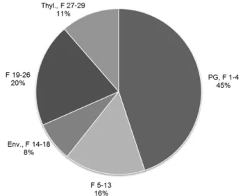
Plastoquinone-9 (PQ-9) is associated with QA and QB sites of PSII but also exists in a free form in thylakoid membranes. Together, these are considered the thylakoid or photoactive PQ pool (Strzalka and Kruk, 1999). However, PQ-9 is also present in a separate pool contained in PGs (Szymanska and Kruk, 2010;Zbierzak et al., 2010) (Fig. 1). Fig. 3. Enrichment of DMPBQ in vte1 PGs. DMPBQ
(2,3-dimethyl-5-phytyl-1,4-benzoquinol), the precursor of c-tocopherol, accumulates in the vte1 mutants. Subplastidial chloroplast frac-tions were isolated from leaves of 8-week-old vte1 mutant plants (Vidi et al., 2006). The distribution of DMPBQ (as a percentage of the total) was measured in pooled chloroplast membrane fractions. Fractions F1–4 and F5–13 contained mostly PGs; F14–18 contained envelopes (Env.); F19–26 contained some envelopes and thylakoids; and F27–29 contained thylakoids (Thyl.).
The highest amount of DMPBQ is present in the fractions enriched in PGs (;45%).
The PG PQ pool is not normally photoactive and does not directly participate in photosynthetic electron flow (Eugeni Piller et al., 2011). It might therefore serve both as a reservoir of antioxidant and to replenish the thylakoid pool (Zbierzak et al., 2010).
In Arabidopsis leaves, the level of plastoquinol dramati-cally increases under high light stress. The reduced form of PQ (PQH2-9) increased 16- and 9- fold in old and young rosette leaves while the total amount of PQ (reduced plus oxidized) increased 8- and 11- fold, respectively (Szymanska and Kruk, 2010). The majority of the plastoquinol under high light conditions is photosynthetically inactive and accumulates in PGs. It is tempting to speculate that the plastoquinol that is irreversibly degraded by ROS in thylakoid membranes is replaced by plastoquinol from PGs (Szymanska and Kruk, 2010). This may partially explain why PQ accumulates to very high levels under high light conditions. NDC1, the NADPH-dependent quinone de-hydrogenase C1, in PG functions to reduce the oxidized proportion of the non-photochemical pool of plastoquinol in PGs. Indeed, the ndc1 mutant had a significantly higher percentage of oxidized PQ than the wild type (Eugeni Piller et al., 2011). Therefore, NDC1 constitutes a unique electron transport pathway separate from cyclic electron flow mediated by the NAD(P)H dehydrogenase (NDH) complex or the PROTON GRADIENT REGULATION 5 (PGR) pathway (Shikanai, 2007; Peng et al., 2010). The NDC1-mediated electron pathway, however, is probably limited by the availability of PQ inside PGs that cannot rapidly be reoxidized as happens in NDH- and PGR5-dependent cyclic electron flow (Eugeni Piller et al., 2011).
Plastochromanol (PC8), derived from PQH2-9 by tocoph-erol cyclase activity, is present in leaves, seeds, and other organs of Arabidopsis plants (Mene-Saffrane and Della-Penna, 2010; Szymanska and Kruk, 2010; Zbierzak et al., 2010). It constitutes 5–10% of the total tocochromanol, although this value may be higher in senescing leaves (Szymanska and Kruk, 2010). Around 50% of the PC8 is present in PGs (Zbierzak et al., 2010). Together with c-tocopherol, PC8 has been shown to be required for effi-cient germination after longer periods of seed quiescence (Mene-Saffrane and DellaPenna, 2010). The levels of PC8 increase under high light stress as well as in ageing leaves. Several studies have demonstrated that PC8 is an efficient singlet oxygen scavenger (Gruszka et al., 2008) and an inhibitor of lipid peroxidation (Olejnik et al., 1997). Its antioxidant activity is comparable with that of tocopherols (Olejnik et al., 1997). This is not surprising because plastochromanol has a chromanol group identical to that of c-tocopherol, differing only in the C40 polyunsaturated solanesyl side chain instead of the phytol. Most probably, its source is the PQH2-9 in the PG pool (Kumar et al., 2005;
Kruk and Trebst, 2008) (Fig. 2) where the tocopherol cyclase is also present (Fig. 1).
In the ndc1 mutant, PC8 was decreased (Eugeni Piller et al., 2011). Most probably, this is linked to the decrease of its direct precursor, PQH2-9, the substrate of VTE1 (Grutter et al., 2006). In the vte1 mutant, PC8 formation
was entirely abolished but the overexpression of VTE1 induced a 2.4-fold increase of PC8 levels. This resulted in the proliferation of PG numbers and increased cluster formation (Kanwischer et al., 2005;Zbierzak et al., 2010).
Implication of PGs in storage and
biosynthesis of phylloquinone via NDC1
Phylloquinone (2-methyl-3-phytyl-1,4-naphthoquinone) or vitamin K1is an prenylquinone composed of a naphthoqui-none ring and a prenyl side chain derived from phytyl-diphosphate (Fig. 2). It is synthesized in all organisms performing oxygenic photosynthesis. In higher plants, phylloquinone occurs in leaves where it serves as an electron carrier in the quinone/semiquinone turnover in PSI (Joyard et al., 2009). The overall stoichiometry of phylloquinone has been estimated at 3 mol of vitamin K1 per 1 mol of PSI. However, only two molecules of phylloquinone are present for each PSI complex. This suggests that a separate pool exists that is not associated with PSI (Lohmann et al., 2006;Brehelin and Kessler, 2008). Interestingly, this is in good agreement with the ;30% of the total phylloquinone located in Arabidopsis PGs (Lohmann et al., 2006) (Fig. 1).
The enzymatic reactions of phylloquinone biosynthesis take place at the inner membrane of the chloroplast envelope (Schultz et al., 1981), but recent evidence in Arabidopsis suggests that peroxisomes may also be impli-cated. In cyanobacteria and red algae, the pathway is catalysed by Men proteins including successively: MenF, MenD, MenH, MenC, MenE, MenB, MenA, and MenG enzymes. The first step of phylloquinone biosynthesis (MenF) implicates the conversion of chorismate to isochor-ismate. In Arabidopsis, this reaction may be catalysed by two genes, ICS1 and ICS2, showing homology with the MenF gene (Ausubel et al., 2001; Gross et al., 2006;
Garcion et al., 2008;Metraux et al., 2008).
The double homozygous ics1 ics2 mutant was completely devoid of phylloquinone, and plants remained smaller and had a pale green or yellowish phenotype compared with the wild type or the single mutants. The two enzymes may form a complex in the chloroplast stroma to facilitate the efficient channelling of intermediates through the pathway (Ausubel et al., 2001;Metraux et al., 2008).
The conversion of isochorismate into o-succinyl-benzoate (OSB) implicates three distinct enzymes (Men D, Men H, and Men C) in cyanobacteria. These functions are encoded by the composite gene PHYLLO in Arabidopsis (Gross et al., 2006). In cyanobacteria, the conversion of OSB to o-succinyl-benzoyl-CoA is catalysed by the ligase MenE. The presence of several MenE homologues in the Arabidop-sis genome makes it difficult to assign the ligase function: it seems likely, however, that the OSB-CoA ligase corresponds to the acyl-activating enzyme 14 (AAE14) (Browse et al., 2008). The aae14 mutant is unable to grow on soil due to the lack of phylloquinone. Recently, it has been demonstrated that MenE/AAE14 is dually targeted to both chloroplasts and peroxisomes (Reumann et al., 2010). In the following
step, OSB-CoA is converted to 1,4-dihydroxy-2-naphthoyl-CoA by an enzyme orthologous to MenB, the naphthoate synthase (NS/ECHId) encoded by a single Arabidopsis gene (Gross et al., 2006;Browse et al., 2008;Babujee et al., 2010). Prior to these studies, neither functional data nor subcellular localization had been reported for a MenB homologue in plants. Recently, NS/ECHId was localized to the Arabidopsis peroxisome (Babujee et al., 2010; Reumann et al., 2010). It has also been proposed that the conversion of 1,4-dihydroxy-2-naphthoyl-CoA to 1,4-dihydroxy-2-naphthoate (DHNA) is catalysed by the 1,4-dihydroxy-2-naphthoyl-CoA (DHNA-CoA) thioesterase and that this enzyme may also be a peroxisomal protein (Reumann, 2004; Reumann et al., 2010). These recent findings suggest that phylloquinone biosynthesis of higher plants is partially compartmentalized in peroxisomes.
The last steps of phylloquinone synthesis involve the attachment of the phytyl chain to DHNA by MenA/ABC4 (Shimada et al., 2005) and the methylation of 2-phytyl-1,4-naphthoquinone by MenG (Lohmann et al., 2006). Genetic analysis of phylloquinone pathway in plants allowed the isolation of the AtmenA mutant of Arabidopsis, which lacks the DHNA phytyltransferase (Shimada et al., 2005). Total absence of vitamin K1 in AtmenA mutant plants results in a drastic reduction in both growth and the accumulation of PSII and PQ-9.
In contrast, growth and photosynthesis were only slightly affected in AtmenG mutant plants raised under normal light conditions. This fact suggests that 2-phytyl-1,4-naphthoquinone can functionally replace phylloqui-none as an electron carrier in PSI. Under high light stress, biochemical and physiological studies of the AtmenG mutant demonstrated a significant decrease in the level of PSI complexes caused by oxidative damage at the PSI reaction centre. This led to lowered PSII efficiency and negatively affected the performance of the entire photo-synthetic electron transfer chain (Lohmann et al., 2006).
Interestingly, a much higher proportion of the 2-phytyl-1,4-naphthoquinone in the AtmenG mutant than of phyllo-quinone in the wild type was present in PGs. A non-targeted lipidomic analysis of ndc1 mutant plants led to the un-expected discovery that, similar to AtmenG, phylloquinone was almost completely absent and that 2-phytyl-1,4-naphthoquinone accumulated instead (Eugeni Piller et al., 2011). Also, AtMenG was normally expressed in ndc1 plants. While this result is difficult to explain, it suggests the implication of NDC1 and PGs in the AtMenG methylation step.
Interestingly, the multifunctional protein PHYLLO and AtMenG gave punctate fluorescence rather than the ring-like fluorescence typical of chloroplast envelope proteins when transiently expressed as green fluorescent protein (GFP) or yellow fluorescent protein (YFP) fusion proteins in Arabidopsis protoplasts. As the punctate fluorescence resembles that of NDC1 it may hint at a PG localization of PHYLLO and AtMenG (Gross et al., 2006;Lohmann et al., 2006; Eugeni Piller et al., 2011). However, PHYLLO and AtMenG were not found in the PG proteome. Possibly they
are only loosely or transiently associated with PGs and lost during the purification procedure.
Together with the presence of phylloquinone and 2-phytyl-1,4-naphthoquinone, the new role for NDC1 suggests a role for PGs in phylloquinone metabolism.
Conclusions
In the last few years several studies demonstrated the important role of PGs in chloroplast lipid metabolism. The determination of the Arabidopsis PG proteome (Vidi et al., 2006;Ytterberg et al., 2006) paved the way to the discovery that PGs do not only store lipids but also actively participate in their synthesis.
The Arabidopsis PG proteome contains 34 proteins, assign-able to three groups: plastoglobulins, metabolic enzymes, and proteins of unknown function. While many of the unknown proteins are predicted to be enzymes, their function still remains to be discovered.
To discover the function of such candidate enzymes in lipid metabolism, a powerful non-targeted lipidomics anal-ysis can be employed to correlate changes in metabolite profiles with enzyme function.
The final biosynthetic steps of tocopherol require VTE1 and VTE4. VTE1 was localized in PGs while VTE4 was located at the inner envelope membrane. This hints at the interesting possibility of prenylquinone metabolite traffick-ing inside the chloroplast.
It is still not known whether PGs are structurally required for proper chloroplast function, but forward genetic screens have the potential to provide answers to this question. Proteome analyses of PGs isolated from plants under various stress or developmental conditions may lead to the identification of more new candidate proteins with impor-tant functions in lipid metabolism. However, what is known for sure now is that PGs are sitting right at the crossroads of the prenylquinone metabolic pathways.
Acknowledgements
FEK thanks the Universite´ de Neuchaˆtel, SystemsX Plant Growth in a Changing Environment and National Center of Competence in Research Plant Survival, and acknowl-edges support from Swiss National Science Foundation Grant 31003A_127380.
References
Austin JR 2nd, Frost E, Vidi PA, Kessler F, Staehelin LA. 2006. Plastoglobules are lipoprotein subcompartments of the chloroplast that are permanently coupled to thylakoid membranes and contain biosynthetic enzymes. The Plant Cell 18, 1693–1703.
Ausubel FM, Wildermuth MC, Dewdney J, Wu G. 2001.
Isochorismate synthase is required to synthesize salicylic acid for plant defence. Nature 414, 562–565.
Babujee L, Wurtz V, Ma C, Lueder F, Soni P, van Dorsselaer A, Reumann S. 2010. The proteome map of spinach leaf peroxisomes indicates partial compartmentalization of phylloquinone (vitamin K1) biosynthesis in plant peroxisomes. Journal of Experimental Botany 61, 1441–1453.
Brehelin C, Kessler F. 2008. The plastoglobule: a bag full of lipid biochemistry tricks. Photochemistry and Photobiology 84, 1388–1394. Brehelin C, Kessler F, van Wijk KJ. 2007. Plastoglobules: versatile lipoprotein particles in plastids. Trends in Plant Science 12, 260–266. Browse J, Kim HU, van Oostende C, Basset GJC. 2008. The AAE14 gene encodes the Arabidopsis o-succinylbenzoyl-CoA ligase that is essential for phylloquinone synthesis and photosystem-I function. The Plant Journal 54, 272–283.
Cheng Z, Sattler S, Maeda H, Sakuragi Y, Bryant DA, DellaPenna D. 2003. Highly divergent methyltransferases catalyze a conserved reaction in tocopherol and plastoquinone synthesis in cyanobacteria and photosynthetic eukaryotes. The Plant Cell 15, 2343–2356.
Cunningham FX Jr, Tice AB, Pham C, Gantt E. 2010. Inactivation of genes encoding plastoglobuli-like proteins in Synechocystis sp. PCC 6803 leads to a light-sensitive phenotype. Journal of Bacteriology 192, 1700–1709.
DellaPenna D, Kobayashi N. 2008. Tocopherol metabolism, oxidation and recycling under high light stress in Arabidopsis. The Plant Journal 55, 607–618.
DeLong JM, Steffen KL. 1997. Photosynthetic function, lipid peroxidation, and alpha-tocopherol content in spinach leaves during exposure to UV-B radiation. Canadian Journal of Plant Science 77, 453–459.
Deruere J, Romer S, d’Harlingue A, Backhaus RA, Kuntz M, Camara B. 1994. Fibril assembly and carotenoid overaccumulation in chromoplasts: a model for supramolecular lipoprotein structures. The Plant Cell 6, 119–133.
Dormann P. 2007. Functional diversity of tocochromanols in plants. Planta 225, 269–276.
Eugeni Piller L, Besagni C, Ksas B, Rumeau D, Brehelin C, Glauser G, Kessler F, Havaux M. 2011. Chloroplast lipid droplet type II NAD(P)H quinone oxidoreductase is essential for prenylquinone metabolism and vitamin K1 accumulation. Proceedings of the National Academy of Sciences, USA 108, 14354–14359.
Falk J, Munne-Bosch S. 2010. Tocochromanol functions in plants: antioxidation and beyond. Journal of Experimental Botany 61, 1549–1566.
Garcion C, Lohmann A, Lamodiere E, Catinot J, Buchala A, Doermann P, Metraux JP. 2008. Characterization and biological function of the ISOCHORISMATE SYNTHASE2 gene of Arabidopsis. Plant Physiology 147, 1279–1287.
Gaude N, Brehelin C, Tischendorf G, Kessler F, Dormann P. 2007. Nitrogen deficiency in Arabidopsis affects galactolipid
composition and gene expression and results in accumulation of fatty acid phytyl esters. The Plant Journal 49, 729–739.
Giacomelli L, Rudella A, van Wijk KJ. 2006. High light response of the thylakoid proteome in arabidopsis wild type and the
ascorbate-deficient mutant vtc2-2. A comparative proteomics study. Plant Physiology 141, 685–701.
Gillet B, Beyly A, Peltier G, Rey P. 1998. Molecular characterization of CDSP 34, a chloroplastic protein induced by water deficit in Solanum tuberosum L. plants, and regulation of CDSP 34 expression by ABA and high illumination. The Plant Journal 16, 257–262. Greenwood AD, Leech RM, Williams JP. 1963. Osmiophilic globules of chloroplasts. 1. Osmiophilic globules as a normal
component of chloroplasts and their isolation and composition in Vicia faba L. Biochimica et Biophysica Acta 78, 148–162.
Gross J, Cho WK, Lezhneva L, Falk J, Krupinska K, Shinozaki K, Seki M, Herrmann RG, Meurer J. 2006. A plant locus essential for phylloquinone (vitamin K1) biosynthesis originated from a fusion of four eubacterial genes. Journal of Biological Chemistry 281, 17189–17196. Gruszka J, Pawlak A, Kruk J. 2008. Tocochromanols, plastoquinol, and other biological prenyllipids as singlet oxygen
quenchers—determination of singlet oxygen quenching rate constants and oxidation products. Free Radical Biology and Medicine 45, 920–928.
Grutter C, Alonso E, Chougnet A, Woggon WD. 2006. A biomimetic chromanol cyclization leading to alpha-tocopherol. Angewandte Chemie International Edition 45, 1126–1130. Havaux M, Eymery F, Porfirova S, Rey P, Dormann P. 2005. Vitamin E protects against photoinhibition and photooxidative stress in Arabidopsis thaliana. The Plant Cell 17, 3451–3469.
Horvath G, Wessjohann L, Bigirimana J, Jansen M, Guisez Y, Caubergs R, Horemans N. 2006. Differential distribution of tocopherols and tocotrienols in photosynthetic and non-photosynthetic tissues. Phytochemistry 67, 1185–1195.
Hundal T, Forsmark-Andree P, Ernster L, Andersson B. 1995. Antioxidant activity of reduced plastoquinone in chloroplast thylakoid membranes. Archives of Biochemistry and Biophysics 324, 117–122. Jones AM, Bennett MH, Mansfield JW, Grant M. 2006. Analysis of the defence phosphoproteome of Arabidopsis thaliana using
differential mass tagging. Proteomics 6, 4155–4165.
Joyard J, Ferro M, Masselon C, Seigneurin-Berny D, Salvi D, Garin J, Rolland N. 2009. Chloroplast proteomics and the compartmentation of plastidial isoprenoid biosynthetic pathways. Molecular Plant 2, 1154–1180.
Kanwischer M, Porfirova S, Bergmuller E, Dormann P. 2005. Alterations in tocopherol cyclase activity in transgenic and mutant plants of Arabidopsis affect tocopherol content, tocopherol composition, and oxidative stress. Plant Physiology 137, 713–723. Kaup MT, Froese CD, Thompson JE. 2002. A role for diacylglycerol acyltransferase during leaf senescence. Plant Physiology 129, 1616–1626.
Kazan K, Manners JM. 2011. The interplay between light and jasmonate signalling during defence and development. Journal of Experimental Botany 62, 4087–4100.
Kessler F, Schnell D, Blobel G. 1999. Identification of proteins associated with plastoglobules isolated from pea (Pisum sativum L.) chloroplasts. Planta 208, 107–113.
Kessler F, Vidi PA. 2007. Plastoglobule lipid bodies: their functions in chloroplasts and their potential for applications. Advances in
Biochemical Engineering Biotechnology 107, 153–172.
Kruk J, Nowicka B. 2010. Occurrence, biosynthesis and function of isoprenoid quinones. Biochimica et Biophysica Acta 1797,
1587–1605.
Kruk J, Trebst A. 2008. Plastoquinol as a singlet oxygen scavenger in photosystem II.. Biochimica et Biophysica Acta 1777, 154–162. Kumar R, Raclaru M, Schusseler T, et al. 2005. Characterisation of plant tocopherol cyclases and their overexpression in transgenic Brassica napus seeds. FEBS Letters 579, 1357–1364.
Lee DG, Ahsan N, Lee SH, Kang KY, Lee JJ, Lee BH. 2007. An approach to identify cold-induced low-abundant proteins in rice leaf. Comptes Rendus Biologies 330, 215–225.
Leggettbailey J, Whyborn AG. 1963. Osmiophilic globules of chloroplasts. 2. Globules of spinach-beet chloroplast. Biochimica et Biophysica Acta 78, 163–174.
Lichtenthaler HK. 1968. Plastoglobuli and the fine structure of plastids. Endeavour 27, 144–149.
Lichtenthaler HK. 1969. Plastoglobuli of spinach—their size and composition during chloroplast degeneration. Protoplasma 68, 315–326.
Lichtenthaler HK. 2007. Biosynthesis, accumulation and emission of carotenoids, alpha-tocopherol, plastoquinone, and isoprene in leaves under high photosynthetic irradiance. Photosynthesis Research 92, 163–179.
Lichtenthaler HK, Peveling E. 1966. Osmiophilic lipid inclusions in the chloroplasts and in the cytoplasm of Hoya carnosa R. Br. Naturwissenschaften 53, 534.
Lichtenthaler HK, Weinert H. 1970. Correlation between
lipoquinone accumulation and plastoglobuli formation in chloroplasts of Ficus-elastica Roxb. Zeitschrift fur Naturforschung Series B25, 619–623.
Locy RD, Chang CC, Nielsen BL, Singh NK. 1996. Photosynthesis in salt-adapted heterotrophic tobacco cells and regenerated plants. Plant Physiology 110, 321–328.
Lohmann A, Schottler MA, Brehelin C, Kessler F, Bock R,
Cahoon EB, Dormann P. 2006. Deficiency in phylloquinone (vitamin K1) methylation affects prenyl quinone distribution, photosystem I
abundance, and anthocyanin accumulation in the Arabidopsis AtmenG mutant. Journal of Biological Chemistry 281, 40461–40472.
Maeda H, Sakuragi Y, Bryant DA, DellaPenna D. 2005. Tocopherols protect Synechocystis sp. strain PCC 6803 from lipid peroxidation. Plant Physiology 138, 1422–1435.
Mene-Saffrane L, DellaPenna D. 2010. Biosynthesis, regulation and functions of tocochromanols in plants. Plant Physiology and
Biochemistry 48, 301–309.
Metraux JP, Garcion C, Lohmann A, Lamodiere E, Catinot J, Buchala A, Doermann P. 2008. Characterization and biological function of the ISOCHORISMATE SYNTHASE2 gene of Arabidopsis. Plant Physiology 147, 1279–1287.
Muh F, Glockner C, Hellmich J, Zouni A. 2011. Light-induced quinone reduction in photosystem II. Biochimica et Biophysica Acta 1817, 44–65.
Munne-Bosch S. 2005. The role of alpha-tocopherol in plant stress tolerance. Journal of Plant Physiology 162, 743–748.
Nordby HE, Yelenosky G. 1985. Change in citrus leaf lipids during freeze–thaw stress. Phytochemistry 24, 1675–1679.
Oksanen E, Sober J, Karnosky DF. 2001. Impacts of elevated CO2 and/or O-3 on leaf ultrastructure of aspen (Populus tremuloides) and birch (Betula papyrifera) in the Aspen FACE experiment. Environmental Pollution 115, 437–446.
Olejnik D, Gogolewski M, NogalaKalucka M. 1997. Isolation and some properties of plastochromanol-8. Nahrung-Food 41, 101–104. Peng L, Yamamoto H, Shikanai T. 2010. Structure and biogenesis of the chloroplast NAD(P)H dehydrogenase complex. Biochimica et Biophysica Acta 1807, 945–953.
Porfirova S, Bergmuller E, Tropf S, Lemke R, Dormann P. 2002. Isolation of an Arabidopsis mutant lacking vitamin E and identification of a cyclase essential for all tocopherol biosynthesis. Proceedings of the National Academy of Sciences, USA 99, 12495–12500. Pospisil P. 2011. Molecular mechanisms of production and scavenging of reactive oxygen species by photosystem II. Biochimica et Biophysica Acta 1817, 218–231.
Pozueta-Romero J, Rafia F, Houlne G, Cheniclet C, Carde JP, Schantz ML, Schantz R. 1997. A ubiquitous plant housekeeping gene, PAP, encodes a major protein component of bell pepper chromoplasts. Plant Physiology 115, 1185–1194.
Reumann S. 2004. Specification of the peroxisome targeting signals type 1 and type 2 of plant peroxisomes by bioinformatics analyses. Plant Physiology 135, 783–800.
Reumann S, Babujee L, Wurtz V, Ma C, Lueder F, Soni P, van Dorsselaer A. 2010. The proteome map of spinach leaf peroxisomes indicates partial compartmentalization of phylloquinone (vitamin K1) biosynthesis in plant peroxisomes. Journal of Experimental Botany 61, 1441–1453.
Rey P, Gillet B, Romer S, Eymery F, Massimino J, Peltier G, Kuntz M. 2000. Over-expression of a pepper plastid lipid-associated protein in tobacco leads to changes in plastid ultrastructure and plant development upon stress. The Plant Journal 21, 483–494.
Schultz G, Ellerbrock BH, Soll J. 1981. Site of prenylation reaction in synthesis of phylloquinone (vitamin K1) by spinach chloroplasts. European Journal of Biochemistry 117, 329–332.
Shikanai T. 2007. Cyclic electron transport around photosystem I: genetic approaches. Annual Review of Plant Biology 58, 199–217. Shimada H, Ohno R, Shibata M, Ikegami I, Onai K, Ohto MA, Takamiya K. 2005. Inactivation and deficiency of core proteins of photosystems I and II caused by genetical phylloquinone and plastoquinone deficiency but retained lamellar structure in a T-DNA mutant of Arabidopsis. The Plant Journal 41, 627–637.
Soll J, Schultz G, Joyard J, Douce R, Block MA. 1985. Localization and synthesis of prenylquinones in isolated outer and inner envelope membranes from spinach chloroplasts. Archives of Biochemistry and Biophysics 238, 290–299.
Strzalka K, Kruk J. 1999. Dark reoxidation of the plastoquinone-pool is mediated by the low-potential form of cytochrome b-559 in spinach thylakoids. Photosynthesis Research 62, 273–279.
Szymanska R, Kruk J. 2010. Plastoquinol is the main prenyllipid synthesized during acclimation to high light conditions in Arabidopsis and is converted to plastochromanol by tocopherol cyclase. Plant and Cell Physiology 5, 537–545.
Tevini M, Steinmuller D. 1985. Composition and function of plastoglobuli. 2. Lipid-composition of leaves and plastoglobuli during beech leaf senescence. Planta 163, 91–96.
Thomson WW, Platt K. 1973. Plastid ultrastructure in barrel cactus, Echinocactus-Acanthodes. New Phytologist 72, 791–797.
Valentin HE, Qi Q. 2005. Biotechnological production and application of vitamin E: current state and prospects. Applied Microbiology and Biotechnology 68, 436–444.
Vidi PA, Kanwischer M, Baginsky S, Austin JR, Csucs G, Dormann P, Kessler F, Brehelin C. 2006. Tocopherol cyclase (VTE1) localization and vitamin E accumulation in chloroplast
plastoglobule lipoprotein particles. Journal of Biological Chemistry 281, 11225–11234.
Vidi PA, Kessler F, Brehelin C. 2007. Plastoglobules: a new address for targeting recombinant proteins in the chloroplast. BMC
Biotechnology 7, 4.
Yang Y, Sulpice R, Himmelbach A, Meinhard M, Christmann A, Grill E. 2006. Fibrillin expression is regulated by abscisic acid response regulators and is involved in abscisic acid-mediated photoprotection. Proceedings of the National Academy of Sciences, USA 103, 6061–6066.
Youssef A, Laizet Y, Block MA, Marechal E, Alcaraz JP, Larson TR, Pontier D, Gaffe J, Kuntz M. 2010. Plant lipid-associated fibrillin proteins condition jasmonate production under photosynthetic stress. The Plant Journal 61, 436–445.
Ytterberg AJ, Peltier JB, van Wijk KJ. 2006. Protein profiling of plastoglobules in chloroplasts and chromoplasts. A surprising site for differential accumulation of metabolic enzymes. Plant Physiology 140, 984–997.
Zbierzak AM, Kanwischer M, Wille C, et al. 2010. Intersection of the tocopherol and plastoquinol metabolic pathways at the