SCIENTIFIC ARTICLE
Extreme hip motion in professional ballet dancers: dynamic
and morphological evaluation based on magnetic
resonance imaging
Frank C. Kolo&Caecilia Charbonnier&Christian W. A. Pfirrmann&
Sylvain R. Duc&Anne Lubbeke&Victoria B. Duthon&Nadia Magnenat-Thalmann&
Pierre Hoffmeyer&Jacques Menetrey&Christoph D. Becker
Received: 16 August 2012 / Revised: 3 October 2012 / Accepted: 29 October 2012 / Published online: 29 November 2012 # ISS 2012
Abstract
Objective To determine the prevalence of femoroacetabular impingement (FAI) of the cam or pincer type based on mag-netic resonance imaging (MRI) in a group of adult female professional ballet dancers, and to quantify, in vivo, the range of motion (ROM) and congruence of the hip joint in the splits position.
Materials and methods Institutional review board approval and informed consent from each volunteer were obtained. Thirty symptomatic or asymptomatic adult female profession-al bprofession-allet dancers (59 hips) and 14 asymptomatic non-dancer adult women (28 hips, control group) were included in the present study. All subjects underwent MRI in the supine position, while, for the dancers, additional images were ac-quired in the splits position. Labral abnormalities, cartilage lesions, and osseous abnormalities of the acetabular rim were assessed at six positions around the acetabulum. A
morpho-logical analysis, consisting of the measurement of theα angle,
acetabular depth, and acetabular version, was performed. For the dancers, ROM and congruency of the hip joint in the splits position were measured.
Results Acetabular cartilage lesions greater than 5 mm were
significantly more frequent in dancer’s hips than in control
hips (28.8 vs 7.1%, p=0.026), and were mostly present at the superior position in dancers. Distribution of labral lesions between the dancers and the control group showed substantially more pronounced labral lesions at the superior, posterosuperior, and anterosuperior positions in dancers (54
lesions in 28 dancer’s hips vs 10 lesions in 8 control hips).
Herniation pits were found significantly more often (p=0.002) in dancer’s hips (n=31, 52.5%), 25 of them being located in a superior position. A cam-type morphology was found for one dancer and a retroverted hip was noted for one control. Fem-oroacetabular subluxations were observed in the splits posi-tion (mean: 2.05 mm).
Conclusion The prevalence of typical FAI of the cam or pincer type was low in this selected population of profes-sional ballet dancers. The lesions’ distribution, mostly
su-perior, could be explained by a“pincer-like” mechanism of
impingement with subluxation in relation to extreme move-ments performed by the dancers during their daily activities. Keywords Hip . Early hip osteoarthritis . Impingements . Dancing . Ballet
Introduction
Professional ballet dancers’ hips are subject to extreme ranges of motion (ROM) during their daily activities. ROM of the hip joint assuming extreme positions, especially while do-ing the splits, has not yet been determined. Furthermore, it is unclear whether the femoral head and acetabulum are congruent in these extreme positions regularly assumed by dancers. Joint incongruence could be a potential cause of early osteoarthritis (OA).
F. C. Kolo (*)
:
S. R. Duc:
C. D. BeckerDepartment of Radiology, University Hospital of Geneva, Rue Gabrielle-Perret-Gentil 4,
1205 Geneva, Switzerland
e-mail: [email protected] C. Charbonnier
:
N. Magnenat-ThalmannMIRALab, University of Geneva, Geneva, Switzerland C. W. A. Pfirrmann
Department of Radiology, University Hospital Balgrist, Zürich, Switzerland
A. Lubbeke
:
V. B. Duthon:
P. Hoffmeyer:
J. Menetrey Department of Orthopaedic Surgery,Femoroacetabular impingement (FAI) occurs when there is an abnormal contact between the proximal femur, typi-cally the anterosuperior femoral head neck junction, and the
acetabular rim. As described previously [1–7], FAI is the
result of femoral or acetabular morphological abnormalities. FAI of the cam or pincer type is believed to be a potential mechanism for the development of early OA for most
non-dysplastic hips [8]. The study of professional ballet dancers’
hips while doing the splits provides us with a potential extreme model of cam/pincer FAI, because of extreme flexion in that position.
Cam/pincer FAI, as well as subluxation (i.e., a loss of joint congruence), could be a potential cause of the devel-opment of hip pain and OA in this selected population with potential stigmata of FAI and/or subluxation in the symp-tomatic dancers. Thus, the purpose of this study was to determine the prevalence of FAI of the cam or pincer type based on magnetic resonance imaging (MRI) in a group of symptomatic and asymptomatic adult female professional ballet dancers. Moreover, this study aimed to quantifying, in vivo, the ROM and congruence of the hip joint in extreme flexion, using MRI and computer-assisted techniques.
Materials and methods Subjects
We conducted a cross-sectional comparative prospective study performing 59 hip MRIs in 30 consecutive symptomatic or asymptomatic adult female professional ballet dancers (mean age, 24.6 years; age range, 18–39 years) and 28 control MRIs in a group of 14 asymptomatic non-dancer adult women (mean age, 27.1 years; age range, 20–34 years). The volun-teers were recruited from March to November 2007. They
were excluded if they reported prior hip surgery or if they presented any usual contraindication of MRI. All dancers had been dancing for more than 10 years and practised for more than 12 h per week. The study was approved by our Institu-tional Review Board and informed consent was obtained from each volunteer.
Outcomes of interest
The following outcomes were evaluated among the dancers and the control group: prevalence of FAI of the cam or pincer type; acetabular cartilage lesions; labral lesions; and herniation pits. For the dancers, the ROM and congruency of the hip joint in extreme flexion were also assessed. MRI and three-dimensional reconstruction
MRI was performed using a 1.5-T system (Avanto; Siemens Medical Solutions, Erlangen, Germany). A flexible surface coil was used. The hips of the dancers and control group were scanned in the supine position. For the dancers,
addi-tional images were acquired in the splits position (Fig.1).
As this selected population of ballerinas comprised profes-sional dancers, many of them having no complaints, no articular contrast media injection was performed because of the invasiveness of this procedure.
In the supine position, a transverse three-dimensional (3D) fast gradient echo sequence Volumetric Interpolated Breath-hold Examination (VIBE), a coronal T1-weighted turbo spin-echo sequence, a coronal intermediate-weighted fast spin-spin-echo sequence with fat saturation, a radial intermediate-weighted fast spin-echo sequence without fat saturation using the long axis of the femoral neck as a rotation center, and a sagittal water excitation 3D double-echo steady-state sequence were performed. While doing the splits, a sagittal water excitation Fig. 1 A ballet dancer in the
splits position before magnetic resonance imaging
3D double-echo steady-state sequence and a transverse 3D fast
gradient echo sequence (VIBE) were achieved. Table1details
the imaging parameters of each MRI sequence.
Using the MR images in the supine position, a virtual 3D model of the hip joint was reconstructed utilizing validated
segmentation software [9, 10]. Thus, for each volunteer,
patient-specific 3D models of the pelvis and femur were obtained. The average [standard deviation] accuracy of this
reconstruction was 1.25 mm (±1 mm) [9,10].
Image analysis
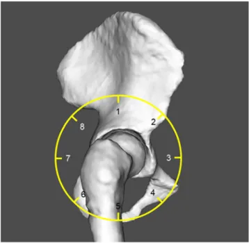
Two experienced musculoskeletal radiologists (with 6 and 14 years’ experience in musculoskeletal radiology respec-tively) analyzed, in a randomized order, all MR images in consensus. The readers were blinded to the clinical evaluation. The acetabular cartilage abnormalities, the labral abnormal-ities, and the acetabular bony contours were assessed qualita-tively at six positions (1, superior; 2, anterosuperior; 3, anterior; 6, posteroinferior; 7, posterior; 8, posterosuperior),
as depicted in Fig.2. Cartilage lesions were considered absent
or present, and the extent of cartilage damage was reported in millimeters. The acetabular labrum was considered normal,
degenerated (abnormal signal intensity), torn (abnormal linear intensity extending to the labral surface), as ossification of the labrum (continuity of the labrum with acetabular bone mar-row), or as a separated ossicle (os acetabuli). The presence of subchondral acetabular or femoral bony abnormalities (e.g., edema, cysts) and the presence of a herniation pit (a round cystic lesion at the anterior aspect of the femoral neck) were also reported.
The evaluation of the wasting of the cervico-cephalic
junction (femoralα neck angle), and the assessment of the
acetabular depth and version were performed by a third reader (with 4 years of experience in musculoskeletal
radi-ology). Theα angle was measured in eight positions around
the femoral neck (see Fig. 2) using radial plane images
centered on the femoral neck axis [11] and superimposed
on the 3D reconstructed bony models (Fig.3a). Theα angle
measurement was performed in accordance with the method
described by Nötzli et al. [12]. Deviation from the normal
geometry was associated with largerα angles (> 55°). The
acetabular depth was evaluated according to the method
detailed by Pfirrmann et al. [2]. The depth was considered
positive and normal if the center of the femoral head was lateral to the line connecting the anterior and posterior
acetab-ular rim (Fig. 3b). Measurement of the acetabular version
was based on the angle between the sagittal direction and lines drawn between the anterior and posterior acetabular
rim, at different heights (Fig. 3c). The angle was
consid-ered positive when inclined medially to the sagittal plane Table 1 Magnetic resonance imaging (MRI) sequences and their
imaging parameters
MRI sequence Imaging parameters
3D fast gradient echo (VIBE) Section thickness 5 mm; no intersection gap; TR/TE ms 4.15/1.69; flip angle, 10°; field of view, 35 cm; matrix 256×256; one signal acquired Coronal T1-weighted turbo
spin-echo
Section thickness 3 mm; no intersection gap; TR/TE ms 565/13; flip angle 180°; field of view, 16 cm; matrix 320×208; one signal acquired Coronal
intermediate-weighted fast spin-echo
Section thickness 3 mm; no intersection gap; TR/TE ms 2180/13; flip angle, 180°; field of view 16 cm; matrix 320×224; two signals acquired Radial intermediate-weighted
fast spin-echo
Section thickness 3 mm; TR/TE ms 2180/13; field of view 16 cm; matrix 384×269; one signal acquired Sagittal water excitation 3D
double-echo steady state
Section thickness 0.6 mm; TR/TE ms 10.74/4.8; flip angle 28°; field of view, 20 cm; matrix 384×307, one signal acquired
Sagittal water excitation 3D double-echo steady state
Section thickness 1.3 mm; no
intersection gap; TR/TE ms 10.74/4.8; flip angle 28°; field of view, 20 cm; matrix 384×384; one signal acquired Transverse 3D fast gradient
echo (VIBE)
Section thickness 5 mm; no intersection gap; TR/TE ms 4.15/1.69; flip angle, 10°; field of view, 35 cm; matrix 256×256; one signal acquired TE echo time, TR repetition time
Fig. 2 Acetabulum divided into eight sectors (position 1, superior; position 2, anterosuperior; position 3, anterior; position 4, anteroinferior; position 5, inferior; position 6, posteroinferior; position 7, posterior; position 8, posterosuperior). The acetabular cartilage abnormalities, the labral abnormalities, and the acetabular bony contours were assessed qualitatively at positions 1–3 and 6–8
(anteversion) and negative when inclined laterally to the sag-ittal plane (retroversion).
Extreme ROM and joint congruency computation
Extreme ROM of the hip joint were calculated using the 3D bony models derived from the dancers’ MRI data and two coordinate systems (one for the femur and one for the
pelvis). We used the definitions proposed by the Standard-ization and Terminology Committee of the International
Society of Biomechanics [13] to report joint motion in an
intra- and inter-subject repeatable way. First, the local axis system in each articulating bone was generated. This was achieved by setting a geometric rule that constructed the pelvic and femoral coordinate systems using selected ana-tomical landmarks defined on the reconstructed surface of Fig. 3 a Definition of theα angle on a radial magnetic resonance (MR)
image (radial intermediate-weighted fast spin-echo sequence without fat saturation, 2,180/13) according to Rakhra et al. [11], illustrating a cam-type morphology (α=85°). The α angle is defined by the angle formed by the line O−O′ connecting the center of the femoral head (O) and the center of the femoral neck (O′) at its narrowest point, and the line O−P connecting O and the point P where the distance between the bony contour of the femoral head and O exceeds the radius (r) of the femoral head. b Definition of the acetabular depth on a transverse oblique MR
image (TrueFISP, 10.74/4.8, flip angle 28°) obtained through the center of the femoral neck according to Pfirrmann et al. [2]. The depth is defined by the distance between the center of the femoral neck (O) and the line AR– PR connecting the anterior (AR) and posterior (PR) acetabular rim. c Computation of the acetabular version based on three-dimensional recon-struction; roof edge (RE) and equatorial edge (EE) are lines drawn between the anterior and posterior acetabular edges, defining the orienta-tion of the acetabular opening proximally and at the maximum diameter of the femoral head respectively (arrows)
Fig. 4 a The pelvic coordinate system (XYZ), the femoral coordinate system (xyz), and the joint coordinate system (e1e2e3) for the right hip joint. Flexion/extension is about the fixed axis of the pelvic body (e1). Internal/external rotation is about the fixed axis of the femoral body (e3) and abduction/adduction is about the floating axis (e2). b Representation of the relative orientation between the hip bone and femur using the pelvic and femoral coordinate systems, while the subject is in the splits position (top view)
the hip and femur bones. These axes then standardized the
joint coordinate system (Fig. 4a). In the neutral position
and orientation, the pelvic and femoral frames were aligned. Thus, given the extracted bone positions from MR images in the splits position, the relative orientation between the hip bone and femur was determined by computing the relative orientation of the femoral frame
to the pelvic frame (Fig. 4b). This was finally expressed
in clinically recognizable terms (flexion/extension, abduc-tion/adduction and internal/external rotation) by represent-ing the relative orientation as three successive rotations. It is important to note that the measurements were per-formed independently of the major anatomical planes (i.e., sagittal, transverse, frontal planes).
Using the method described in Gilles et al. [14], the
congruency of the hip joint in extreme flexion was
com-puted as follows: the hip–joint center (HJC) position was
first estimated in the reference neutral posture. This was determined based on the simulation of a circumduction motion pattern applied to the 3D bony models, while enforcing a constant inter-articular distance corresponding
to the reference distance in the neutral posture. For this simulated motion (involving rotation and translations), the HJC was estimated to be the least moving femoral point in the pelvic frame. The 3D bony models were then registered to extract joint poses from MR images in the splits position. Finally, femoroacetabular trans-lations were measured with reference to the previously estimated HJC.
Statistical analysis
The outcomes of interest were evaluated in 59 hips (29 bilateral and 1 unilateral) of 30 dancers and in 28 hips of 14 control subjects of similar age and sex. For categor-ical variables, odds ratios (OR) and their 95% confidence intervals (CI) were calculated and a p value was obtained using the Chi-squared or Fisher’s exact test. For contin-uous variables, mean values and SD were calculated, as well as p values using the Mann–Whitney U test. The statistical software package SPSS, version 15.0, was employed.
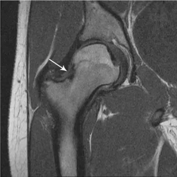
Fig. 5 Sagittal true Fast Imaging with Steady-state Precession (FISP) (10.74/4.8; 28° flip angle) magnetic resonance image shows a posterosupe-rior acetabular cartilage defect associated with a subchondral cyst (arrow)
Fig. 6 a Coronal intermediate-weighted magnetic resonance (MR) image (2,180/13) with fat saturation shows an incomplete tear of the anterosuperior la-brum (arrow). b Coronal intermediate-weighted MR im-age (2,180/13) with fat satura-tion shows areas of high signal intensity inside the superior part of the labrum (arrow) indi-cating degenerative changes
Fig. 7 Coronal intermediate-weighted image (2,180/13) with fat satu-ration. Note the herniation pit at the superior position of the femoral head–neck junction (arrow)
Results Imaging data
Based on the assessment of the MRI scans, three types of
lesions were found in the dancers’ hips: acetabular cartilage
thinning associated with subchondral cysts (Fig.5),
degen-erative labral lesions (Fig. 6), and herniation pits in the
superior position (Fig.7).
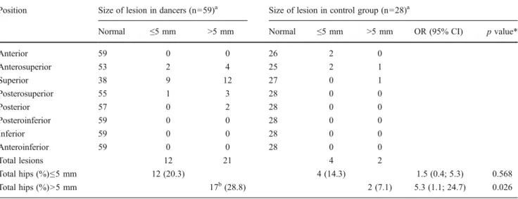
Acetabular cartilage lesions >5 mm were significantly more frequent in dancer’s hips than in control hips (28.8 vs 7.1%, p=0.026). In dancers, they were mostly present at
the superior position (Table2). Distribution of labral lesions
between the dancers and the control group in six positions
around the acetabulum (Table3) showed substantially more
pronounced labral lesions in the superior, posterosuperior, and anterosuperior positions in dancers (54 lesions in 28 dancer’s hips vs 10 lesions in 8 control hips). Fibrocystic
changes (herniation pits, Table4) were found significantly
more often (p=0.002) in dancer’s hips (n=31, 52.5%), 25 of them being located in the superior position. In the control group, pits were found in 5 hips (17.9%), 4 at the anteroin-ferior position and 1 at the anterior position. Osseous bump formation at the femoral neck was observed for one dancer only. Subchondral acetabular cysts were noted for two dancer’s hips, one being located in the posterior and 1 in the posterosuperior positions.
Results of the morphological measurements revealed that the dancers’ and control group hips were normal, except for one dancer in whom a cam morphology was found in relation to detected osseous bump formation, and for one patient in the control group in whom a retroverted
hip was noted. Table 5 summarizes the results of our
morphological analysis.
ROM and joint congruency data
As reported in Gilles et al. [14], the 59 hip MR images of
dancers taken while doing the splits showed a mean femo-roacetabular subluxation of 2.05 mm (range 0.63–3.56 mm). We did not observe any privileged direction of femoroace-tabular translations. For the ROM, the angles showed low SDs, suggesting that movements were repeated similarly across dancers. No significant left–right differences were
noted. Table6reports the computed ROM and subluxation
of the hip joint in extreme flexion.
Discussion
According to the clinical examination performed by two experienced orthopedic surgeons, the majority of dancers
complained of hip pain while dancing only [15]. Fifty-five
percent of the dancers had pain and lesions on MRI, while 35 % had no pain and lesions visible on MRI. Some dancers (5 %) had pain, but no lesions were visible on MRI. We therefore concluded that no clear correlation between
clini-cal and radiologiclini-cal findings could be made [15]. As
dem-onstrated by the morphological analysis and distribution of lesions in dancers’ hips, typical FAI is low in this selected population of professional ballet dancers. Indeed, an abnor-mal morphology of the cam type was found in only one hip, where the characteristic findings expected in cam-type FAI were observed: an osseous bump at the anterosuperior Table 2 Acetabular cartilage lesions
Position Size of lesion in dancers (n=59)a Size of lesion in control group (n=28)a
Normal ≤5 mm >5 mm Normal ≤5 mm >5 mm OR (95% CI) p value*
Anterior 59 0 0 26 2 0 Anterosuperior 53 2 4 25 2 1 Superior 38 9 12 27 0 1 Posterosuperior 55 1 3 28 0 0 Posterior 57 0 2 28 0 0 Posteroinferior 59 0 0 28 0 0 Inferior 59 0 0 28 0 0 Anteroinferior 59 0 0 28 0 0 Total lesions 12 21 4 2 Total hips (%)≤5 mm 12 (20.3) 4 (14.3) 1.5 (0.4; 5.3) 0.568 Total hips (%)>5 mm 17b(28.8) 2 (7.1) 5.3 (1.1; 24.7) 0.026
*p values obtained with the use of Fisher’s exact test
a
Data are the number of hips
b
T able 3 Labral lesions Position Labrum condition in dancers (n = 59) a Labrum condition in control group (n = 28) a OR (95 % CI) p value Normal Degeneration T ear Ossification Ossicle Normal Degeneration T ear Ossification Ossicle Anterior 52 3 3 1 0 28 0 0 0 0 Anterosuperior 37 7 1 3 2 0 2 2 3 3 0 0 Superior 20 18 19 2 0 12 9 3 4 0 Posterosuperior 35 10 13 1 0 23 1 4 0 0 Posterior 53 2 3 1 0 28 0 0 0 0 Posteroinferior 53 2 3 1 0 28 0 0 0 0 Inferior 57 2 0 0 0 28 0 0 0 0 Anteroinferior 59 0 0 0 0 28 0 0 0 0 T otal lesions 34 54 8 1 3 1 0 4 T otal hips (%) degeneration 24 (40.7) 12 (42.9) 0.847* Hips with ≥ 2 lesions (%) 1 1 (18.6) 1 (3.6) T otal hips (%) tear 28 (47.5) 8 (28.6) 2.3 (0.9; 5.9) 0.095* Hips with ≥ 2 lesions (%) 12 (20.3) 1 (3.6) T otal hips (%) ossification 2 (3.4) 4 (14.3) 0.082** Hips with ≥ 2 lesions (%) 2 (3.4) 0 * p values obtained with the use of Chi-squared test ** p values obtained with the use of Fisher ’s exact test a Data are the number of hips
femoral head–neck junction and labro-cartilaginous lesions located along the anterosuperior part of the acetabulum. Moreover, when analyzing the MR images acquired in the splits position, it is interesting to note that the herniation pits were located exactly at the contact zone between the ante-rosuperior femoral head–neck junction and the acetabulum,
as expected in the case of cam-type FAI (Fig.8).
Despite the absence of articular contrast media injection, which could lower the sensitivity and specificity of cartilag-inous and labral detection, hip lesions of the acetabular labrum and cartilage, as well as the herniation pits, were, for the majority of dancers, statistically more pronounced in the superior position around the acetabular rim compared with the group of asymptomatic non-dancer female volun-teers. Acetabular cartilage lesions >5 mm were significantly more frequent in dancers (28.8 vs 7.1%, p=0.026) and were mostly present in the superior position. Distribution of labral lesions between the dancers and the control group in six positions around the acetabulum showed substantially more
pronounced labral lesions at the superior, posterosuperior, and anterosuperior positions in dancers (54 lesions in 28 dancers’ hips vs 10 lesions in 8 control hips). Herniation pits
were found significantly more often (p=0.002) in dancers’
hips (n=31, 52.5%), 25 of them being located in the supe-rior position. This pattern of lesion distribution has, to our knowledge, not been reported in typical FAI of the cam or pincer type. In the absence of a focal or global acetabular over-coverage, such as a prominent posterior acetabular wall, acetabular retroversion, coxa profunda or protrusio acetabuli, the explanation for the presence of these lesions seems to be their correlation with extreme motion assumed by the dancers’ hips during their daily activities. These
extreme positions seem to be responsible for a“pincer-like”
mechanism of impingement, with linear contact between the superior or posterosuperior acetabular rim and the femoral
head–neck junction. This mechanism has been
demonstrat-ed by Charbonnier et al. [16,17], who assessed,
dynamical-ly, dancers’ hip joint motions. Dynamic data were collected by these authors, while the professional dancers were performing six dancing movements: arabesque, développé devant, développé à la seconde, grand écart facial, grand écart latéral, and grand plié. Visualization of the hip motion and functional evaluation were based on dancer-specific 3D models obtained by the segmentation of MRI data and the use of optical motion capture. The authors demonstrated that Table 4 Herniation pits
*p values obtained with use of Fisher’s exact test
aData are the number of hips
Herniation pits in dancers (n=59)a Herniation pits in control group (n=28)a Position Absent Present Absent Present OR (95 % CI) p value*
Anterior 57 2 27 1 Anterosuperior 57 2 28 0 Superior 34 25 28 0 Posterosuperior 55 4 28 0 Posterior 59 0 28 0 Posteroinferior 58 1 28 0 Inferior 59 0 28 0 Anteroinferior 58 1 24 4 Total lesions 35 5 Total hips (%) 31 (52.5) 5 (17.9) 5.1 (1.7; 15.2) 0.002
Table 5 ! Angle (degrees) in eight positions around the femoral head, acetabular depth (mm), and version (degrees)
Measure Dancers Control group p value* α Angle (anterior) 45.5±5.3 47.5±4 0.018 α Angle (anterosuperior) 46.7±6.7 46.0±4.9 0.863 α Angle (superior) 40.2±4.8 46.6±4.4 <0.001 α Angle (posterosuperior) 38.3±3.6 43±6.7 α Angle (posterior) 39.9±4.6 40.2±4.8 α Angle (posteroinferior) 38.3±3.6 48.7±6.9 α Angle (inferior) 40.2±3.6 51.2±6.3 α Angle (anteroinferior) 40.1±4.3 44.7±5.4 Acetabular depth 7.5±1.7 8.7±2.1 Acetabular version 7.5±4.1 5.9±5 Data are mean ± standard deviation
*p values obtained with Mann–Whitney U test
Table 6 Range of movement (ROM; degrees) according to our refer-ential and subluxation (mm) in the splits position
Measure Minimum Mean ± SD Maximum
Flexion 109 133±10 158.5
Abduction 17 32±7 49
IR/ER 0/14.5 17.5±13/0 41.5/0
Subluxation 0.63 2.05±0.74 3.56 ER external rotation, IR internal rotation
for almost all the aforementioned movements assessed, the impingement, in other words the abnormal contact between the proximal femur and acetabular rim, was located mainly in the superior or posterosuperior quadrant of the acetabu-lum. From a morphological point of view, this mechanism is also supported by the fact that some dancers presented cortical irregularity in the superolateral part of the femoral
neck (Fig.9).
As reported in Gilles et al. [14], the MRI data acquired
with the dancers in the splits position showed for the 59 hips a mean femoroacetabular subluxation of 2.05 mm (range
0.63–3.56 mm). The magnitude of subluxation during the
dancing movements assessed by Charbonnier et al. [16,17]
was even greater (peak value = 6.32 mm). We can thus suppose that the lost of joint congruency exposes the dancers’ hips cartilage to stress, which also favors cartilage lesions. Nevertheless, we must note that we did not find contrecoup lesions in the anteroinferior acetabular cartilage,
as could be expected in a “pincer-like” mechanism of
im-pingement with subluxation. Finally, it is worth mentioning that those extreme movements, such as the splits position performed by the dancers in the magnet bore, imply a combination of abduction, flexion, and rotation.
Two study limitations need to be stated: the radiological analysis was based on hip MRI and not MR arthrography, and the consensus reading of the cases. In spite of these limitations, the results of our study demonstrated interesting findings, which can be summarized as follows. The preva-lence of typical FAI of the cam or pincer type was low in this selected population of professional ballet dancers;
how-ever, a “pincer-like” mechanism of impingement seems to
occur in relation to extreme movements performed by the dancers during their daily activities. This mechanism could explain the acetabular labral and cartilage lesions, as well as the herniation pits, predominantly found in the superior acetabular quadrant. Furthermore, femoroacetabular sublux-ations were observed while doing the splits. On the basis of the evidence, we believe that extreme hip motion in this selected population could be a potential risk factor for the development of early hip OA.
Conflicts of interest The authors declare that they have no conflict of interest.
References
1. Ganz R, Parvizi J, Beck M, Leunig M, Nötzli H, Siebenrock KA. Femoroacetabular impingement: a cause for osteoarthritis of the hip. Clin Orthop Relat Res. 2003;417:112–20.
2. Pfirrmann CWA, Mengiardi B, Dora C, Kalberer F, Zanetti M, Hodler J. Cam and pincer femoroacetabular impingement: Char-acteristic MR arthrographic findings in 50 patients. J Radiol. 2006;240(3):778–85.
3. Beck M, Kalhor M, Leunig M, Ganz R. Hip morphology influen-ces the pattern of damage to the acetabular cartilage: femoroace-tabular impingement as a cause of early osteoarthritis of the hip. J Bone Joint Surg Br. 2005;87:1012–8.
4. Ito K, Minka 2nd MA, Leunig M, Werlen S, Ganz R. Femoroace-tabular impingement and the cam-effect: a MRI based quantitative anatomical study of the femoral head–neck offset. J Bone Joint Surg Br. 2001;83:171–6.
5. Lavigne M, Parvizi J, Beck M, Siebenrock KA, Ganz R, Leunig M. Anterior femoroacetabular impingement: Part I: Techniques of joint preserving surgery. Clin Orthop Relat Res. 2004;418:61–6. Fig. 8 Reformatted TrueFISP (10.74/4.8; 28° flip angle) magnetic
resonance image in a dancer in the splits position. Note the herniation pit located at the contact zone between the anterosuperior femoral head–neck junction and the acetabulum (arrow)
Fig. 9 Coronal T1-weighted (565/13) magnetic resonance image. Note the cortical irregularity in the superolateral part of the femoral neck (arrow)
6. Leunig M, Beaulé PE, Ganz R. The concept of femoroacetabular impingement. Clin Orthop Relat Res. 2009;467:616–22. 7. Reynolds D, Lucac J, Klaue K. Retroversion of the acetabulum: a
cause of hip pain. J Bone Joint Surg Br. 1999;81:281–8. 8. Wagner S, Hofstetter W, Chiquet M, et al. Early osteoarthritic
changes of human femoral head cartilage subsequent to femoro-acetabular impingement. Osteoarthritis Cartilage. 2003;11:508–18. 9. Gilles B, Moccozet L, Magnenat-Thalmann N. Anatomical mod-elling of the musculoskeletal system from MRI. MICCAI '06, Part II. LNCS, Springer Berlin Heidelberg, 4190, pp. 289–296, 2006. 10. Schmid J, Kim J, Magnenat-Thalmann N. Robust statistical shape
models for MRI bone segmentation in presence of small field of view. Med Image Anal. 2011;15:155–68.
11. Rakhra KS, Sheikh AM, Allen D, Beaulé PE. Comparison of MRI alpha angle measurement planes in femoroacetabular impinge-ment. Clin Orthop Relat Res. 2009;467(3):660–5.
12. Nötzli HP, Wyss TF, Stöcklin CH, Schmid MR, Treiber K, Hodler J. The contour of the femoral head–neck-junction as a predictor for
the risk of anterior impingement. J Bone Joint Surg Br. 2002;84:556–60.
13. Wu G, Siegler S, Allard P, et al. ISB recommendation on defini-tions of joint coordinate system of various joints for the reporting of human joint motion - part I: Ankle, hip and spine. J Biomech. 2002;35(4):543–8.
14. Gilles B, Kolo FC, Magnenat-Thalmann N, et al. MRI-based assess-ment of hip joint translations. J Biomech. 2009;42(9):1201–5. 15. Duthon VB, Charbonnier C, Kolo FC, et al. Correlation of clinical
and MRI findings in hips of elite female ballet dancers. Arthros-copy. 2012; In Press.
16. Charbonnier C, Kolo FC, Duthon VB, et al. Assessment of con-gruence and impingement of the hip joint in professional ballet dancers: A motion capture study. Am J Sports Med. 2011;39 (3):557–66.
17. Charbonnier C, Kolo FC, Duthon VB et al. Professional dancer’s hip: a motion capture study. Trans Orthop Res Soc. New Orleans, Louisiana, March 2010.