HAL Id: hal-03152856
https://hal.archives-ouvertes.fr/hal-03152856
Submitted on 25 Feb 2021
HAL is a multi-disciplinary open access
archive for the deposit and dissemination of
sci-entific research documents, whether they are
pub-lished or not. The documents may come from
teaching and research institutions in France or
abroad, or from public or private research centers.
L’archive ouverte pluridisciplinaire HAL, est
destinée au dépôt et à la diffusion de documents
scientifiques de niveau recherche, publiés ou non,
émanant des établissements d’enseignement et de
recherche français ou étrangers, des laboratoires
publics ou privés.
Automated atrial fibrillation source detection using
shallow convolutional neural networks
Isac Do Nascimento Lira, Pedro Marinho R. de Oliveira, Walter Freitas,
Vicente Zarzoso
To cite this version:
Isac Do Nascimento Lira, Pedro Marinho R. de Oliveira, Walter Freitas, Vicente Zarzoso.
Auto-mated atrial fibrillation source detection using shallow convolutional neural networks. Computing in
Cardiology, Sep 2020, Rimini, Italy. �10.22489/CinC.2020.385�. �hal-03152856�
Automated Atrial Fibrillation Source Detection Using Shallow Convolutional
Neural Networks
Isac N. Lira
1, Pedro Marinho R. de Oliveira
2, Walter Freitas Jr.
1, Vicente Zarzoso
21
Universit´e Cˆote d’Azur, CNRS, I3S Laboratory, Sophia Antipolis, France
2Universidade Federal do Cear´a, Brazil
Abstract
Atrial fibrillation (AF) is the most frequent sustained ar-rhythmia diagnosed in clinical practice. Understanding its electrophysiological mechanisms requires a precise anal-ysis of the atrial activity (AA) signal in ECG recordings. Over the years, signal processing methods have helped cardiologists in this task by noninvasively extracting the AA from the ECG, which can be carried out using blind source separation (BSS) methods. However, the robust automated selection of the AA source among the other sources is still an open issue. Recently, deep learning architectures like convolutional neural networks (CNNs) have gained attention mainly by their power of automat-ically extracting complex features from signals and clas-sifying them. In this scenario, the present work proposes a shallow CNN model to detect AA sources with an auto-mated feature extraction step overcoming the performance of other methods present in the literature.
1.
Introduction
The most frequent sustained arrhythmia diagnosed in clinical practice, atrial fibrillation (AF) is a supraventric-ular tachyarrhythmia characterized by an uncoordinated and irregular atrial activation [1]. The electrophysiologi-cal mechanisms underlying AF are not totally understood, which makes this cardiac condition gather increasing at-tention from researchers and cardiologists in the past few years.
Signal processing methods have helped cardiologists to better manage this cardiac rhythm disturbance by nonin-vasively extracting the atrial activity (AA) signal from the standard 12-lead electrocardiogram (ECG). In particular, the AA extraction from multilead AF ECGs accepts a blind source separation (BSS) formulation [2] and several tech-niques to solve BSS problems were reported in the liter-ature as useful tools in noninvasive AA extraction for AF analysis [2]–[5]. In particular, the block term decomposi-tion (BTD), a tensor-based BSS method, have proved to be
an important AA extraction tool, overcoming the matrix-based techniques [4], [5].
In the challenging case of AF ECGs, techniques to solve BSS problems separate the original recording in several sources, where, typically, at least one of these sources con-tains the AA. After separating the source signals, it is nec-essary to select the AA source estimate among the other sources. Atrial source selection requires visual inspection to achieve optimality, as an optimal automated method for this task is still an open challenge. However, machine learning algorithms have provided an improved accuracy compared to other automated methods [7]. In this context, deep learning techniques have proved to be very efficient in many tasks like image detection and classification, mainly due to its ability to perform an automated feature extrac-tion [8].
In the previous work [7] standard machine learning models were applied to detect AA sources using hand-crafted features. In order to improve this process, the present work proposes a framework that combines the BTD technique with a shallow convolutional neural net-work (CNN) in order to tackle the task of detecting AA sources from ECG segments with automatically extracted features.
2.
Data Description and Pre-processing
2.1.
Database
The analyzed data during the experiments are com-posed by 116 random segments of 58 12-lead ECG record-ings from 58 persistent patients, each one with duration of approximately 1 second. These recordings belong to a database provided by the Cardiology Department of Princess Grace Hospital Center, Monaco. The recordings are acquired at a 977 Hz sampling rate and are prepro-cessed by a zero-phase forward-backward type-II Cheby-shev bandpass filter with cutoff frequencies of 0.5 and 40 Hz, in order to suppress high-frequency noise and baseline wandering.
2.2.
AA Source Extraction
Signal processing techniques that solve BSS problems separate the observed ECG signal matrix Y in a linear com-bination of a mixing matrix M and a source matrix S:
Y = MS ∈ R.K×N (1)
In the present case of study, Y ∈ RK×Nis the AF ECG
data matrix, composed of K signals (leads) and N sam-ples, M ∈ RK×Ris the mixing matrix, modeling the
prop-agation of the R cardiac electrical sources from the heart to the K leads in the body surface, and S ∈ RR×N is the
source matrix that contains R sources, mainly atrial, ven-tricular and noise sources.
After some mathematical manipulations, the ECG data matrix can be transformed into a tensor that admits a BTD model [5]. This tensor factorization technique is based on a third-order tensor Y built from Hankel matrices that are constructed from each row of the observed data matrix. The tensor is then decomposed as [6]:
Y =
R
X
r=1
Er◦ c.r (2)
where ◦ represents the outer product, c.ris a nonzero
vec-tor, Eris a Hankel matrix built from each source and R is
the number of sources.
This tensor-based BSS technique is computed using the recently proposed algorithm called constrained alternating group lasso [4] and applied to ECG segments generating 509 sources that are visually labeled as 122 AA sources, 273 ventricular activity (VA) sources and 114 unknown (UNK) sources.
The scheme proposed in this work aims to distinguish only AA sources from the remaining sources. In this way, the VA and UNK sources are grouped into a single class, called non-AA sources, which configures a binary classifi-cation problem to be solved.
Initially, the data is randomly split into train and vali-dation sets with a ratio of 80% for the training samples. The signals are normalized with respect to the mean µt
and variance σt obtained from the training set Xtrain =
{x0, . . . , xT}, where xi is a vector representing an ECG source signal. Considering v the concatenation of all vec-tors from Xtrain, one can compute the overall µt and σt
as: µt= 1 V V X i=1 vi (3a) σt= 1 V V X i=1 (vi− µt)2 (3b)
where vi is the i-th entry of v, and V = |v| denotes the
cardinality of v. The transformed signal components are then computed as:
zi=
xi− µt
σt
, for i = 1, . . . , |x|. (4)
This normalization operation is assigned to a batch nor-malization layer in the CNN model.
2.3.
Data Augmentation
Due to the low number of segments available, the train-ing of deep learntrain-ing models can suffer from overfitttrain-ing. To overcame this problem, a window slicing (WS) based method is applied to augment the data and consequently provides more samples to the training process. This method was first introduced in [11] also in the context of time series classification using CNN and it has proved use-ful to increase model performance. It affects the training as well as the prediction phase.
For a given ECG segment and its class (xi, yi), a
win-dow with size W < |xi| is applied to extract a subsignal
xi,j. The window is moved by S ≤ W samples to obtain a new signal xi,j+1 and the process is repeated until the original segment is completely split.
By applying the WS strategy during the training step, each signal xi generates a set of subsignals Xi =
{(xi,0, yi), . . . , (xi,N, yi)}, all of them sharing the same
label yi. For the prediction phase, we propose to estimate
the value of the class probability ˜yiby averaging the model
scores of the subsignals, as described in Equation (5).
˜ yi= 1 N N X j=0 ˜ yi,j (5)
where N is the number of the generated subsignals from xiand ˜yi,jthe model prediction for xi,j. Just after the data
augmentation process, a random oversampling (ROS) step is applied over the minority class (AA source) in order to balance the training data.
3.
CNN Model Selection
3.1.
Convolutional Neural Networks
The CNN is a Deep Learning model initially designed for multi-dimensional data like images. The main com-ponents of a CNN are the convolutional layers, the pool-ing layers and fully connected layers. Durpool-ing the convolu-tional operation, a bank of filters is applied over the whole input signal using the same weights and it generates acti-vations for each receptive field that are combined to form a feature map [10]. Each set of weights are optimized by a
gradient algorithm to detect specific type of features along the input signal.
Along with the convolutional operation, the pooling lay-ers perform a reduction in the feature space and combine similar features [14]. For example, the max-pooling kernel slides the feature space getting the maximum value from small regions.
3.2.
Architecture Optimization
To find a suitable CNN configuration for the task of AA source detection, a Bayesian algorithm is applied using BoTorch [13], a framework used for optimization tasks. We compare different shallow CNN architectures chang-ing the followchang-ing paramenters: number of hidden nodes, training epochs, convolutional/pooling layers, kernel size, kernel stride and batch size.
The maximal number of convolutional layers is set to 3 which keeps the CNN model simpler and much shal-lower than the common CNN models found in the liter-ature. This reduces the number of trainable weights, thus avoiding overfitting. Along with the model architecture parameters, the augmentation settings (window size and stride) are also optimized. Furthermore, the upper bound value for the window size is limited by the length of the shortest extracted ECG source.
Let L be the number of convolutional layers and ki,l
the size of the convolutional kernel i in the layer l. The constraint ki,l ≤ ki,l+1 is imposed on each layer l ∈
{1, . . . , L − 1}. Another constraint requires all kernels from layer l to have the same size Kl.
Similarly, the stride Slfor the kernels have to follow the
inequality Sl ≤ Sl+1. By doing that, it is produced an
increasing reduction in the feature space.
A final constraint is defined to have an increasing num-ber of channels in consecutive layers which allows the model to capture more complex features from the signals.
3.3.
Model Training and Evaluation
The weights for the shallow CNN models are optimized using the Adam optimizer with a learning rate being se-lected by the Bayesian algorithm. Their values are within the range [10−4, 10−3]. Each model is evaluated on the validation set with respect to the Area Under the ROC Curve (AUC) aiming to find the model that provides the maximum possible score.
In this work, we consider the AA sources as being the positive class, and the non-AA sources as the negative one. The sensitivity and specificity metrics are used to measure the model performance for each class individually. The sensitivity is defined as:
SEN = TP
TP + FN (6) where T P are the true positive samples and F N the False Negatives. Similarly, the specificity is computed by (7) using the T N as the number of true negative samples and F P the quantity of false positive samples:
SPE = TN
TN + FP. (7) Finally, the accuracy is computed to measure the overall model precision.
4.
Experimental Results
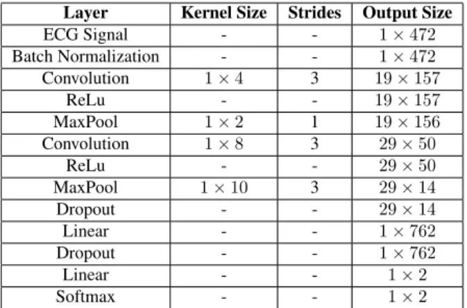
After running 100 trials, the best architecture is chosen with an AUC validation score of 97.5%. The best parame-ters for the CNN are in the Table 1. The must appropriate batch size shown is 124 samples, and for the augmentation window size the best value is 472 with a stride percentage of 21% resulting in an absolute stride of 99 samples. Table 1. Optimized parameters for the shallow CNN ar-chitecture.
Layer Kernel Size Strides Output Size
ECG Signal - - 1 × 472 Batch Normalization - - 1 × 472 Convolution 1 × 4 3 19 × 157 ReLu - - 19 × 157 MaxPool 1 × 2 1 19 × 156 Convolution 1 × 8 3 29 × 50 ReLu - - 29 × 50 MaxPool 1 × 10 3 29 × 14 Dropout - - 29 × 14 Linear - - 1 × 762 Dropout - - 1 × 762 Linear - - 1 × 2 Softmax - - 1 × 2
The model evaluation is performed applying a 10-fold cross validation (CV) to compute ACC and AUC. Not all data are used in the evaluation; instead, the CV folds are computed only over the training data, since the CNN archi-tecture is selected on the validation set. The average AUC and ACC achieved are 96.3% and 93.6%, respectively and the results across the CV folds are plotted in a box plot in Figure 1.
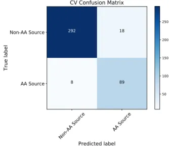
Additionally the model performance is represented in a confusion matrix in Figure 2 whose values are based on the CV folds. From the matrix, the obtained sensitivity and specificity metrics are 91.75% and 94.19%, respectively.
5.
Conclusions
In order to improve the process of detecting AA sources in ECG segments, the present work proposes a framework
to perform an automated source detection. The solution joins a tensor-based BSS method with a shallow CNN model with an optimized structure. The model requires less training weights than common deep learning models and it also has the advantage of automatically extracting features from ECG sources, thus avoiding handcrafted fea-tures and achieving promising results.
Further works may include the application of transfer learning using ECG source spectrograms to increase the detection performance.
Acknowledgments
Pedro Marinho R. de Oliveira is funded by a PhD schol-arship from the IT Doctoral School (EDSTIC) of the Uni-versit´e Cˆote d’Azur.
References
[1] C. T. January, L. S. Wann, H. Calkins, L. Y. Chen, J. E. Cigarroa, et al., “2019 AHA/ACC/HRS Focused Update of the 2014 AHA/ACC/HRS Guideline for the Management of Patients With Atrial Fibrillation”, Journal of the American College of Cardiology, vol. 74, no. 1, pp. 104- 132, 2019. [2] J. J. Rieta, F. Castells, C. S´anchez, V. Zarzoso, and J.
Mil-let, “Atrial activity extraction for atrial fibrillation analysis using blind source separation”, IEEE Trans. Biomed. Eng., vol. 51, no. 7, pp. 1176-1186, Jul. 2004.
[3] F. Castells, J. J. Rieta, J. Millet, and V. Zarzoso, “Spatiotem-poral blind source separation approach to atrial activity es-timation in atrial tachyarrhythmias”, IEEE Trans. Biomed. Eng., vol. 52, no. 2, pp. 258-267, Feb. 2005.
[4] J. H. de M. Goulart, P. M. R. de Oliveira, R. C. Farias, V. Zarzoso, and P. Comon “Alternating group lasso for block-term tensor decomposition with application to ECG source separation”. IEEE Trans. Sig. Proc., vol. 68, pp. 2682–2696, 2020.
[5] P. M. R. de Oliveira and V. Zarzoso, “Block term decom-position of ECG recordings for atrial fibrillation analysis: Temporal and inter-patient variability”, Journal of Comm. Inf. Syst., vol. 34, no. 1, pp. 111-119, 2019.
[6] L. De Lathauwer, “Blind separation of exponential polyno-mials and the decomposition of a tensor in rank-(lr, lr, 1)
terms”, SIAM Journal on Matrix Analysis and Applications, vol. 32, no. 4, pp. 1451–1474, 2011.
[7] P. M. R. de Oliveira, V. Zarzoso and C.A.R. Fernandes, “Source classification in atrial fibrillation using a machine learning approach”, in Proc. CinC-2019, 46th Computing in Cardiology Conference, Biopolis, Singapore, Sep. 8-11, pp. 1-4, 2019.
[8] Litjens, Geert, et al. ”A survey on deep learning in medical image analysis.” Medical image analysis 42 (2017): 60-88. [9] Le Guennec, Arthur, Simon Malinowski, and Romain Tave-nard. ”Data augmentation for time series classification us-ing convolutional neural networks.” 2016.
[10] Johnson, Justin M., and Taghi M. Khoshgoftaar. ”Survey on deep learning with class imbalance.” Journal of Big Data 6.1 (2019): 27. 86 88 90 92 94 96 98 100 Score AUC ACC M et ri c CV scores
Figure 1. Box plot of the AUC and ACC metrics over CV.
No n-AA S ourc e AA S ourc e Predicted label Non-AA Source AA Source Tr ue la be l 292 18 8 89 CV Confusion Matrix 50 100 150 200 250
Figure 2. CV confusion matrix.
[11] Cui, Zhicheng, Wenlin Chen, and Yixin Chen. ”Multi-scale convolutional neural networks for time series classifica-tion.” arXiv preprint arXiv:1603.06995 (2016).
[12] Van Hulse, Jason, Taghi M. Khoshgoftaar, and Amri Napolitano. ”Experimental perspectives on learning from imbalanced data.” Proceedings of the 24th international conference on Machine learning. 2007.
[13] Balandat, Maximilian, et al. ”Botorch: Programmable bayesian optimization in pytorch.” arXiv preprint arXiv:1910.06403 (2019).
[14] LeCun, Yann, Yoshua Bengio, and Geoffrey Hinton. ”Deep learning.” nature 521.7553 (2015): 436-444.
Address for correspondence: Pedro Marinho R. de Oliveira
Universit´e Cˆote d’Azur, CNRS, I3S Laboratory 06903 Sophia Antipolis Cedex