HAL Id: hal-00097211
https://hal.archives-ouvertes.fr/hal-00097211
Submitted on 23 May 2014
HAL is a multi-disciplinary open access
archive for the deposit and dissemination of
sci-entific research documents, whether they are
pub-lished or not. The documents may come from
teaching and research institutions in France or
abroad, or from public or private research centers.
L’archive ouverte pluridisciplinaire HAL, est
destinée au dépôt et à la diffusion de documents
scientifiques de niveau recherche, publiés ou non,
émanant des établissements d’enseignement et de
recherche français ou étrangers, des laboratoires
publics ou privés.
1-kHz table-top ultrashort hard x-ray source for
time-resolved x-ray protein
Adeline Bonvalet, Adeline Darmon, Jean-Christophe Lambry, Jean-Louis
Martin, Patrick Audebert
To cite this version:
Adeline Bonvalet, Adeline Darmon, Jean-Christophe Lambry, Jean-Louis Martin, Patrick Audebert.
1-kHz table-top ultrashort hard x-ray source for time-resolved x-ray protein. Optics Letters, Optical
Society of America - OSA Publishing, 2006, 31 (18), pp.2753. �10.1364/OL.31.002753�. �hal-00097211�
1 kHz tabletop ultrashort hard x-ray source for
time-resolved x-ray protein crystallography
Adeline Bonvalet, Adeline Darmon, Jean-Christophe Lambry, and Jean-Louis Martin
Laboratoire d’Optique et Biosciences CNRS UMR 7645–INSERM U696–Ecole Polytechnique, 91 128 Palaiseau Cedex, France
Patrick Audebert
Laboratoire pour l’Utilisation des Lasers Intenses, UMR 7605 CNRS–CEA–Ecole Polytechnique–Université Pierre et Marie Curie, 91 128 Palaiseau Cedex, France
Received April 14, 2006; revised July 3, 2006; accepted July 4, 2006; posted July 10, 2006 (Doc. ID 69980); published August 25, 2006
We describe a compact, reliable, and high-average-power femtosecond x-ray source and its first application to diffraction on protein crystal. The setup relies on a homemade Ti: sapphire system delivering12 mJ at a 1 kHz repetition rate, associated with a small vacuum chamber especially designed for laser-plasma inter-action and x-ray applications. This device allows the generation of5 ⫻ 109photons/ s / sr at 8 keV and
opti-mized x-ray irradiation of the studied sample, which can be placed close to the source. We present the dif-fraction pattern of a protein crystal in a divergent beam geometry, which is a first step to a subpicosecond x-ray diffraction experiment. © 2006 Optical Society of America
OCIS codes: 320.5550, 320.7090, 340.7480, 350.5400.
The generation of ultrashort hard x-ray sources is of prime interest for a broad range of applications in bi-ology, chemistry, and physics, since it should extend three-dimensional structure determination to the femtosecond time scale. In recent years, several ex-periments aimed to follow structural evolution in simple crystals by using the ultrashort x-ray pulses emitted by a laser-plasma source1–5 or other techniques.6,7But the low flux of these sources makes diffraction experiments on more demanding diffrac-tors such as protein crystals more difficult.8 There-fore laser-plasma sources offer a great advantage over other x-ray sources, since they should allow tem-poral resolution down to the period of atomic vibra-tion, typically of the order of 100 fs. To reach this ob-jective, ultrashort x-ray sources must become compact and reliable and exhibit high average power. Moreover, their potential must be better utilized by developing a new diffraction method that takes ad-vantage of laser-plasma source divergence.9
Ultrafast monochromatic x-ray pulses can be gen-erated by the interaction of ultrashort laser pulses of adjusted intensity with condensed matter.10–13 Re-cently, detailed experimental14–16 and theoretical17 studies have characterized the dependence of the emission over laser and target characteristics. Laser parameters such as pulse duration, focal intensity, polarization, incidence angle, and intensity contrast ratio play an important role in x-ray yield. Targets have to move fast enough to present a virgin surface at each laser shot while remaining precisely aligned. In addition, the concept of a robust high-repetition-rate laser-plasma source requires particular atten-tion to the debris generated by the laser–target inter-action.
In this Letter we describe an ultrashort hard x-ray source relying on a simple and unique 12 mJ, 1 kHz Ti:sapphire amplifier and present the diffraction
pat-tern of a protein crystal exposed to a divergent beam. The whole setup has been carefully designed to gather the key features for time-resolved crystallog-raphy, such as reliability, high average power, and optimized flux on the studied sample.
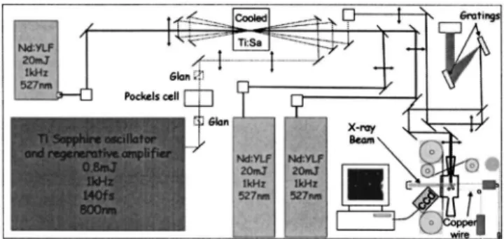
The experimental setup is shown in Fig. 1. The la-ser system delivers 12 mJ, 160 fs pulses at a 1 kHz repetition rate, with high spatial quality, a measured level of amplified spontaneous emission 109 lower
than the main pulse, and contrast with intrinsic nanosecond and picosecond prepulses measured to be 5 ⫻ 106and 5 ⫻ 104, respectively. The system relies on
a commercial oscillator and regenerative amplifier (Hurricane, Spectra-Physics), delivering 0.8 mJ and seeding a homemade amplifier pumped by three frequency-doubled diode-pumped Nd:YLF lasers (two Jades, Thales-Laser; one Evolution-30, Positive Light). To limit thermal lensing due to large pump fluence onto the amplifying crystal (Crystal Systems,
10 mm⫻ 10 mm⫻ 10 mm, 90% absorption at
527 nm), we have designed a simple
liquid-nitrogen-Fig. 1. Design of the experimental setup. The beams of the two 20 W Nd:YLF lasers are in two different quasi-horizontal planes. Two Glan–Laser polarizing prisms and a Pockels cell (Medox, Thales Laser) reduce the nanosecond prepulses issued from leakage in the regenerative amplifier.
September 15, 2006 / Vol. 31, No. 18 / OPTICS LETTERS 2753
cooled cryostat that yields a decrease in the focal-length power from f = 49 cm at 300 K to f = 9.6 m at 100 K while using 63 W of total pump power.18 We obtained a very simple and compact 4-pass amplifier that routinely delivers 18 mJ with excellent spatial quality. Temporal compression of the pulses is achieved in a two-grating compressor (120 mm ⫻ 140 mm, groove density 2000 lines / mm), leading to pulses of 12 mJ energy and 160 fs duration.
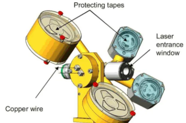
The x-ray source consists of a copper wire running through a small vacuum chamber (see Fig. 2). The wire, issued from a spool, is first flattened and moved by a motorized rolling mill to expose a flat and fresh surface to each laser shot. It then crosses the cham-ber through two Teflon guides that maintain an air pressure of 100 Pa and is guided by two free-rotating bearings. The wire tension is controlled by a pair of toothed wheels that pulled the wire out from the chamber. The jitter of the wire motion is still reduced by a small metallic finger positioned between the bearings. The laser beam is focused with an 18 cm focal-length lens and hits the wire with an incidence angle of 60°. The laser intensity on the target is esti-mated to 3 ⫻ 1016W / cm2, which is thought to be
op-timum for Cu-K␣ x-ray yield.17X-rays are collected through a 17 mm diameter beryllium window placed 16 mm from the target. The deposition of debris on the beryllium window is prevented by a 12.5 mm wide plastic band continuously moving in front of it 共1 m / min兲. The laser beam entrance window is also protected by a similar running band system, with a slower speed 共25 mm/ h兲, and a band tilt that opti-mizes the laser beam transmission. The spectrum is measured by analyzing the deposited energy of indi-vidual x-ray photons on a thermoelectrically cooled CCD camera (Andor Technology, DY434-FI-962),19 that was previously calibrated with an Fe55 source
emitting K␣radiation at 5.9 keV. The emitted laser-plasma x-ray spectrum consists of a broad continuum and two narrow features at 8.05 and 8.91 keV corre-sponding to the characteristic K␣ and K lines of copper, respectively. If necessary, the Kphotons can be filtered with a thin Ni foil. The Cu K␣ yield was evaluated by considering the CCD efficiency at 8 keV (given by the manufacturer), and integration of the measured spectra over the K␣line and is found to be 5 ⫻ 109photons/ s / sr. The size of the x-ray emitting
spot has been determined in the horizontal plane by using a knife-edge technique and is 20m FWHM.
It is larger than the laser spot
(7m FWHM), which may be due to either the jitter in the wire motion or the spreading of fast electrons in the target.
We performed an x-ray diffraction experiment on a protein crystal with a 1 kHz laser-plasma source. Conventional methods for single-crystal diffraction data collection are based on a collimated beam. How-ever, collimating an x-ray beam greatly decreases the flux available on the sample, making diffraction on weak diffractors such as protein crystal difficult. Here we have applied a new method first demon-strated by Ho et al.,9in which a stationary crystal is
exposed to a beam with a large two-dimensional con-vergence or dicon-vergence. We recorded the diffraction pattern of a lysozyme crystal, an enzyme widely dis-tributed in animals and plants. The setup relies sim-ply on a stationary crystal exposed to a divergent beam. The sample is mounted on a goniometric head placed at the outside of the vacuum chamber just be-hind the beryllium window, thus being perfectly pro-tected from the debris. The x-ray beam diameter is reduced by a lead pinhole to the size of the crystal (a cube with 400m sides), and the source angular spread is thus 1°. This value is easily adjusted
by changing the hole. The CCD camera
(1024⫻ 1024 pixels of 13m ⫻ 13m) is placed 2.5 cm behind the sample, off the direction of the di-rect x-ray beam. Acquisition consists of summing im-ages of exposition time equal to 20 s. This exposition time is chosen so that the probability of having more than one photon per pixel is negligible.20 Conse-quently, each image is filtered to eliminate photons whose energy is above the Cu K lines, and the final image is then obtained by summing all the images. This basic filtering method can be further improved by use of event recognition techniques.21 Figure 3 shows the image obtained after only 50 min of acqui-sition. The resulting diffraction pattern resembles
Fig. 2. (Color online) Vacuum chamber of the x-ray laser-plasma source.
Fig. 3. Diffraction pattern from a lysozyme crystal. 2754 OPTICS LETTERS / Vol. 31, No. 18 / September 15, 2006
that of a summation of precession images, while the use of a divergent beam with a stationary crystal al-lows simultaneous data collection over the range of the divergence angle. The main effect of beam divergence–convergence on the diffraction spots is tangential elongation, known as a Kossel line.9 For each spot the elongation is a function of the part of the angular spread of the source that can participate in the diffraction of the spot. Unfortunately, we ob-served that some high-energy photons get through our digital filtering by error, but their impacts are not larger than one or two pixels, and they do not change the evaluation of the diffraction pattern.
To collect a “complete” data set, one should record the diffraction pattern for different rotation angles of the crystal. But we underline that, for the same pho-ton flux, the divergent beam geometry associated with the lack of x-ray optics allows us to significantly decrease the exposure time required to obtain a com-plete data set compared with that of the conventional oscillation method. Moreover, our objective is to study the structural modifications of a protein as it executes its function and not to determine an un-known structure. We will concentrate on specific lo-cations in the protein, so a limited number of reflec-tions will be necessary to determine local movements of the atoms.
In summary, we have presented an experimental setup for recording diffraction images of protein crys-tal with an ultrashort laser-plasma source. The whole setup lies on a single 3 m ⫻ 1.5 m optical table, which ensures the stability of a system producing 5 ⫻ 109hard x-ray photons/s/sr. The use of a divergent geometry with a sample located close to the source al-lows simultaneous data collection over the range of the divergence angle and optimized photon flux, de-creasing the required exposure time. The signal-to-noise ratio we obtained in 50 min with a lyzozyme crystal reaches 103 for the most intense diffraction
spots. Subpicosecond time-resolved diffraction ex-periments are in progress.
The authors are grateful to Jean-Marc Sintes and Marcel Bierry for the realization of the vacuum chamber, Amplitude Technologies for the laser con-trast measurement with a Sequoia third-order auto-correlator, and Marcel Knossow for providing protein crystal. This work was supported by the Region Ile-de-France under contract E.1300. A. Bonvalet’s e-mail address is [email protected].
References
1. C. Rischel, A. Rousse, I. Uschmann, P. A. Albouy, J. P. Geindre, P. Audebert, J. C. Gauthier, E. Förster, J. L. Martin, and A. Antonnetti, Nature 390, 490 (1997).
2. C. W. Siders, A. Cavalleri, K. Sokolowski-Tinten, C. Toth, T. Guo, M. Kammler, M. H. von Hoegen, K. R. Wilson, D. von der Linde, and C. P. J. Barty, Science 286, 1340 (1999).
3. C. Rose-Petruck, R. Jimenez, T. Guo, A. Cavalerri, C. W. Siders, F. Raski, J. A. Squier, B. C. Walker, K. Wilson, and C. J. Barty, Nature 398, 310 (1999). 4. M. Bargheer, N. Zhavoronkov, Y. Gritsai, J. C. Woo, D.
S. Kim, M. Woerner, and T. Elsaesser, Science 306, 1772 (2004).
5. K. Kinoshita, H. Harano, K. Yoshii, T. Ohkubo, A. Fukasawa, K. Nakamura, and M. Uessaka, Laser Part. Beams 19, 125 (2001).
6. P. Chen, I. V. Tomov, and P. M. Rentzepis, J. Phys. Chem. 104, 10001 (1996).
7. F. Schotte, M. Lim, T. A. Jackson, A. V. Smirnov, J. Soman, J. S. Olson, G. N. Phillips Jr., M. Wulff, and P. A. Anfinrud, Science 300, 1944 (2003).
8. D. Boschetto, C. Rischel, I. Uschmann, J. Perez, S. Fourmaux, D. Hulin, E. Förster, and A. Rousse, J. Appl. Crystallogr. 36, 348 (2003).
9. J. X. Ho, J. R. Ruble, T. R. McInnis, D. C. Carter, H. Huang, and W. M. Gibson, Acta Crystallogr. 58, 2087 (2002).
10. Y. Jiang, T. Lee, W. Li, G. Ketwaroo, and C. G. Rose-Petruck, Opt. Lett. 27, 963 (2002).
11. R. J. Tompkins, I. P. Mercer, M. Fettweis, C. J. Barnett, D. R. Klug, Lord G. Porter, I. Clark, S. Jackson, P. Matousek, A. W. Parker, and M. Towrie, Rev. Sci. Instrum. 69, 3113 (1998).
12. G. Korn, A. Thoss, H. Stiel, U. Vogt, M. Richardson, T. Elsaesser, and M. Faubel, Opt. Lett. 27, 866 (2002). 13. E. Fill, J. Bayerl, and R. Tommasini, Rev. Sci. Instrum.
73, 2190 (2002).
14. D. C. Eder, G. Pretzler, E. Fill, K. Eidmann, and A. Saemann, Appl. Phys. B: Photophys. Laser Chem. 70, 211 (2000).
15. Y. Hironaka, K. G. Nakamura, and K.-I. Kondo, Appl. Phys. Lett. 77, 4110 (2000).
16. J. Kutzner, M. Silies, T. Witting, G. Tsilimis, and H. Zacharias, Appl. Phys. B: Photophys. Laser Chem. 78, 949 (2004).
17. Ch. Reich, P. Gibbon, I. Uschmann, and E. Förster, Phys. Rev. Lett. 84, 4846 (2000).
18. S. Backus, R. Bartels, S. Thompson, R. Dollinger, H. C. Kapteyn, and M. M. Murnane, Opt. Lett. 26, 465 (2001).
19. S. J. Pestehe, G. J. Tallents, Y. Abou Ali, E. Turcu, M. Powers, and W. Shaikh, Central Laser Facility Annual Report 1999/2000, 217 (2000).
20. S. Cornaby, A. Reyes-Mena, H. K. Pew, P. W. Moody, T. Hughes, A. Stradling, and L. V. Knight, J. X-Ray Sci. Technol. 9, 85 (2001).
21. A. Owens, T. Mineo, K. J. MacCarthy, and A. Wells, Nucl. Instrum. Methods Phys. Res. A 346, 353 (1994). September 15, 2006 / Vol. 31, No. 18 / OPTICS LETTERS 2755