HAL Id: tel-00442206
https://tel.archives-ouvertes.fr/tel-00442206
Submitted on 18 Dec 2009
HAL is a multi-disciplinary open access
archive for the deposit and dissemination of sci-entific research documents, whether they are pub-lished or not. The documents may come from
L’archive ouverte pluridisciplinaire HAL, est destinée au dépôt et à la diffusion de documents scientifiques de niveau recherche, publiés ou non, émanant des établissements d’enseignement et de
Andreas Stadler
To cite this version:
Andreas Stadler. La Dynamique Moléculaire dans les Globules Rouges. Biophysique [physics.bio-ph]. Université Joseph-Fourier - Grenoble I, 2009. Français. �tel-00442206�
THESE
Andreas STADLER
Pour obtenir le titre de
Docteur en Sciences
de l’Université Joseph Fourier – Grenoble 1
Spécialité : Biophysique
Molecular Dynamics in Red Blood Cells
La Dynamique Moléculaire dans les Globules
Rouges
Composition du jury:
Prof. Dr. Alessandro Paciaroni
Rapporteur
PD. Dr. Wolfgang Doster
Rapporteur
Prof. Dr. Judith Peters
Président
Prof. Dr. Gerhard Artmann
Examinateur
Dr. Moeava Tehei
Examinateur
Dr. Frank Gabel
Examinateur
Prof. Dr. Georg Büldt
Directeur de thèse
Dr. Giuseppe Zaccai
Directeur de thèse
Prof. Dr. Fritz Parak
Invité
Thèse préparée à l’Institut Laue-Langevin, Grenoble,
et au Forschungszentrum Jülich
Dedicated to
Estela Suarez Garcia,
Helga and Max Stadler
Acknowledgements
The thesis would not have been possible without the help of a number of people.
First of all, I would like to thank my supervisors Dr. Giuseppe Zaccai and Prof. Dr. Georg Büldt for their friendly guidance, continuous support and their openness for discussion throughout my thesis. I always had the opportunity to explore my own ideas some of which gave very interesting results.
I want to thank the members of the jury: Alessandro Paciaroni and Wolfgang Doster for kindly having accepted to be referee, Judith Peters for being president, Moeava Tehei, Frank Gabel, Gerhard Artmann, Fritz Parak for being part of the jury and Georg Büldt and Giuseppe Zaccai for being directors of my thesis.
Special thanks go to Moeava Tehei, Elisa Fabiani, Marion Jasnin, Frank Gabel and Martin Weik for inspiring discussions, the good atmosphere in the group and the nice moments spent together at workshops and conferences. In particular, I want to thank Moeava who helped me a lot during my first neutron scattering experiments and who was always open for discussion.
I also want to thank Gerhard Artmann, Ilya Digel, Kay Zerlin, Peter Kayser and Dariusz Porst for their help with sample preparation and organisation. I always spent a nice time in the lab in Jülich and the pool party is simply unforgettable. I also want to thank Birgit Gehrmann for all the help and support in many administrative issues.
I want to thank Jan Peter Embs, Fanni Juranyi, Tobias Unruh, Franz Demmel, Judith Peters and Francesca Natali who helped me as local contacts during the neutron experiments.
I want to thank Katy Wood, Alexander Grünwald, Beate Brüning, Tinka Spehr, Lola Ruiz Martin, Navid Qureshi, Audrey Schollier, Estelle Mossou, Clara Gonzalez Jimenez, Marc Laver and Amy Dee for all the nice moments spent together at ILL and in Grenoble.
und Carolina für die gemeinsam verbrachte Zeit.
Besonderer Dank gilt meiner Freundin Estela für ihre Gedult, ihre Unterstützung und ihre Liebe während all dieser Zeit.
1. Introduction Générale/ General Introduction………....…………....………....1
2. Hemoglobin and Red Blood Cells………....………9
2.1 Biological Background………....……..9
2.2 Body Temperature Transition of Hemoglobin………....……11
2.3 References………...12
3. Neutron Scattering to Study Biomolecular Dynamics………....………….15
3.1 Protein and Water Dynamics………....…………...15
3.2 Properties, Production and Detection of Neutrons………....…………..22
3.3 Scattering Processes and Observational Limits………...………...25
3.4 Elements of Neutron Scattering Theory………....………..29
3.5 Connecting Theory with Experiments………...……….…....………….33
3.6. Elastic and Quasielastic Incoherent Neutron Scattering...35
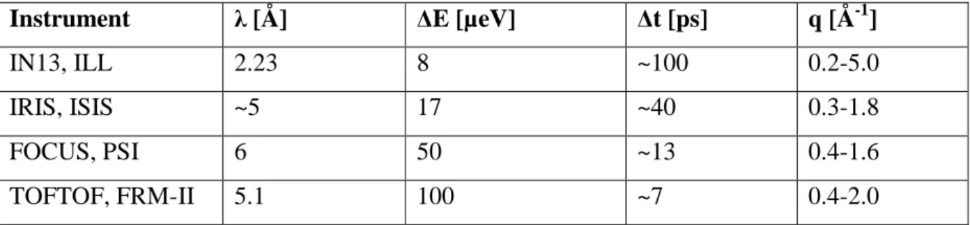
3.7 Instruments Used for Incoherent Neutron Spectroscopy...38
3.8 Mean Square Displacements and Force Constants obtained from Elastic Incoherent Neutron Scattering...41
3.9 Models for Quasielastic Neutron Scattering...44
3.9.1 Long Range Translational Diffusion………...….44
3.9.2 Rotational Diffusion………...………..46
3.9.3 Diffusion within Confined Space...47
3.10 References...50
4. Hemoglobin Dynamics in Red Blood Cells: Correlation to Body Temperature...55
4.1 Abstract...58
4.2 Introduction...58
4.3 Materials and Methods...62
4.3.1 Sample preparation...62
4.4 Results...68
4.4.1 Neutron Scattering...68
4.4.2 Global Motions...70
4.4.3 Internal Motions...75
4.4.4 Dynamic light scattering...80
4.5 Discussion...80 4.5.1 Global Motions...80 4.5.2 Internal Motions...87 4.6 Conclusion...90 4.7 Acknowledgements...91 4.8 References...91
5. From Powder to Solution: Hydration Dependence of Human Hemoglobin Dynamics Correlated to Body Temperature...97
5.1 Abstract...100
5.2 Introduction...101
5.3 Material and Methods...104
5.3.1 Sample preparation...104
5.3.2 Neutron scattering experiments...104
5.3.3 Quasielastic Neutron Scattering Analysis...105
5.3.4 Elastic Neutron Scattering Analysis...107
5.4 Results...108
5.4.1 Quasielastic Neutron Scattering...108
5.4.2 Elastic Neutron Scattering...116
5.5 Discussion...117
5.6 Conclusion...120
5.7 Acknowledgments...121
6.4 Discussion...135 6.5 Acknowledgment...135 6.6 Supporting Information...136 6.6.1 Sample preparation...136 6.6.2 Neutron experiments...137 6.6.3 Data analysis...138
6.6.4 Surface Area Calculations...143
6.7 References...144
7. Conclusions Générales et Perspectives /General Conclusion and Perspectives……..147
8. Abbreviations...159
9. References...161
10. Appendix...171
10.1 From shell to cell: neutron scattering studies of biological water dynamics and coupling to activity...173
10.2 Dynamics of apomyoglobin in the α-to-β transition and of partially unfolded aggregated protein...191
1. Introduction Générale
Cette thèse a été effectuée à l’Institut Laue-Langevin, Grenoble, et au Centre de Recherche Jülich, Allemagne, sous la direction du Dr. Joseph Zaccai et du Prof. Dr. Georg Büldt. Le sujet de la thèse concerne l’étude de la dynamique de l’hémoglobine et de l’eau in-vivo dans les globules rouges comparée à la dynamique de l’hémoglobine isolée en fonction de l’hydratation, mesurée par diffusion incohérente quasiélastique et élastique de neutrons. Le manuscrit est basé sur trois articles scientifiques. Deux sont déjà publiés, respectivement dans
Biophysical Journal et Journal of the American Chemical Society, le troisième a été soumis
pour publication à Biophysical Journal en janvier 2009.
Tous les organismes vivants sont formés par des cellules. Une thématique de recherche importante vise à comprendre les processus moléculaires qui ont lieu dans les cellules. Le but de la biophysique moléculaire est de comprendre la fonction, la structure, la dynamique et l’interaction avec le solvant des systèmes biomoléculaires comme les protéines, l’ADN, ou les membranes au niveau atomique. Les protéines sont des macromolécules qui sont produites dans les cellules et qui sont responsables d’une grande partie des fonctions dans l’organisme. Le génome humain contient l’information pour des dizaines de milliers de protéines différentes. Chacune a sa ou ses fonctions spécifiques. Quelques protéines fonctionnent comme des enzymes et catalysent des réactions chimiques. D’autres forment le tissu et des structures comme les muscles, la peau ou le cytosquelette. Des protéines spécialisées transforment l’énergie solaire en énergie chimique via la photosynthèse. D’autres encore transportent et stockent des substances de façon vivement régulée, comme c’est le cas de l’hémoglobine avec l’oxygène. Les protéines font souvent partie de grands assemblages avec d’autres protéines, l’ADN, l’ARN, des sucres, ou des lipides. La connaissance de la structure au niveau atomique est nécessaire pour comprendre comment les protéines peuvent réaliser toutes leurs fonctions. Néanmoins, les protéines ne sont pas des objets statiques mais leur structure est animée par des fluctuations permanentes. Elles sont des objets mous et
déformables. La dynamique et les fluctuations ont une importance fondamentale pour la fonction biologique.
La technique de diffusion incohérente des neutrons est très bien adaptée pour étudier la dynamique des biomolécules dans la gamme de temps de la picoseconde à la nanoseconde et la gamme de longueur de l’Ångstrom. La section efficace de diffusion incohérente de l’hydrogène est d’un ordre de grandeur plus élevé que celles des autres éléments qui constituent normalement la matière biologique, ainsi que celle du deutérium. Les atomes d’hydrogène sont distribués quasi-uniformément dans les protéines. Pratiquement, un atome sur deux dans une protéine est un atome d’hydrogène. En conséquence, la diffusion incohérente de neutrons mesure la dynamique moyenne des protéines. La technique de diffusion quasiélastique de neutrons est particulièrement bien adaptée pour mesurer la dynamique de l’eau dans la gamme de temps correspondant aux mouvements rapides de l’eau volumique à ceux plus lents de l’eau interfaciale. Le système biologique choisi pour cette thèse est l’hémoglobine dans les globules rouges. L’hémoglobine est le constituant macromoléculaire principal des globules rouges. La protéine transporte, avec une régulation extrêmement fine, l’oxygène des poumons aux tissus. Les globules rouges sont des cellules plutôt simples, comme ils ne possèdent pas de noyau ou d’organelles et ils ne sont pas capables de synthétiser des protéines. Des expériences d’aspiration de globules rouges humains individuels utilisant des micropipettes ont montré qu’il existe une transition de passage à la température physiologique (Artmann et al. 1998). Des expériences de viscosimétrie avec des solutions d’hémoglobine très concentrées ont montré qu’il y a une transition colloïdale d’un gel à un état fluidique également à la température physiologique (Kelemen et al. 2001). Des expériences de dichroïsme circulaire et de diffusion dynamique de la lumière ont été effectuées sur l’hémoglobine de différentes espèces à températures physiologiques différentes. Elles ont montré que la température de transition est corrélée directement à la température physiologique (Digel et al. 2006 ; Zerlin et al. 2007). Le but de la thèse était d’étudier par diffusion des neutrons s’il y a une transition dans le comportement dynamique de l’hémoglobine ou du solvant autour de la température du corps. Une série d’expériences de diffusion de neutrons ont été faites sur des globules rouges entiers et comparées à des expériences sur des poudres d’hémoglobine hydratées et sur l’hémoglobine en solution concentrée.
Suite à l’introduction générale, le deuxième chapitre de la thèse présente une introduction à l’hémoglobine et aux globules rouges. Les expériences sur la transition de l’hémoglobine à la température physiologique sont résumées dans ce chapitre.
Le troisième chapitre décrit la diffusion des neutrons appliquée à l’étude de la dynamique des biomolécules. La dynamique des protéines en générale et les propriétés de la dynamique de l’eau dans les systèmes biologiques sont présentées. Les propriétés du neutron et les méthodes de production et de détection des neutrons sont données. Des éléments de la théorie de diffusion de neutrons qui sont nécessaires pour la compréhension de la thèse sont résumés. Les méthodes de la diffusion élastique et quasiélastique de neutrons et les instruments sont décrits. Finalement, les concepts de déplacement carré moyen et constante de force obtenus par diffusion élastique de neutrons sont expliqués et les modèles utilisés pour interpréter les expériences quasiélastiques sont résumés.
Le quatrième chapitre est basé sur un article publié dans Biophysical Journal. Il présente une étude sur la dynamique de l’hémoglobine dans les globules rouges mesurée par diffusion quasiélastique de neutrons.
Hemoglobin Dynamics in Red Blood Cells: Correlation to Body Temperature (2008) Stadler A. M., I. Digel, G. M. Artmann, J. P. Embs, G. Zaccai, and G. Büldt. Biophysical Journal 95: 5449–5461
Dans l’étude, la dynamique interne de l’hémoglobine et la diffusion macromoléculaire globale ont été mesurées. L’expérience a montré qu’il y a une transition dans la géométrie des mouvements internes à 36.9°C. À des températures plus élevées que la température du corps, la dynamique des chaînes latérales des acides amimés occupent des volumes plus grands que prévu par l’effet normal de la température. La diffusion macromoléculaire globale a été interprétée avec des concepts théoriques pour des particules colloïdales.
Le cinquième chapitre présente un article basé sur la dynamique de l’hémoglobine en fonction de l’hydratation. L’article a été soumis au Biophysical Journal.
From Powder to Solution: Hydration Dependence of Human Hemoglobin Dynamics Correlated to Body Temperature. Stadler A. M., I. Digel, J. P. Embs, T. Unruh, M. Tehei, G. Zaccai, G. Büldt, and G. M. Artmann.
L’influence de l’hydratation sur la dynamique de l’hémoglobine a été mesurée par diffusion élastique et quasiélastique de neutrons. Les temps de résidence entre les sauts locaux, de
l’ordre de quelques picosecondes, sont réduits en solution concentrée et augmentés en poudre d’hémoglobine hydratée. La transition dans la géométrie des mouvements internes à la température du corps a été trouvée dans la solution concentrée mais non dans la poudre hydratée. Ceci indique que les mouvements rapides impliqués dans la transition sont activés seulement à partir d’un certain niveau d’hydratation. Un résultat annexe est que les poudres hydratées ne pourraient pas montrer tous les aspects dynamiques dans l’ordre de quelques picosecondes, qui pourraient être nécessaires pour la fonction biologique.
Le sixième chapitre est basé sur un article scientifique publié dans Journal of the American
Chemical Society qui décrit des expériences sur la dynamique de l’eau cellulaire dans les
globules rouges.
Cytoplasmic Water and Hydration Layer Dynamics in Red Blood Cells (2008) Andreas M. Stadler, Jan P. Embs, Ilya Digel, Gerhard M. Artmann, Tobias Unruh, Georg Büldt, and Giuseppe Zaccai. Journal of the American Chemical Society 130 (50): 16852–16853
La dynamique de l’eau dans les globules rouges a été mesurée par diffusion quasiélastique de neutrons. Des spectromètres de neutrons à résolution en temps de 40, 13 et 7 picosecondes ont été combinés pour couvrir l’échelle de temps des mouvements de l’eau volumique et de l’eau interfaciale. Une fraction d’eau cellulaire d’environ 90% est caractérisée par un coefficient de diffusion translationnel similaire à l’eau volumique. Une partie de l’eau cellulaire (~10%) montre par contre une dynamique significativement ralentie. Le ralentissement a été attribué à l’interaction avec la surface de l’hémoglobine. La fraction de l’eau ralentie correspond à environ la moitié des molécules d’eau dans la première couche d’hydratation.
1. General Introduction
The present PhD thesis was carried out at the Institut Laue-Langevin, Grenoble, and at the Research Centre Jülich, Germany, under the joint supervision of Dr. Giuseppe Zaccai and Prof. Dr. Georg Büldt. The subject concerns incoherent quasielastic and elastic neutron scattering experiments on the dynamics of hemoglobin and water in whole red blood cells
in-vivo, and as a comparison, hemoglobin dynamics in response to hydration. The document is
based on three scientific articles. Two of them are published in Biophysical Journal and
Journal of the American Chemical Society, respectively. The third one has been submitted for
publication to Biophysical Journal, in January 2009.
All living organisms consist of cells. One subject of research lies in a detailed understanding of the molecular processes that occur in cells. Molecular biophysics focuses on the investigation of biological systems at the atomic level, such as for example proteins, membranes, DNA and their interactions with the surrounding solvent. Proteins are responsible for a large variety of different functions. The human genome contains the information for several tens of thousands of different proteins. Each one has got one or more specific functions. Some proteins work as enzymes and catalyse chemical reactions. Others form tissues and structures such as muscles, skin or the cytoskeleton. Specialised proteins convert solar energy via photosynthesis into chemical energy, or transport and store substances in a sophisticated way, as it is the case for hemoglobin that carries oxygen. Proteins are often part of complex macromolecular assemblies that consist of other proteins, DNA, RNA, sugar or lipid molecules. The knowledge of its structure at the atomic level is necessary to understand how a protein can fulfil its specific task. However, proteins are not static structures but they are soft and deformable. The dynamics and fluctuations of proteins are of fundamental importance for biological function.
Incoherent neutron scattering is a particular well suited technique to study biomolecular dynamics in the picosecond to nanosecond time and Ångstrom length scale. The incoherent scattering cross section of hydrogen is one order of magnitude larger than all other elements that usually occur in biological matter, and deuterium. Hydrogen atoms are uniformly distributed in proteins and constitute nearly every second atom. Neutron scattering therefore probes average protein dynamics. The technique of quasielastic incoherent neutron scattering is especially useful for the study of the dynamics of water in the time scales of fast motions of bulk water to slower dynamics of interfacial water. The biological system that was studied in the thesis is hemoglobin in red blood cells. Hemoglobin is the main macromolecular constituent of red blood cells. The protein transports oxygen from the lungs to the tissues in a highly coordinated and regulated way. Red blood cells are simple cells as they do not contain a nucleus or organelles, and are not able to synthesize proteins. Micropipette experiments on single red blood cells identified a passage transition at body temperature (Artmann et al. 1998). Viscosity measurements of hemoglobin solutions at high concentration showed that there equally occurs a colloidal gel to fluid transition of hemoglobin solutions at body temperature (Kelemen et al. 2001). Circular dichroism and dynamic light scattering experiments further revealed that the temperature transition of hemoglobin is directly correlated to the body temperature of different species (Digel et al. 2006; Zerlin et al. 2007). The aim of the thesis was to study if a transition in the dynamics of hemoglobin or solvent occurs at body temperature. This was achieved in a series of incoherent neutron scattering experiments on whole red blood cells that were compared to results of hydrated hemoglobin powder, and concentrated hemoglobin solution samples.
Following the general introduction, the second chapter of the thesis gives an introduction to hemoglobin and red blood cells. Recent experiments on the temperature transition of hemoglobin at body temperature are summarized.
The third chapter deals with neutron scattering applied to the study of biomolecular dynamics. Protein dynamics and the dynamic properties of water in biological systems are presented. The general properties of the neutron and methods of neutron production and detection are described. The elements of neutron scattering theory that are necessary for the further understanding of the thesis are summarized. The methods of elastic and quasielastic
elastic incoherent neutron scattering are explained and typical models that are used for quasielastic neutron scattering are summarized.
The fourth chapter is based on a published article in Biophysical Journal about hemoglobin dynamics in red blood cells measured with quasielastic incoherent neutron scattering:
Hemoglobin Dynamics in Red Blood Cells: Correlation to Body Temperature (2008) Stadler A. M., I. Digel, G. M. Artmann, J. P. Embs, G. Zaccai, and G. Büldt. Biophysical Journal 95: 5449–5461
Internal protein dynamics and global macromolecular diffusion were separated. The experiments revealed a change in the geometry of internal protein dynamics above 36.9°C. Above this temperature amino acid side-chain dynamics occupy larger volumes than expected from normal temperature dependence. Global macromolecular diffusion was interpreted favourable with theoretical concepts for short-time self-diffusion of non-charged hard sphere colloids.
The fifth chapter presents an article about hemoglobin dynamics as response to hydration. The article has been submitted to Biophysical Journal.
From Powder to Solution: Hydration Dependence of Human Hemoglobin Dynamics Correlated to Body Temperature. Stadler A. M., I. Digel, J. P. Embs, T. Unruh, M. Tehei, G. Zaccai, G. Büldt, and G. M. Artmann.
The influence of hydration on hemoglobin dynamics was studied with elastic and quasielastic incoherent neutron scattering. The residence times of localized jumps in the order of a few picoseconds were found to be significantly reduced in concentrated solution compared to fully hydrated powder. The body temperature transition in protein dynamics was found in the concentrated solution sample but was absent in the fully hydrated powder, indicating that picosecond protein dynamics responsible for the transition is activated only at a sufficient level of hydration. A collateral result from the study is that fully hydrated protein powder samples do not accurately describe all aspects of protein picosecond dynamics that might be necessary for biological function.
The sixth chapter deals with the dynamic properties of cellular water in red blood cells in the form of a scientific article that was published in Journal of the American Chemical Society.
Cytoplasmic Water and Hydration Layer Dynamics in Red Blood Cells (2008) Andreas M. Stadler, Jan P. Embs, Ilya Digel, Gerhard M. Artmann, Tobias Unruh, Georg Büldt, and Giuseppe Zaccai. Journal of the American Chemical Society 130 (50): 16852–16853
The dynamics of water in human red blood cells was measured with quasielastic incoherent neutron scattering. Neutron spectrometers with time resolutions of 40, 13, and 7 ps were combined to cover time scales of bulk water dynamics to reduced mobility interfacial water motions. A major fraction of around 90% of cell water is characterized by a translational diffusion coefficient similar to bulk water. A minor fraction of around 10% of cellular water exhibits reduced dynamics. The slow water fraction was attributed to dynamically bound water on the surface of hemoglobin, which accounts for approximately half of the hydration layer.
The seventh chapter gives a summary of the performed work and presents an outlook on future perspectives.
2. Hemoglobin and Red Blood Cells
2.1 Biological Background
The main natural environment of proteins is within the cell, and protein function necessarily is adapted to these conditions. In bacteria and eukaryotic cells macromolecular interactions are likely to be influenced by the high cellular protein concentration (Hall and Minton 2003; Minton 2001; Zimmerman and Minton 1993). This effect, called ‘crowding’, results from high volume occupancy and steric hindrance of the protein molecules between each other. The free distance between macromolecules in the cell is in the order of a few Ångstroms (Krueger and Nossal 1988) and intercellular water is in close vicinity to the protein surfaces. There is much interest and discussion about similarities and differences in structure and dynamics of cytoplasmic and bulk water (Ball 2008). Little is still known on how protein dynamics is influenced by the crowded cytoplasmic environment and the special properties of cellular water.
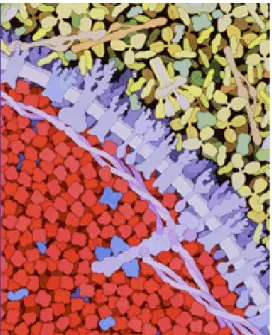
The main protein constituent of red blood cells is hemoglobin (92% of dry weight) (see
Figure 1). Its biological function is to carry oxygen from the lungs to the tissues. The X-ray
structure of hemoglobin is shown in Figure 2. The protein is a tetramer with a molecular weight of around 64 kDa. It consists of two α-chains and two β-chains, each having 141 and 146 amino acid residues, respectively. The α-chains contain seven and the β-chains eight helices (Perutz 1987). Each chain carries one heme group, in a pocket, to which oxygen and several other small molecules can bind reversibly. The concentration of hemoglobin in the red blood cells is around 330 mg/ml (Krueger and Nossal 1988).
Figure 1: Illustration of a red blood cell in the lower half of the picture with surrounding
blood serum proteins in the upper half. The red blood cell is densely filled with hemoglobin, drawn in red. The cell wall with membrane proteins is coloured purple. (Goodsell 2000).
Figure 2: Hemoglobin is a tetrameric protein. It consists of two α- and two β-chains. Every
chain carries one heme molecule in a pocket to which oxygen binds reversible. (PDB code 1G09)
2.2 Body Temperature Transition of Hemoglobin
Hemoglobin at high concentration shows a variety of interesting effects. Micropipette experiments with aspirated single human red blood cells revealed a sudden change in the behaviour of the cells from blockage of the pipette below TPipette=36.4 ± 0.4 °C to easy
passage above. The transition temperature TPipette of cell passage was surprisingly very close
to human body temperature. During the micropipette experiment water is pressed out of the red blood cell and the hemoglobin concentration rises to values of more than 500 mg/ml (Kelemen et al. 2001).
Viscosity measurements were performed on human hemoglobin solutions between 330 mg/ml and 500 mg/ml to study the flow properties at such concentrations (Artmann et al. 1998). The experiments found a sharp drop in viscosity in Arrhenius plots, at concentrations higher than 450 mg/ml at body temperature. The drop was absent at the physiological concentration of 330 mg/ml. The results were interpreted as a colloidal phase transition in highly concentrated hemoglobin solution from a gel-like to a fluid state at body temperature (Artmann et al. 1998; Kelemen et al. 2001). In the fluid-like state, above body temperature, hemoglobin exhibits pronounced aggregation, whereas at lower temperatures hemoglobin aggregation is suppressed (Artmann et al. 1998).
Further studies with circular dichroism investigated changes of the protein secondary structure around the transition temperature. A partial loss of alpha-helical content was found at a temperature TCD=37.2 ± 0.6 °C for human hemoglobin (Artmann et al. 2004). The loss of
hemoglobin alpha-helical structure at a specific temperature TCD was also observed for
hemoglobin molecules of a large variety of different species (Digel et al. 2006; Zerlin et al. 2007). Amazingly, the transition temperatures TCD were directly correlated to the body
temperature of the animals ranging from 34 °C, for the duck-billed platypus, to 42 °C for a bird, the spotted nutcracker. It was excluded that the partial loss of protein structure at TCD
results from irreversible protein denaturation which occurs at distinctly higher temperatures. Independently of these experiments, two-dimensional infrared correlation spectroscopy suggested a structural perturbation stage of bovine hemoglobin between 30 °C and 44 °C (Yan et al. 2004). The observed perturbations were assigned to hydrogen-bonded extended chains that connect the helices. It was concluded that the passage transition of red blood cells is caused by hemoglobin molecules and that the observed small structural changes of
hemoglobin at TCD might be the cause for the drop of viscosity at TPipette (Kelemen et al.
2001). It has been speculated that partial unfolding of the α-helical structure at body temperature goes in hand with an increase in surface hydrophobicity that promotes protein aggregation (Digel et al. 2006), which causes a drop in colloidal osmotic pressure (Artmann et al. 2008).
2.3 References
Artmann GM, Burns L, Canaves JM, Temiz-Artmann A, Schmid-Schonbein GW, Chien S, Maggakis-Kelemen C (2004) Circular dichroism spectra of human hemoglobin reveal a reversible structural transition at body temperature. Eur. Biophys. J. 33:490-6 Artmann GM, Kelemen C, Porst D, Büldt G, Chien S (1998) Temperature transitions of
protein properties in human red blood cells. Biophys. J. 75:3179-83
Artmann GM, Zerlin KF, Digel I (2008) Hemoglobin Senses Body Temperature. In: Artmann GM, Chien S (eds) Bioengineering in Cell and Tissue Research, vol IV. Springer Verlag, Berlin, Heidelberg, pp 415-447
Ball P (2008) Water as an Active Constitutent in Cell Biology. Chem. Rev. 108:74-108 Digel I, Maggakis-Kelemen C, Zerlin KF, Linder P, Kasischke N, Kayser P, Porst D, Temiz
Artmann A, Artmann GM (2006) Body temperature-related structural transitions of monotremal and human hemoglobin. Biophys. J. 91:3014-21
Goodsell DS (2000) http://mgl.scripps.edu/people/goodsell/illustration/public
Hall D, Minton AP (2003) Macromolecular crowding: qualitative and semiquantitive successes, quantitative challenges. Biochim. Biophys. Acta 1649:127-139
Kelemen C, Chien S, Artmann GM (2001) Temperature transition of human hemoglobin at body temperature: effects of calcium. Biophys. J. 80:2622-30
Krueger S, Nossal R (1988) SANS studies of interacting hemoglobin in intact erythrocytes. Biophys. J. 53:97-105
Minton AP (2001) The Influence of Macromolecular Crowding and Macromolecular Confinement on Biochemical Reactions in Physiological Media. J. Biol. Chem. 276:10577-10580
Yan YB, Wang Q, He HW, Zhou HM (2004) Protein thermal aggregation involves distinct regions: sequential events in the heat-induced unfolding and aggregation of
hemoglobin. Biophys. J. 86:1682-90
Zerlin KF, Kasischke N, Digel I, Maggakis-Kelemen C, Temiz Artmann A, Porst D, Kayser P, Linder P, Artmann GM (2007) Structural transition temperature of hemoglobins correlates with species' body temperature. Eur. Biophys. J. 37:1-10
Zimmerman SB, Minton AP (1993) Macromolecular Crowding: Biochemical, Biophysical and Physiological Consequences. Ann. Rev. Biophys. Biomol. Struct. 22:27-65
3. Neutron Spectroscopy to Study Biomolecular
Dynamics
The chapter starts with a presentation of protein and water dynamics in general. Physical concepts and equations that are necessary for the understanding of the theory of incoherent neutron scattering are schematized. Inelastic and elastic scattering processes and incoherent and coherent neutron scattering are presented. The application of elastic and quasielastic incoherent neutron scattering to measure biomolecular dynamics is explained at the end of the chapter.
3.1 Protein and Water Dynamics
First studies on protein dynamics used sperm whale myoglobin as a model system. It was the first protein of which the structure could be solved by X-ray crystallography (Kendrew et al. 1960). The protein serves as a scaffold for the iron containing heme-group that is responsible for oxygen binding and transport. The crystal structure showed clearly that there are no permanent channels in the protein structure through which the oxygen molecule could migrate from the surrounding solvent to its binding place deep in the protein. Only fluctuations of the protein structure can allow the rapid opening and closing of channels in the protein structure for the purpose of oxygen binding indicating that knowledge of both protein structure and dynamics is necessary for a complete understanding of protein function.
Ligand rebinding in myoglobin was measured with optical absorption in the range of 10-6 to 103 seconds after photo flash dissociation (Austin et al. 1975). In total, up to four different rebinding processes were found that depend differently on temperature. Between around 180 K to 280 K all four processes could be found; below around 180 K only the first rebinding process occurs. The rebinding processes were identified with four different activation energy barriers that the ligand has to pass before rebinding. It was concluded that the first energy barrier corresponds to direct rebinding of the ligand to the iron atom. Nonexponential rebinding rates were found for the first process below 180 K. The results
imply that the first energy barrier is in fact a distribution of activation energies. The authors explained this by the assumption that myoglobin does not exist in only one fixed structure but it possesses many slightly different conformational states. Below 180 Keach protein is frozen and trapped in one of the conformational states. Above 180 K the protein can interchange between the different conformations. The differences between many different structural conformations need to be rather small as they could not be seen with X-ray crystallography (Frauenfelder et al. 1979). These experiments led to the picture of conformational substates of proteins (Frauenfelder et al. 1988). In the native state a protein does not exist in a single static structure. Instead it fluctuates between a large ensemble of slightly different conformational structures. The free enthalpy of all substates is the same (or very close) and the different conformations are separated by activation energy barriers. The sampling of the conformational substates leads to the entropic stabilisation of proteins.
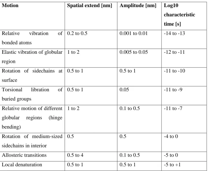
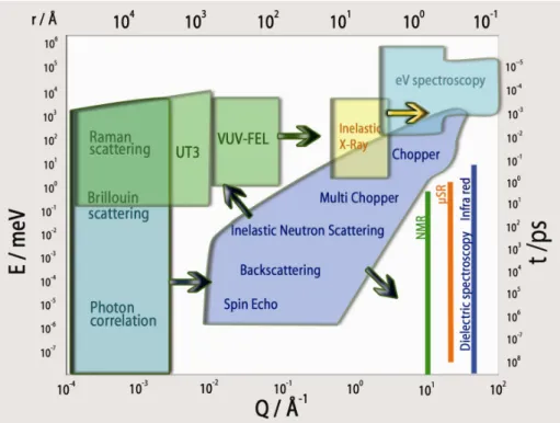
Motions in biological macromolecules and proteins occur over a very broad range of time scales: from fast vibrations and electron transfer processes in the order of some femtoseconds, to protein folding events in the order of some seconds. The amplitudes of motion lie between around 0.1 Å for fast atomic vibrations up to several 10 Å for collective motions. A short overview of time and length scales of motions in biological macromolecules is given in
Table 1: Typical time and length scales of internal motion in proteins modified after
(McCammon and Harvey 1987)
Motion Spatial extend [nm] Amplitude [nm] Log10
characteristic time [s]
Relative vibration of bonded atoms
0.2 to 0.5 0.001 to 0.01 -14 to -13
Elastic vibration of globular region 1 to 2 0.005 to 0.05 -12 to -11 Rotation of sidechains at surface 0.5 to 1 0.5 to 1 -11 to -10 Torsional libration of buried groups 0.5 to 1 0.05 -11 to -9
Relative motion of different globular regions (hinge bending) 1 to 2 0.1 to 0.5 -11 to -7 Rotation of medium-sized sidechains in interior 0.5 0.5 -4 to 0 Allosteric transitions 0.5 to 4 0.1 to 0.5 -5 to 0 Local denaturation 0.5 to 1 0.5 to 1 -5 to +1
First experiments on protein dynamics came from Mößbauer spectroscopy studies of myoglobin. The technique specifically probes the dynamics of the iron atom in the heme group with very high resolution. Motions in the time scale up to 140 ns are detected. The results showed that the mean square displacements of the iron atom increase linearly with temperature up to around 180 K. Above 180 K the mean square displacements increase with a steeper gradient. This was interpreted in terms of supplemental anharmonic motions that contribute to the dynamics above 180 K (Parak et al. 1982). The results obtained with Mößbauer spectroscopy were interpreted with a model of an overdamped Brownian oscillator (Knapp et al. 1982). Below the dynamical transition temperature of 180 K proteins are trapped in conformational substates and perform harmonic oscillations. Above 180 K the proteins switch by thermal activation to a more flexible state, in which internal quasi-diffusive motions are possible.
The nature of the hydration shell of myoglobin was investigated with infrared spectroscopy and calorimetry (Demmel et al. 1997; Doster et al. 1986). At low temperature the hydration water turns into an amorphous, disordered state with strongly reduced dynamics. The experiments revealed a glass-like transition of the hydration water at around 200 K which is close to the dynamical transition temperature of myoglobin. The authors emphasized the importance of the glass-like transition of the solvent near 200 K for the dynamical transition of myoglobin. They concluded that the cooperativity of the hydrogen bonding network provides the intimate coupling between solvent and protein dynamics (Doster et al. 1986). The important role of the solvent on the dynamical transition was further demonstrated in a Mößbauer experiment that investigated myoglobin dynamics in a sucrose- water solvent (Lichtenegger et al. 1999). The dynamic transition of myoglobin was shifted to 240 K due to the higher viscosity of the solvent.
Incoherent neutron scattering is dominated by hydrogen atom motions as their incoherent scattering cross section is one order of magnitude bigger than that of all other elements which usually occur in biological matter, and deuterium (Sears 1992). The technique probes average protein dynamics because hydrogen atoms are uniformly distributed in the natural abundance protein. The time and length scales of molecular motions that are accessible are determined by the energy resolution and the scattering vector range of the spectrometer, respectively. On the ps-Å time-length scale, hydrogen atoms that are covalently bound to amino acid side-chains reflect the dynamical behaviour of the bigger chemical units (Reat et al. 1998; Smith 1991; Wood et al. 2008).
The dynamical transition at about 180 K was revealed to exist for the motion of all hydrogen atoms in myoglobin by energy resolved incoherent neutron scattering (Doster et al. 1989). The dynamical transition was also found by neutron scattering in a membrane protein (Ferrand et al. 1993). Below 180 K the dynamics of myoglobin are similar to a harmonic solid. At 180 K additional non-vibrational motions set on that were attributed to jumps of the hydrogen atoms between two states (Doster et al. 1989). The jump motion was described with a double well model for two states separated with a temperature independent distance d and a free energy difference. The amplitude of the jump distance was found to be d=1.5 Å which
Doster 1990; Doster et al. 1989, 1990). The fast β-process was attributed to localised jumps with correlation times of ~0.5ps, the slower process was brought into connection with the α-process that occurs in liquids and polymers (Doster et al. 1989). The onset of the α-α-process occurs at 240 K and was attributed to collective excitations that are induced by the melting of glassy water in the hydration shell (Demmel et al. 1997; Doster et al. 1986; Doster et al. 1989). Model independent mean square displacements were obtained in the Gaussian approximation that included the contributions of the α-, the β-process and vibrational motions (Doster et al. 1989). In that way, the amplitudes of the α-process were obtained as a function of temperature. The slow α-process is absent in dehydrated powders. Recently, the β-process was attributed to localised jumps of amino acid side-chains (Doster 2008). The shape of the inelastic spectra of the α- and β-process could be described quantitatively with predictions of mode coupling theory (Doster et al. 1990). It was concluded that a cage effect of nearest neighbours is responsible for the spectral features. However, the difference between protein dynamics and glass physics was pointed out, as the structure of a protein imposes restrictions to the possible movements in the long time limit, amino acid side chains are attached to the protein backbone for example (Doster et al. 1990). In analogy to the glass transition, the onset of the slower α-process at 240 K was defined as the dynamical transition temperature and the
α-process was identified as the elementary step of diffusion. The onset at 180 K due to fast
jumps between states of different energy is a precursor of the slower α-process (Doster 2008).
Depending on the energy resolution and scattering vector range of the neutron spectrometer, a third inflection in the thermal displacements, which is hydration-independent, was observed at a temperature between 100 and 150 K. It has been attributed to methyl group rotations (Cornicchi et al. 2006; Doster and Settles 2005; Roh et al. 2006; Roh et al. 2005). These local jumps contribute to the sampling of a large number of conformational substates that are responsible for the entropic stabilization of proteins (Fraunfelder et al. 1991).
A model for protein dynamics related to the Doster et al. (1989) double well used in this work was proposed by Bicout and Zaccai (Bicout and Zaccai 2001). Protein dynamics is interpreted with a picture of a quasi-harmonic average potential well for the complex macromolecular force field (Bicout and Zaccai 2001; Zaccai 2000). The model assumes that protein dynamics can be described by two states: local fluctuations around the equilibrium positions with small amplitude and larger amplitude fluctuations in a cage formed by neighboring molecules. The two states are separated by a free energy difference and the transition from the low amplitude
state to the large amplitude state is activated by temperature. The dynamical transition is defined as the deviation of the mean square displacements from low temperature linear behaviour. In the model this occurs when a small fraction (10%) of the total population of particles in the small amplitude state changes into the large amplitude state.
Following the Bicout and Zaccai model, protein flexibility was defined as the amplitude of atomic motions u2 which corresponds to the width of the quasi-harmonic average potential well. Protein thermal stability would correspond to the depth of the well (Tehei et al. 2001; Tehei and Zaccai 2007). A mean effective force constant <k’> can be obtained from the dependence of the <u2> as function of temperature. This mean effective force constant, called
resilience, describes the shape of the well (Zaccai 2000). Many conformational substates exist within the average well and are sampled by localized jump-diffusion (Fraunfelder et al. 1991). The geometry and activation energy of localized jumps can be determined by QENS. From elastic incoherent neutron scattering (EINS) mean square displacements <u2> of the thermal
cloud of atomic motions can be determined. Quasielastic neutron scattering (QENS) on the other hand, enables to distinguish between vibrational and diffusive components (Bee 1988). This technique allows the quantification of internal diffusion coefficients, residence times and the determination of the average geometry of motions.
Both protein function and dynamics are closely linked to sufficient hydration. Enzymes are non-functional in the dry state and catalytic function is only possible above a sufficient threshold hydration level of around 0.2g H2O/ g protein (Rupley and Careri 1991). In a membrane protein, anharmonic processes are only activated above a hydration level of around 0.35g H2O/ g protein (Ferrand et al. 1993). Recent work in literature points out that it is the onset of translational diffusion of the solvent molecules that enables the protein dynamical transition (Tarek and Tobias 2002; Tournier et al. 2003; Wood et al. 2007). The dynamical properties of water that is in close contact to protein surfaces are therefore of high importance for the understanding of protein function.
The cytoplasmic environment in cells is very crowded and protein concentrations range up to ~400 mg/ml (Ellis and Minton 2003). Distances between macromolecules are in the order of
Funel et al. 1996; Dellerue and Bellissent-Funel 2000) compared to that of bulk water (Teixeira et al. 1985). The dynamics of water in contact with hydrophilic and hydrophobic peptides at high concentration was found to be reduced compared to bulk water (Russo et al. 2005). Molecular dynamics simulations showed that mostly water molecules in protein surface cavities exhibit strongly reduced dynamics (Makarov et al. 2000). Water in cells was therefore assumed to show different dynamics than bulk water. Recent work on water dynamics in cells proved the contrary: Jasnin et al. measured the dynamics of cellular water in
Escherichia coli with neutron scattering (Jasnin et al. 2008). The authors reported that the
average translational and rotational diffusion coefficients of cell water show similar behaviour to bulk water. The residence times of cell water were twice as long which was attributed to the longer times spent in the hydration shell. Persson and Halle measured water dynamics in E.
coli with NMR spectroscopy (Persson and Halle 2008). They concluded that around 85% of
cell water has similar dynamics to bulk water, and around 15% of cell water interacts with the surface of biomolecules, being slowed down by a factor of around 15 ± 3 compared to bulk water.
The halophilic cell is a special case. The cell accumulates large amounts of KCl in the molar level in its cytoplasm and halophilic proteins are only stable at such high salt concentrations. Solvent interactions of halophilic proteins are particularly strong and halophilic proteins bind both water and salt ions in their hydration shell (Madern et al. 2000). A previous experiment on water dynamics in the halophilic organism Haloarcula marismortui revealed a major cellular water fraction of around 76% with 250 times slower dynamics than bulk water (Tehei et al. 2007). The slow water fraction at 300 K showed characteristic signs for confined motions in a restricted volume. That study also revealed that cell water dynamics in E. coli is not governed by such a slow water fraction (Tehei et al. 2007). Tehei et al. therefore suggested that an ordering effect of water molecules, KCl and halophilic proteins is responsible for the large slow water fraction in H. marismortui (Tehei et al. 2007).
Fast bulk-like dynamics of water are in the order of some picoseconds, whereas interfacial water motions are slowed down and are in the order of some ten picoseconds. Quasielastic neutron scattering is a well suited technique for the study of the microscopic nature of water dynamics.
3.2 Properties, Production and Detection of Neutrons
The neutron is a nuclear particle that decays into a proton, an electron and an electronantineutrino with a half-life of 887.6±3 s (Mampe et al. 1989) (ß decay mediated via the weak interaction). The energy of 0.77 MeV that is produced during the decay is shared between the proton, the electron and the antineutrino.
Mev e p n→ + + e+0.77 − + υ
Neutrons can be described as waves with the wave vector k
r defined by λπ 2 = k r , ( 3.1 )
where
λ
is the wave length of the neutron.The momentum pr of the neutron is given by
k v m p n r h r r = = , ( 3.2 )
with the mass of the neutron mn and its velocity v
r
. The kinetic energy of the neutron is
n 2 2 kin m 2 k E = h . ( 3.3 )
Several properties of the neutron are summarized in Table 2.
Table 2: Properties of the neutron
Mass mn=1.675·10-27 kg
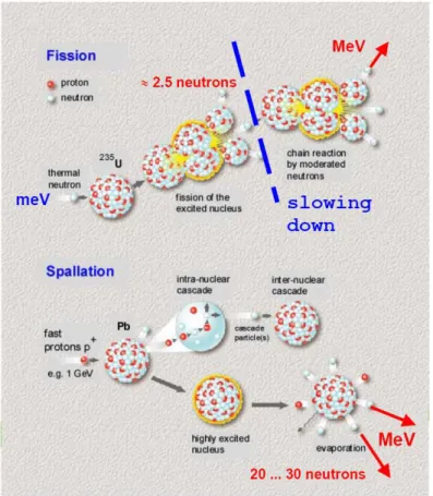
Although ~50% of all matter consists of neutrons, it is difficult to extract bound neutrons from the atomic nuclei. Free neutrons need to be produced by nuclear reactions. A high flux is mandatory for scientific investigations using neutron beams. Research neutron sources either use nuclear fission or spallation for the production of high flux neutron beams. Both nuclear fission and spallation are schematically presented in Figure 1. Examples for reactor neutron sources are the High Flux Reactor at the Institut Laue-Langevin (Grenoble, France) and the Research Reactor Heinz Maier-Leibnitz at the Technical University Munich (Garching, Germany). Neutron spallation sources are the ISIS pulsed spallation source at the Rutherford Appleton Laboratory (Didcot, United Kingdom) and the continuous spallation source SINQ at Paul Scherrer Institut (Villigen, Switzerland).
The produced neutrons have energies in the range of some MeV. This energy is too high for neutron scattering experiments. In reactor sources, the neutron energy has also to be reduced in order to maintain the chain reaction. Therefore, the kinetic energy of the neutrons needs to be changed by thermal equilibration with moderator elements that are kept at different temperatures. In the moderators the neutrons exchange their kinetic energy with the moderator atoms through collisions. At thermal equilibrium the velocities of the neutrons follow a Maxwell distribution determined by the temperature of the moderator. So called thermal neutrons are produced with moderators at ambient temperature, while cold neutrons are obtained from mainly liquid hydrogen or deuterium moderators at ~25 K.
A thermal neutron produced by a moderator at T=293 K has got in average a kinetic energy of E=25 meV, a wavelength of λ=1.8 Å and a velocity of 2200 m/s.
Figure 1: Schematic presentation of the nuclear fission and spallation process (Richter 2003).
Neutrons are produced in reactors through the fission of Uranium235. The excited Uranium decays into a series of fission products, including in average ~2.5 neutrons with energies in the order of some MeV. These high energy neutrons need to be moderated such that they can maintain the nuclear chain reaction. In spallation sources, protons are accelerated to high energies and brought to collision with heavy metal targets. The excited target nuclei evaporate in average ~20-30 neutrons with energies in the order of some MeV.
Neutrons are detected indirectly via nuclear reactions that produce charged particles. The charged particles can then be detected by appropriate detectors such as for example proportionality counters or photo multipliers.
Proportionality counters are filled with a gas that contains 3He. The helium isotope absorbs neutrons efficiently. The products of the nuclear reaction are charged protons or tritons with energies in the order of one MeV and e-. Scintillation counters are enriched with 6Li and ZnS. Neutron absorption leads to fluorescence radiation that can be detected with photo multipliers.
3.3 Scattering Processes and Observational Limits
We now consider the scattering of a neutron by a sample. Let k
r
and 'k
r
be the wave vectors of the incident and scattered neutron, respectively. A scheme of the scattering process is given in
Figure 2.
Figure 2: Scheme of the scattering process of neutrons with a sample. An incident neutron
enters from the left and is scattered into the solid angle d
Ω
. The incident neutron has got the wave vector kr
, the scattered neutron the wave vectork'
r
. The direction of k'
r
is determined by the scattering angle 2
θ
and the azimuthal angleφ
. The sample is indicated as a box at the centre of the coordinate system and the wave vector of the incident neutron beam is parallel to the z-axis.The difference in kinetic energy E between the incident and scattered neutron is
(
2 2)
2 ' 2m k k E n − = h , ( 3.4 )E is also called energy transfer. Positive values of E correspond to the situation when the
neutron deposits energy in the sample. In the case of negative values of E, the neutron gains
If E=0 than the process is called elastic scattering. If there occurs exchange of energy between the neutron and the sample with E ≠0, then the event is called inelastic scattering.
The difference in momentum between the incident and scattered neutron is
' k k q r h r h r h = − , ( 3.5 )
with the so called scattering vector qr. Sometimes, in analogy with crystallography, the scattering vector is defined as q k k
r r r
− = ' .
In the case of elastic scattering k = k’ and the scattering vector q can be written as
( )
λ θ π sin 4 = = q q r , ( 3.6 )with the scattering angle 2θ as defined in Figure 2.
For inelastic scattering processes it is k ≠k' and equation ( 3.6 ) is not valid anymore. The
connection q k k' r h r h r h = − can be written as ) 2 cos( k ' k 2 k ' k q2 = 2+ 2 −
θ
. ( 3.7 )With the energy transfer E it is
2 2 2 2 ' h E m k k = − n . ( 3.8 )
This can be written finally as
2 2 2 2 ) 2 cos( 2 2 2k m E k k m E q = − n −
θ
− n , ( 3.9 )which describes the fundamental kinematic connection for neutrons between scattering vector
q, energy transfer E and scattering angle 2
θ
. This relation gives the limiting boundaries of the accessible q-E space for any neutron spectrometer. The connection between q, E and 2θ
according to equation ( 3.9 ) is plotted exemplarily in Figure 3 for neutrons with the incident energy Ekin=3.14 meV. It is important to notice that the scattering vector q changes as a function of E at a fixed scattering angle 2θ
.Figure 3: The connection between scattering vector q, energy transfer E and scattering angle
2θ. In this plot the kinetic energy of the incident neutron is Ekin=3.14 meV, which corresponds
to a neutron wave length of λ=5.1Å. The maximum energy transfer E is determined by the kinetic energy of the neutron that it can deposit in the sample; it is indicated by the vertical dotted line at E=3.14 meV. The energy transfer in the case of elastic scattering is E=0, which is shown by the vertical dotted line at E=0. The solid horizontal line indicates the scattering vector q=1.7Å-1, as shown in Figure 4.
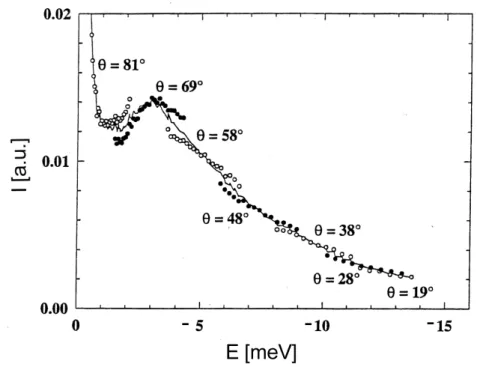
If the incident neutrons have a fixed kinetic energy and neutron scattering shall be measured at constant scattering vector q as a function of energy transfer E, then the scattering angle value of the detector position has to be varied according to equation ( 3.9 ) (see Figure 3).
Figure 4: Different scattering angles need to be connected to cover a broad energy transfer
range at constant scattering vector q=1.7Å-1. It is schematically illustrated how experimental data measured at different detector positions are joined together (Settles 1996). At energy transfer values close to E=0 it is not necessary to merge different detector positions.
3.4 Elements of Neutron Scattering Theory
A neutron scattering experiment with angular and energy resolution measures the double differential scattering cross section
dE d
d2 Ω σ
, which is the number of neutrons that are scattered per second into the solid angle d
Ω
in the direction of the scattering vector k'r
with an energy in the interval between E and E+dE, normalised by the incident neutron flux
Φ
.The incident neutron wave can be written as a plane wave with
r k i e r r r r = ) (
ψ
( 3.10 )The scattered neutron wave ψ' r(r) can be written at sufficient large distance as a sum of the transmitted wave and a spherical wave multiplied with the scattering amplitude f
( )
Ω( )
Ω + = f r e e r r k i r k i r r r r r ' ) ( ' ψ . ( 3.11 )The neutron interacts with the sample via nuclear and magnetic forces. The nuclear interaction depends on the element, the isotope and the orientation of the spin of the neutron and the spin of the nucleus of the sample.
The magnetic interaction is neglected in the following and only the nuclear interaction is further considered. The wave length of thermal neutrons is in the order of some 10-10 m, the diameter of the nucleus of an atom is around 10-15 m which is much smaller than the neutron wave length. The phenomenological assumption of a point like interaction potential (Fermi pseudo potential) has been proven to be very successful in the theory of neutron scattering. However, this assumption is only valid to describe scattering of cold and thermal neutrons and fails completely for high energy neutrons.
( )
r R b m r V n r r h r − ⋅ ⋅ = 2π
2δ
) ( , ( 3.12 )with the scattering length b that is a complex number. The real part of b describes the scattering strength, the imaginary part of b the absorption.
The further evaluations of the double differential scattering cross section take into account the Fermi pseudo potential and the so called Born approximation that is a first order approximation of the scattering process. Only single scattering is considered. The double differential scattering cross section finally can be written as
[
i
q
r
(
0
)
] [
exp
i
q
r
(
t
)
]
e
dt
exp
b
b
k
'
k
2
1
dE
d
d
i t 'j , j 'j j 'j j 2 ωπ
σ
+∞ − ∞ −>
−
<
=
Ω
∑ ∫
r
r
r
r
h
. ( 3.13 )The indices j and j’ account for the atoms in the sample. The scattering length bj depends on
the element, the isotope and the orientation of the spins. The position of the atom j at time t=0 is rrj(0); its position is rrj(t) at time t.
The sum in equation ( 3.13 ) can be split into two parts,
[
] [
]
[
i
q
r
] [
i
q
r
t
]
e
dt
b
k
k
dt
e
t
r
q
i
r
q
i
b
k
k
dE
d
d
t i j j j inc t i j j j j coh ω ωπ
π
σ
− ∞ + ∞ − < − +∞ ∞ − <>
−
+
>
−
=
Ω
∑ ∫
∑ ∫
)
(
exp
)
0
(
exp
'
2
1
)
(
exp
)
0
(
exp
'
2
1
2 ' , ' 2 2r
r
r
r
h
r
r
r
r
h
, ( 3.14 )with the so called coherent scattering length bcoh
2 2
coh b
b = , ( 3.15 )
2 2
2
inc b b
b = − . ( 3.16 )
The coherent scattering length is the average scattering length of the element. The incoherent scattering length is the standard deviation from the average.
The first term in equation ( 3.13 ) is called coherent scattering, while the second term is the
incoherent scattering. The double sum in the first term of equation ( 3.14 ) contains cross
terms of the different atoms j and j’; the second term only contains the self term and runs over the different atom as a single sum of j. The double differential scattering cross section can be written shortly as inc coh d dE d dE d d dE d d Ω + Ω = Ω σ σ σ 2 2 2 . ( 3.17 )
Coherently scattered neutrons from different nuclei can interfere with each other. These interferences contain both information of structure and collective excitations of the sample. An ideal system would give purely coherent scattering if all atoms in the sample of the same element have the average scattering length of the element <b> and do not deviate from the average. Deviation from the average occurs, for example, with variation in the spin state of the nuclei.
Incoherent scattering contains information about the dynamics of the individual atoms. It can be interpreted as a superposition of neutron waves that were scattered from the same nucleus at different times. The time range is determined by the energy resolution of the instrument. The physical reason for the existence of incoherent scattering is the random distribution of scattering nuclei that have a deviation from the average scattering length <b>.
Scattering cross sections
σ
inc,σ
coh and scattering lengths binc, bcoh are connected by therelations:
2
4 inc
inc
π
b2
4 coh
coh
π
bσ
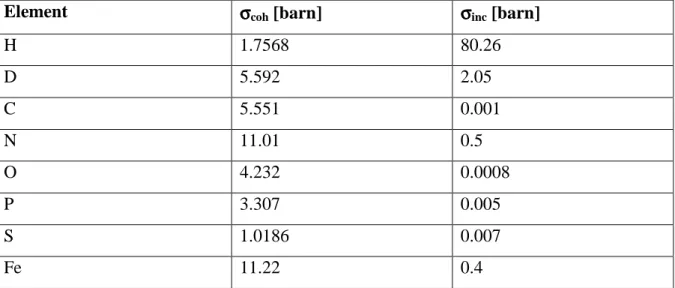
= . ( 3.19 )The incoherent scattering cross section of hydrogen is the largest of all elements that usually occur in biological matter (see Table 3). The reason for this behaviour is that the scattering length for different spin orientations between the neutron and the proton spin differs rather strongly. Incoherent scattering of natural abundance proteins is dominated by hydrogen atoms. The incoherent scattering cross section of hydrogen is around 40 times larger than that of deuterium. Therefore, hydrogen/ deuterium labelling is made possible. Scattering of natural abundance proteins in D2O solvent is dominated by the protein signal, whereas scattering of deuterated proteins hydrated with H2O allows to focus on the signal of the solvent. The unit of the scattering cross section is the barn with 1 barn = 10-24 cm2.
Table 3: Coherent and incoherent scattering cross sections for elements occurring in
biological systems (Sears 1992).
Element σσσσcoh [barn] σσσσinc [barn]
H 1.7568 80.26 D 5.592 2.05 C 5.551 0.001 N 11.01 0.5 O 4.232 0.0008 P 3.307 0.005 S 1.0186 0.007 Fe 11.22 0.4
3.5 Connecting Theory with Experiments
The Van Hove pair correlation function describes the microscopic structure and dynamics of the sample (Van Hove 1954). Van Hove showed mathematically in his article how the theoretical pair correlation function can be related to measurable neutron scattering quantities. The work paved the ground for all the models on microscopic motion that came afterwards. The pair correlation function is defined as
[
]
∑
+ − = N j j j j r t r r N t r G ' , '( ) ) 0 ( 1 ) , (rδ
r r r . ( 3.20 )The pair correlation function contains the probability to find a scattering particle at time t at the position rr(t), when it was at time t=0 at the position rr(0). N is the number of atoms.
The self term of the pair correlation function is
[
]
∑
+ − = N j j j s r r (0) r (t) N 1 ) t , r ( G rδ
r r r . ( 3.21 )The intermediate scattering function
) , ( ) , ( ) , (q t I q t I q t I r = coh r + inc r ( 3.22 )
is obtained by a space Fourier transformation of the Van Hove pair correlation function. The incoherent part of the intermediary scattering function Iinc(qr,t) is related with the self-term
) t , r (
Gs r . The coherent part Icoh(qr,t) is connected with the pair correlation function G(rr,t):
[
] [
]
∑
∫
= − = − 'j , j 'j j r q icoh expiqr (0) exp iqr (t) N 1 r d e ) t , r ( G ) t , q ( I r r r rr rr r r ( 3.23 )
[
] [
]
∑
∫
= − = − j j j r q i sinc expiqr (0) exp iqr (t) N 1 r d e ) t , r ( G ) t , q ( I r r r rr rr r r . ( 3.24 )
The intermediary scattering functions can be calculated nowadays from molecular dynamics simulations. Measured neutron data can be compared to these simulations and molecular events that are responsible for the measured processes can be eventually identified.
The scattering function S(qr,ω) in energy space is obtained by a time Fourier transformation of the intermediate scattering function
∫
− = I q t e dt q S iωt π ω ( , ) 2 1 ) , (r r . ( 3.25 )The scattering function S(qr,ω) can also be divided into a coherent part and an incoherent part
) , q ( S ) , q ( S ) , q ( S r
ω
= coh rω
+ inc rω
. ( 3.26 )The experimentally accessible double differential scattering cross section is connected to the scattering function with
inc inc coh coh q S k k N q S k k N dE d d ) , ( ' 4 ) , ( ' 4 2
ω
π
σ
ω
π
σ
σ
= + Ω . ( 3.27 )Measured incoherent neutron scattering data in the form of
inc dE d d Ω σ 2
can be corrected with computer programs for the term
k k'
and the scattering function S(q,ω)inc is containing