Accepted Manuscript
Title: A retrospective study on equine herpesvirus type-1 associated myeloencephalopathy in France (2008-2011) Author: Gaby van Galen Agnes Leblond Pierre Tritz Ludovic Martinelle St´ephane Pronost Claude Saegerman
PII: S0378-1135(15)00265-5
DOI: http://dx.doi.org/doi:10.1016/j.vetmic.2015.07.003
Reference: VETMIC 7022
To appear in: VETMIC Received date: 16-4-2015 Revised date: 26-6-2015 Accepted date: 4-7-2015
Please cite this article as: Galen, Gaby van, Leblond, Agnes, Tritz, Pierre, Martinelle, Ludovic, Pronost, St´ephane, Saegerman, Claude, A retrospective study on equine herpesvirus type-1 associated myeloencephalopathy in France (2008-2011).Veterinary Microbiology http://dx.doi.org/10.1016/j.vetmic.2015.07.003
This is a PDF file of an unedited manuscript that has been accepted for publication. As a service to our customers we are providing this early version of the manuscript. The manuscript will undergo copyediting, typesetting, and review of the resulting proof before it is published in its final form. Please note that during the production process errors may be discovered which could affect the content, and all legal disclaimers that apply to the journal pertain.
SHORT COMMUNICATION
A retrospective study on Equine Herpes Virus type-1 associated myeloencephalopathy in France (2008-2011)
Gaby van Galen 1,*,+, Agnes Leblond 2,3, Pierre Tritz 3,4,5, Ludovic Martinelle 1, Stéphane Pronost 3,6,7
, Claude Saegerman 1,3
1. Research Unit of Epidemiology and Risk Analysis applied to veterinary science (UREAR-ULg), Department of Infectious and Parasitic diseases, Faculty of Veterinary Medicine, University of Liege, Liege, Belgium
2. UR 346 Animal Epidemiology INRA Theix, Vetagrosup, Equine Department, University of Lyon, Lyon, France
3. Réseau d’Epidémio-Surveillance en Pathologie Equine (RESPE), Mondeville, France
4. Veterinary clinic of Faulquemont, Faulquemont, France
5. Committee of infectious diseases of the French equine veterinary association (association vétérinaire equine francaise - AVEF)
6. Frank Duncombe Laboratory-LABEO, Caen, France
7. Normandie Université, Unité Risques Microbiens (U2RM), 14000 Caen, France
* Corresponding author:
E-mail address: gaby@equinespecialists.eu or gaby@sund.ku.dk
+
Large Animal Clinic, Internal Medicine and Surgery Faculty of Health and Medical Sciences
University of Copenhagen Højbakkegaard Allé 5 2630 Taastrup
Denmark
Abstract
Diagnosis of equine herpesvirus-1 associated myeloencephalopathy (EHM) can be troublesome, but early recognition and knowledge of risk factors are essential for prevention and control. The objectives for this study are to 1) describe EHM in France, 2) improve clinical recognition, 3) identify risk factors. Through epidemiosurveillance of acute neurological cases (all considered to be potentially infectious cases) in France (2008-2011), 26 EHM cases were identified and 29 EHM negative control cases. EHM cases were described and compared to controls with univariate, multivariate and classification and regression tree analysis. EHM cases had a 46% fatality rate and were frequently isolated cases. Most showed ataxia, paresis and a cauda equina syndrome, yet presence of other neurological signs was variable. Statistical analysis identified the following variables to be significantly associated to EHM compared to controls: introduction of a new horse to the herd, cauda equina syndrome, larger herd size, saddle horses and month of occurrence. The presence of many isolated cases, and less typical and variable clinical presentations emphasize the difficulty in diagnosing EHM. Nevertheless, history and clinical examination of acute neurological cases can be valuable in recognizing EHM early as well in order to select those cases that need further laboratory testing and infection control measures. Moreover, with a different study format and geographic location, risk factors were found to be similar to previous studies, therefore strengthening their significance to the spread of EHM.
Keywords: neurology, EHV, EHM, horse Introduction
Neurological disorders caused by equine herpesvirus-1 (EHV-1) are called equine herpesvirus-1 associated myeloencephalopathy (EHM) and this is considered a contagious emerging syndrome (Lunn et al., 2009; Kydd et al., 2010; Traub-Dargatz et al., 2013). Currently, early recognition of suspected cases and close monitoring of high-risk horses represent the most reliable measures for preventing EHM outbreaks and limiting the consequences (Lunn et al., 2009; Pusterla et al., 2009). Although clinical signs of EHM are often perceived as well described (Kydd et al., 2010) and clinical diagnosis therefore straightforward, its clinical recognition can be troublesome, especially when isolated cases are considered rather than outbreaks. The diffuse and multifocal distribution of the lesions in the central nervous system (Lunn et al., 2009; Pusterla et al., 2009) can cause considerable variability in clinical presentation (van der Meulen et al., 2003; Pusterla et al., 2009). Laboratory analyses therefore remain indispensable, but take up precious time. New tools for improved clinical recognition of EHM would therefore be highly valuable.
Geographical region appears to be associated with EHM development (Goehring et al., 2006; Lunn et al., 2009) and risk factors could therefore potentially differ between countries. There are only few reports available in literature on EHM in France (Pronost et al., 2010b; Pronost et al., 2012) and none describe risk factors. Moreover, epidemiological studies are often restricted to a single outbreak, thereby limiting their potential at identifying risk factors unrelated to a specific outbreak. More data establishing possible risk factors are required (Lunn et al., 2009).
This study aims at improving the current understanding of EHM by: 1) describing EHM in France, 2) improving early clinical recognition of EHM by identifying variables that are specifically related to EHM rather than to other equine acute neurological diseases, and 3) the identification of host and management related factors.
Data collection
In France, a passive epidemiological surveillance program is implemented by the “Réseau d’Epidémio-Surveillance en Pathologie Equine” (RESPE; http://www.respe.net) to detect and monitor emerging and infectious neurological equine diseases, one of which is EHM. Veterinarians throughout France are asked to report all horses with acute neurological signs (all are considered potentially infectious), and to fill in a detailed standardized questionnaire for each case. Laboratory analysis was offered free of charge. There is owner informed consent.
Retrieved data from reported cases and definitions
Detailed information on season, and demographic, management, clinical and laboratory data were retrieved from the reporting veterinarians with use of standardized questionnaires. Cauda equina syndrome was defined as presence of a single or a combination of clinical signs related to lumbosacral cord pathology, i.e. an abnormal tonus, reflexes and sensibility of tail, anus and/or perineum, inability to urinate and/or defecate and urinary incontinence. Urinary retention caused by upper motor neuron lesions was also included in this definition, as reflection of diagnostic difficulties under field conditions.
EHM cases
Reported cases with acute neurological signs (regardless of further clinical and specific neurological signs) and with a positive test result for EHV-1 were considered EHM cases. EHV-1 positive testing was based on recent reviews (Lunn et al., 2009; Pusterla et al., 2009): 1) for ante mortem testing, a positive polymerase chain reaction (PCR) on nasal swabs, blood and / or cerebrospinal fluid, 2) for post mortem testing, histology with characteristic lesions in the spinal cord (vasculitis), or positive PCR from nervous tissue, or 3) seroconversion for EHV on acute and convalescent serum (4-fold increase)..
Control cases
The control group included reported cases that: 1) presented with acute neurological signs, and therefore were clinically suspect to have infectious neurological diseases amongst which EHM, and 2) were considered not to be infected with EHV-1 following diagnostic laboratory testing. They were considered EHV negative based on: 1) absence of seroconversion for EHV on paired sera or on a single serum taken > 1 week after disease onset, and/or 2) negative PCR (control cases should not have a positive PCR, but a negative PCR test was considered inadequate to rule out EHM) and/or 3) confirmation or high suspicion of another disease leading to neurological signs. To reduce bias as much as possible, the latter criterium was minimally based on clinical signs and mainly based on additional diagnostic testing.
Laboratory testing
Depending on the samples sent by the reporting veterinarian, complement fixation testing on blood and real-time PCR (on blood, cerebrospinal fluid, nasal swab and/or tissue) were performed as previously described (Pronost et al., 2012; Slater, 2014).
Statistical methods
To compare the EHM cases versus control cases, frequency variables were assessed by odds ratio (OR), categorical variables were assessed by Fisher’s exact test and quantitative variables were assessed by a two-sample Wilcoxon rank-sum test. All variables with a P-value < 0.10 in those univariate analyses were entered in a multivariate logistic regression. In addition, to assess collinearity, a backward elimination of variables was performed. Variables that induced a modification of OR of >20% were retained in final analysis. Goodness of fit was assessed using the Hosmer–Lemeshow goodness-of-fit test (StataCorp, 2012). Furthermore, classification and regression tree (CART) analysis (Saegerman et al., 2011) were performed to compare history and clinical signs or only clinical signs between groups.
Results Description
Out of the 219 neurological cases reported to the RESPE from 2008 to 2011, 26 cases fulfilled the inclusion criteria for EHM cases, and 29 for control cases. All EHM cases were considered EHV positive based on a positive PCR for EHV-1 on blood, nasal swab, cerebrospinal fluid and/or nervous tissue. The two cases that underwent a full post-mortem examination furthermore showed typical histology following post-mortem. Table 1 and 2 summarize signalment and history. EHM cases were Selle Francais (8), Thoroughbreds (4), Arabian horses (3), Spanish breed (1), French trotter (1), barb horse (1), poney (1), welsh (1), KWPN (1), Quarter Horse (1), unspecified saddle horse breeds (4). Three EHM cases were diagnosed in 2008, 7 in 2009, 11 in 2010 and 5 in 2011, and most (17) during winter months. No significant seasonal difference was observed between groups (P = 0.47). Some cases occurred together in the same herd or were related to each other, but half of them were unrelated to other cases (13/26). Clinical signs and their comparison between groups are described in Table 3, and details on neurological signs of EHM cases in Table 4. Apart from the fact that most EHM cases showed ataxia or paresis and a cauda equina syndrome, the remainder of the clinical picture was found to be variable. The mortality rate of the EHM group was 46%.
Control horses (29) were considered to be EHM negative based on absent seroconversion on paired sera (13), low antibody titre on a single serum taken > 1 week after the onset of clinical signs (2), and/or confirmation or high suspicion of another disease (17; Table 5).
Statistical analysis
Introduction of a new horse to the herd, EHV vaccination (but not if EHV vaccination occurred less than 6 months ago), and cauda equina syndrome were variables with a significantly higher association to EHM horses than controls. The herd size was significantly larger for EHM than for controls (Table 2 and 3). On the multivariate analysis only the introduction of a new horse to the
herd (OR = 14.64; 95% CI: 1.32-161.93; P = 0.03) and cauda equina syndrome (OR = 28.49; 95% CI: 1.23-427.07; P = 0.015) could be retained. The CART analysis showed that when variables of history and the clinical exam were used, herd size, month of occurrence and introduction of a new horse in the herd were the best predictors for EHM, and this with a sensitivity of 65% and specificity of 52% for the decision tree. When only the clinical exam was taken in consideration, presence of cauda equina syndrome was the best predictor with a sensitivity of 88% and specificity of 41%.
Discussion
This study provides clinical and epidemiological data on French EHM cases over a period of 4 years. The number of French EHM cases is believed underestimated, because of underreporting of this unnotifiable disease and the associated diagnostic challenges. From these results, it can be suggested that EHM occurs in the form of isolated cases at least as often as in the form of an outbreak, highlighting the need for improved tools for clinical recognition. Besides the fact that most EHM cases showed ataxia and/or paresis and a cauda equina syndrome, the clinical picture of EHM in the reported cases was variable. Some cases were reported with cerebral signs of abnormal behaviour; this is not the most typical expression of EHM, nevertheless it has been previously described (van der Meulen et al., 2003). This highly variable clinical picture, the atypical neurological expressions and the high number of isolated cases are all likely to be a result of the surveillance format used, where all acute neurological cases were considered as potentially infectious, reported and tested, therefore including EHM cases that might otherwise remain unidentified.
Not unexpectedly, cauda equina syndrome was the only clinical variable significantly more present in EHM horses than in controls. The definition of cauda equina syndrome in this study was rather large, including urinary retention due to both lower motor neuron and upper motor neuron lesions. While probably better reflecting the field conditions, this leads to an overestimation of
control cases with a cauda equina syndrome and therefore underestimation of its statistical power and specificity as a predictor for EHM. Nonetheless, its odds ratios are still high. Following the results of this study, it is 28 times more likely that a veterinarian is dealing with EHM than another neurological disease when he or she is called to see a horse with acute neurological symptoms and identifies a cauda equina syndrome. Also in the CART analysis, cauda equina syndrome was considered a main predictor for EHM and showed a good sensitivity. This makes it potentially a very useful clinical indicator as a first screening tool for syndromic surveillance for EHM. Of course after clinical suspicion, laboratory analysis remains necessary.
Age and sex were not statistically different between groups in the current study but it should be noted that, similar to studies performed in The Netherlands (Goehring et al., 2006), and in the USA (Henninger et al., 2007) EHM was not associated with young age. Nonetheless, EHM has been reported in horses of all ages (Greenwood and Simson, 1980; Friday et al., 2000). Breed was different between groups in the present study, with more saddle horses in the EHM group. Breed has been previously identified to be a risk factor for EHM (Goehring et al., 2006; Barbic et al., 2012), with Haflingers, Fjord horses, Icelandic horses and archetypical pony breeds less frequently affected by EHM (Goehring et al., 2006).
Introduction of horses to a herd before development of EHM outbreaks is commonly reported (van Maanen et al., 2001; van der Meulen et al., 2003; Goehring et al., 2006; Henninger et al., 2007), and was identified as a risk factor for EHM in this study by all different statistical means. Following our results, it is 14-15 times more likely to be EHM than another neurological disease when this variable is present in the history of an acute neurological case.
EHV vaccination was not uncommon in all groups and the findings of this study cannot support it to be a risk factor for EHM. Although by univariate analysis EHV vaccination was identified as a risk factor, it could not be retained in the multivariate analysis and more importantly failed as a risk factor in the univariate analysis when only those horses were taken into account where EHV vaccination was performed within the last 6 months before EHM developed. EHV
vaccination was reported previously to be associated with EHM (Henninger et al., 2007; Traub-Dargatz et al., 2013), but EHM also develops in populations where none or few horses are vaccinated (Goehring et al., 2006) and vaccination status could have been potentially confounded with increasing age (Lunn et al., 2009). The current study shows that age is unlikely to be a confounding factor since vaccinated EHM cases were younger than those unvaccinated (12.0 ± 4.2
versus 14.6 ± 3.6 years, respectively).
In this study, the variables significantly different between groups have been identified by statistical comparison of EHM cases to a control group. Both groups potentially have infectious neurological diseases due to the presence of acute neurological signs, and therefore those variables have diagnostic potential. At the same time the significantly different variables between groups can be appreciated as risk or protective factors for attracting EHM compared to another acute neurological disease. This type of control group has not been used previously for risk analysis of EHM.
Limitations include the retrospective nature of this work, the relatively limited case numbers and reporting by different veterinarians mostly under field conditions. However, potential bias has been reduced to a minimum by the use of a structured network, standardized clinical forms and case classification by an expert committee.
Conclusion
This study is the first to provide a thorough description of French EHM cases, including outbreaks and a high percentage of isolated cases. Although the clinical picture can be variable, the history and clinical examination of acutely neurologically affected horses can potentially be a valuable help to recognize EHM cases early, and to select those cases that would need further laboratory testing and immediate infection control measures. In addition, risk factors for EHM were identified and although in a different geographic location and study setup they were largely in accordance with other studies, therefore strengthening their significance in the spread of EHM.
Acknowledgements and funding
All French veterinarians reporting cases to the RESPE are gratefully acknowledged. Christel MARCILLAUD-PITEL and Charlène DAIX are gratefully acknowledged for their help with data collection, and Julie Storme for her valuable assistance in manuscript editing. Laboratory Franc Duncombe, Caen France is acknowledged for their analytical work. This study was funded by the RESPE.
References
Barbic, L., Lojkic, I., Stevanović, V., Bedekovic, T., Starešina, V., Lemo, N., Lojkic, M., Madic, J., 2012. Two outbreaks of neuropathogenic equine herpesvirus type 1 with breed-dependent clinical signs. Vet Rec 170, 227.
Burgess, B.A., Tokateloff, N., Manning, S., Lohmann, K., Lunn, D.P., Hussey, S.B., Morley, P.S., 2012. Nasal Shedding of Equine Herpesvirus-1 from Horses in an Outbreak of Equine Herpes Myeloencephalopathy in Western Canada. J Vet Intern Med 26, 384-392.
Friday, P.A., Scarratt, W.K., Elvinger, F., Timoney, P.J., Bonda, A., 2000. Ataxia and paresis with equine herpesvirus type 1 infection in a herd of riding school horses. J Vet Intern Med 14, 197-201.
Goehring, L., van Winden, S.C., van Maanen, C., Sloet van Oldenruitenborgh-Oosterbaan, M.M., 2006. Equine herpesvirus type 1-associated myeloencephalopathy in The Netherlands: a four-year retrospective study (1999-2003). J Vet Intern Med 20, 601-607.
Goehring, L., Landolt, G.A., Morley, P.S., 2010a. Detection and management of an outbreak of equine herpesvirus type 1 infection and associated neurological disease in a veterinary teaching hospital. J Vet Int Med 24, 1176-1183.
Goehring, L.S., Wagner, B., Bigbie, R., Hussey, S.B., Rao, S., Morley, P.S., Lunn, D.P., 2010b. Control of EHV-1 viremia and nasal shedding by commercial vaccines. Vaccine 28, 5203-5211.
Greenwood, R.G., Simson, A.R., 1980. Clinical report of a paralytic syndrome affecting stallions, mares and foals on a thoroughbred studfarm. Equine Vet. J. 12, 113-117.
Henninger, R.W., Reed, S.M., Saville, W.J., Allen, G.P., Hass, G.F., Kohn, C.W., Sofaly, C., 2007. Outbreak of neurologic disease caused by equine herpesvirus-1 at a university equestrian center. J Vet Intern Med 21, 157-165.
Kydd, J.H., Slater, J., Osterrieder, N., Antczak, D.F., Lunn, D.P., 2010. Report of the Second Havemeyer EHV-1 workshop, Steamboat Springs, Colorado, USA, September 2008. Equine Vet J 42, 572-575.
Lunn, D.P., Davis-Poynter, N., Flaminio, M.J.B.F., Horohov, D.W., Osterrieder, K., Pusterla, N., Townsend, H.G.G., 2009. Equine Herpesvirus-1 consensus statement. J Vet Intern Med 23, 450-461.
Pronost, S., Cook, R.F., Fortier, G., Timoney, P.J., Balasuriya, U.B.R., 2010a. Relationship between equine herpesvirus-1 myeloencephalopathy and viral genotype. Equine Vet J 42, 672-674.
Pronost, S., Legrand, L., Pitel, P.H., Wegge, B., Lissens, J., Freymuth, F., Richard, E., Fortier, G., 2012. Outbreak of equine herpesvirus myeloencephalopathy in France: a clinical and molecular investigation. Transbound Emerg Dis 59, 256-263.
Pronost, S., Leon, A., Legrand, L., Fortier, C., Miszczak, F., Freymuth, F., Fortier, G., 2010b. Neuropathogenic and non-neuropathogenic variants of equine herpesvirus 1 in France. Vet Microbiol 145, 329-333.
Pusterla, N., Wilson, W.D., Madigan, J.E., Ferraro, G.L., 2009. Equine herpesvirus-1 myeloencephalopathy: A review of recent developments. Vet J 180, 279-289.
Saegerman, C., Porter, R.S., Humblet, M.F., 2011. The use of modelling to evaluate and adapt strategies for animal disease control. Rev Sci Tech 30, 555-569.
Slater, J. 2014. Equine Herpes viruses, In: Sellon, Long (Eds.) Equine infectious diseases. Elsevier, Missouri, 151-168.
Traub-Dargatz, J.L., Pelzel-McCluskey, A.M., Creekmore, L.H., Geiser-Novotny, S., Kasari, T.R., Wiedenheft, A.M., Bush, E.J., Bjork, K.E., 2013. Case-control study of a multistate equine herpesvirus myelopencephalopathy outbreak. J Vet Intern Med 27, 339-346.
van der Meulen, K., Vercauteren, G., Nauwynck, H., Pensaert, M., 2003. A local epidemic of equine herpesvirus 1-induced neurological disorders in Belgium. Vlaams Diergeneeskundig Tijdschrift 72, 366-372.
van Maanen, C., Sloet van Oldenruitenborgh-Oosterbaan, M.M., Damen, E.A., Derksen, A.G., 2001. Neurological disease associated with EHV-1-infection in a riding school: clinical and virological characteristics. Equine Vet J 33, 191-196.
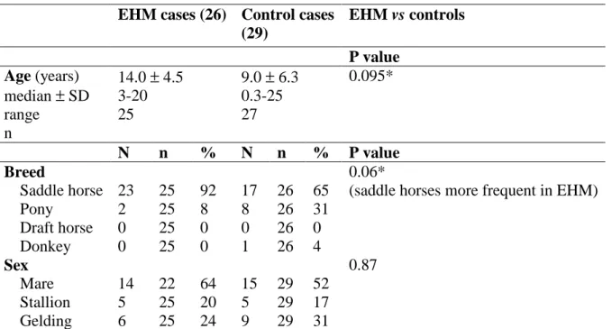
Table 1. Signalment of horses with equine herpesvirus-1 associated myeloencephalopathy (EHM)
and herpes virus negative control groups, and univariate statistical comparison between EHM and control groups.
EHM cases (26) Control cases (29) EHM vs controls P value Age (years) median ± SD range n 14.0 ± 4.5 3-20 25 9.0 ± 6.3 0.3-25 27 0.095* N n % N n % P value Breed 0.06*
Saddle horse 23 25 92 17 26 65 (saddle horses more frequent in EHM)
Pony 2 25 8 8 26 31 Draft horse 0 25 0 0 26 0 Donkey 0 25 0 1 26 4 Sex 0.87 Mare 14 22 64 15 29 52 Stallion 5 25 20 5 29 17 Gelding 6 25 24 9 29 31
Legend: EHM = equine herpesvirus-1 associated myeloencephalopathy; SD = standard deviation;
specific parameter; * variable selected for multivariate analysis based on
Table 2. History variables of horses with equine herpesvirus-1 associated myeloencephalopathy
and control horses, and univariate statistical comparison between groups.
Leg end: EH M = equi ne herp esvir us-1 asso ciated myeloencephalopathy; SD = standard deviation; N = number of horses with a positive response; n = number of horses with a response for this specific parameter; OR = odds ratio; CI95%
= confidence interval 95%;
EHV = equine herpes virus; NU = not used for statistical comparison; * variable selected for multivariate analysis based on P < 0.10; # variable significantly different between groups.
EHM cases (26)
Control cases (29) EHM vs controls P value
Herd size (number of horses) median ± SD range n 42.5 ± 33.7 2-100 22 20.0 ± 32.7 1-150 22 0.002*# N n % N n % P value Herd activity 1 Riding school 9 24 38 11 28 39 Training centre 0 24 0 1 28 4 Breeding facility 5 24 21 7 28 25 Pleasure riding / home 6 24 29 8 28 29
P value OR CI95%xxx Vaccination status
EHV vaccinated 16 23 70 10 28 36 0.02*# 4.11 1.27-13.36 EHV vaccination < 6 months before disease 10 21 48 7 27 26 0.12 2.60 0.71-8.75 Tetanus vaccinated 21 22 95 21 28 75 0.08* 7.00 0.79-61.98 Factors related to viral spread
Other sick horses since 3 months 13 20 65 10 26 38 0.16 2.31 0.72-7.38 New horse introduced in herd 14 20 70 5 24 21 0.002*# 8.87 2.25-35.00 Horse moved during last month 6 21 29 7 26 27 0.90 1.09 0.30-3.92 Motif to call veterinarian (multiple answers possible)
Hyperthermia 4 20 20 2 26 8 0.23 2.77 0.49-18.36 Ataxia, paresis 10 20 50 7 26 27 0.11 2.71 0.79-9.31 Recumbence 5 20 25 2 26 8 NU NU NU Lameness 0 20 0 3 26 12 NU NU NU Other / aspecific neurological signs 3 20 15 9 26 35 NU NU NU Other motif 2 20 10 7 26 27 NU NU NU
Table 3. Clinical variables of horses with equine herpesvirus-1 associated myeloencephalopathy
and control horses and univariate statistical comparison between groups.
EHM cases (26) Control cases (29)
EHM vs controls
P value (Wilcoxon rank-sum
test) Rectal temperature (°C) median ± SD range n 38.7 ± 1.5 35.0-40.8 21 38.1 ± 1.0 37.0-40.5 21 0.89 N n % N n % P value OR CI95%xxx Fever (>38.5°C) 11 21 52 7 21 33 0.22 2.20 0.63-7.66 Respiratory signs 8 18 44 10 24 42 0.86 1.12 0.32-3.85 Abnormal posture 5 21 24 4 23 17 0.60 1.48 0.34-6.48 Recumbence 10 22 45 8 26 31 0.30 1.88 0.57-6.12 Abnormal consciousness$ 7 16 44 15 25 60 0.25 0.50 0.15-1.62 Abnormal behaviour$ 12 22 55 17 25 68 0.35 0.56 0.17-1.85 Abnormal head position 2 18 11 3 24 13 0.89 0.88 0.13-5.87 Cranial nerve affection$ 8 21 38 15 25 60 0.14 0.41 0.12-1.35 Abnormalities cervical
area
11 19 58 21 26 81 0.10 0.33 0.09-1.27 Ataxia / weakness$ 11 12 92 20 23 87 0.96 1.05 0,15-7.13 Cauda equina syndrome$ 13 15 87 11 23 48 0.02*# 6.16 1.41-27.02
Death 11 24 46 6 22 27 0.20 2.26 0.66-7.76
Legend: EHM = equine herpesvirus-1 associated myeloencephalopathy; SD = standard deviation;
N = number of horses with a positive response; n = number of horses with a response for this specific parameter; OR = odds ratio; CI95% = confidence interval 95%; EHV = equine herpesvirus; * variable selected for multivariate analysis based on
P < 0.10; # variable significantly different between groups; $ see Table 4 for more details on this
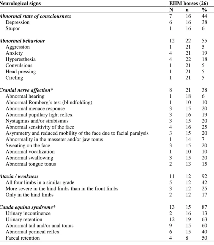
Table 4. Detailed description of neurological signs of horses with equine herpesvirus-1 associated
myeloencephalopathy.
Neurological signs EHM horses (26)
N n %
Abnormal state of consciousness 7 16 44
Depression 6 16 38 Stupor 1 16 6 Abnormal behaviour 12 22 55 Aggression 1 21 5 Anxiety 4 21 19 Hyperesthesia 4 22 18 Convulsions 1 21 5 Head pressing 1 21 5 Circling 1 21 5
Cranial nerve affection* 8 21 38
Abnormal hearing 1 18 6
Abnormal Romberg’s test (blindfolding) 1 10 10
Abnormal menace response 3 15 20
Abnormal pupillary light reflex 3 16 19
Nystagmus and/or strabismus 3 15 20
Abnormal sensitivity of the face 4 16 25
Asymmetry and reduced mobility of the face due to facial paralysis 3 15 20
Abnormality in the masseter and/or jaw tonus 1 14 7
Sweating on the face 3 15 20
Abnormal vocalization 1 10 10
Abnormal swallowing 3 15 20
Abnormal tongue tonus 2 13 15
Ataxia / weakness 11 12 92
All four limbs in a similar grade 5 12 42
More severe in the hind limbs than in the front limbs 3 12 25
Only in the hind limbs 2 12 17
Cauda equina syndrome* 13 15 87
Urinary incontinence 2 16 13
Urinary retention 12 19 63
Abnormal tail and/or anal tonus 9 15 60
Abnormal perineal reflex 6 15 40
Faecal retention 4 8 50
Legend: EHM = equine herpesvirus-1 associated myeloencephalopathy;
N = number of horses with a positive response; n = number of horses with a response for this specific parameter; * horses showed different combinations.
Table 5. Diseases diagnosed in horses from the equine herpes virus negative control group.
Diagnosis Number of horses from control
group Horses where a diagnosis or a very high suspicion was
reached *
17
Equine motor neuron disease (EMND) 5
Hepatoencephalopathy 2
Cervical spinal cord compression 3
Hyperkalemic periodic paralysis (HYPP) 1
Tetanus 1
Borna virus myeloencephalopathy 1
Bacterial encephalomyelitis 1
Fungal encephalomyelitis 1
Cranial nerve affection due to guttural pouch empyema 1
Spinal melanoma 1
Horses without definitive diagnosis but negative for equine herpes virus
13
Highly suspected to have cervical compression or (encephalo-) myelitis
4 Highly suspected to have a brain lesion or (myelo-)
encephalitis
7 Uncertain origin of single cranial nerve dysfunction 1
Total 29
Legend: * in addition to the fact that another neurological disease was diagnosed, some of these