Journal of Neurochemistry
Raven Press, Ltd., New York
© 1995 International Society for Neurochemistry
Thiamine Deficiency in Cultured Neuroblastoma Cells:
Effect on Mitochondrial Function and Peripheral
Benzodiazepine Receptors
L. Bettendorff, *G. Goessens, ~F. Sluse, P. Wins, M. Bureau, J. Laschet, and T. Grisar
Laboratories ofNeurochemistry and *Cellular and Tissular Biology, University of Liège, Liège;and j~Laboratory of Bioenergetics, University of Liège, Sart-Tilman, Belgium Abstract: When neuroblastoma cells were transferred to
a medium of low (6 nM) thiamine concentration, a 16-fold decrease in total intracellular thiamine content occurred within 8 days. Respiration and ATP levels were only slightly affected, but addition of a thiamine transport hibitor (amprolium) decreased ATP content and in-creased lactate production . Oxygen consumption be-came low and insensitive to oligomycin and uncouplers. At least 25% of mitochondria were swollen and electron translucent. Cell mortality increased to 75% within 5 days. [3H]PK 11195, a specific ligand of peripheral Ben-zodiazepine receptors (located in the outer mitochondrial membrane) binds to the cells with high affinity (Ko = 1 .4 0.2 nM) . Thiamine deficiency leads to an increase in both B ,ax and Kp . Changes in binding parameters for peripheral Benzodiazepine receptors may be related to structural or permeability changes in mitochondrial outer membranes . In addition to the high-affinity (nanomolar range) binding site for peripheral Benzodiazepine ligands, there is a low-affinity (micromolar range) saturable bind-ing for PK 11195. At micromolar concentrations, periph-eral benzodiazepines inhibit thiamine uptake by the cells. Altogether, our results suggest that impairment of oxida-tive metabolism, followed by mitochondrial swelling and disorganization of cristae, is the main cause of cell mortal-ity in severely thiamine-deficient neuroblastoma cells. Key Words: Thiamine-Thiamine deficiency-Energy metabolism-Mitochondria-Peripheral Benzodiazepine receptors-Neuroblastoma cells.
J. Neurochem . 64, 2013-2021 (1995) .
The brain is particularly sensitive to thiamine defi-ciency ( see Haas, 1988, for review) . As the coenzyme thiamine diphosphate (TDP) is required for oxidative decarboxylation of pyruvate and a-ketoglutarate, it can be expected that thiamine deficiency will impair oxida-tive energy metabolism. As brain consumes a dispro-portionate amount of the body's oxygen, impairment of oxidative metabolism can be expected to be particu-larly harmful. Moreover, the synthesis of important amino acids and neurotransmitters may be diminished if the availability of key intermediates of the Krebs
2013
cycle decreases (Butterworth and Héroux, 1989 ; Page et al., 1989 ) .
The use of cultured neuronal cells, such as neuro-blastoma cells, in the study of thiamine deficiency has the advantage that the effects on neurotransmitter syn-thesis need not be considered, as the cultured cells can usually live without producing such molecules . Schwartz et al. (1975) and Schwanz and McCandless (1976) have studied the effects of thiamine deficiency on energy metabolism (especially glycolysis ) in gli-oma and neuroblastgli-oma cell lines, but they were unable to quantify the remaining thiamine content of the cells. Though these authors report a strong decrease in py-ruvate dehydrogenase activity, levels of high-energy phosphate compounds (ATP and ADP) were not de-creased, except for phosphocreatine . Thiamine defi-ciency did not result in increased pyruvate and lactate production, except when the antimetabolite pyrithia-mine was added. In the absence of pyrithiapyrithia-mine, cul-tured malignant cells (especially glioma) can live and divide, albeit more slowly, even when external thia-mine concentrations are extremely low ( Schwartz et al., 1975) . This can be explained, at least partially, by the presence of a high-affinity thiamine carrier in neuroblastoma as well as glioma cells (Bettendorff,
1994; Bettendorff and Wins, 1994) .
Recently, Leong et al. (1994) reported that binding of [3H ] PK 11195, a selective ligand for peripheral-type Benzodiazepine receptors (PBRs), was increased in the brain of rats with pyrithiamine-induced thiamine deficiency. These receptors are localized in the mito-Resubmitted manuscript received October 4, 1994; revised manu-script received November 9, 1994; accepted November 9, 1994.
Address correspondence and reprint requests to Dr. L. Bettendorff at University of Liège, Laboratory of Neurochemistry ; 17 place De-cour, B-4020 Liège, Belgium.
Abbreviations used: CCCP, carbonyl cyanide m-chlorophenylhy-drazone ; PBR, peripheral Benzodiazepine receptor; TD, thiamine deficient; TDA, thiamine deficient and treated with amprolium; TDP, thiamine diphosphate ; TTP, thiamine diphosphate; URC, uncoupled respiratory control.
2014
chondrial outer membrane (Anholt et a1., 1986) and may be involved in mitochondrial respiratory control (Hirsch et al., 1988a) . PBRs are believed to be associ-ated with proteins controlling the transport of ions and other metabolites into mitochondria ( Kinnally et al., 1993 ) . Furthermore, it has been claimed that PBRs, were more abundant in glial than in neuronal cells (Syapin and Skolnick, 1979; Starosta-Rubinstein et al., 1987) ; estimation of PBR levels has been suggested as a probe to estimate neuronal damage (Benavides et al., 1987 ) . Thus, the observation of Leong et al. (1994) can be explained either by gliosis after neuronal death or through more specific changes in the number, struc-ture, and oxidative activity of mitochondria. In this study, we investigate the latter possibility by compar-ing control and thiamine-deficient (TD) neuro-blastoma cells .
MATERIALS AND METHODS Chemicals
Thiamine, carbonyl cyanide m-chlorophenylhydrazone (CCCP), amprolium, oligomycin, and rotenone were pur-chased from Sigma. ['aC ]Thiamine (24 mCi/mmol) , ['aC ]
-KSCN, and [methyl-3H]thymidine (5.0 Ci/mmol) were
from Amersham. [3H]Ro 15-4513 (29 Ci/mmol),
[N-methyl-3H]PK 11195 (81.7 Ci/mmol), [~H]flunitrazepam (74.6 Ci/mmol), [3H]H20 (1 mCi/g), and ['aC]inulin (3 mCi/g) were from NEN. Diazepam, clonazepam (Ro 5-4023/002), and Ro 15-1788 were given by Hoffmann-La Roche. Ro 5-4864 was from Fluka Chemie AG (Buchs, Switzerland) and PK-11195 was from Research Biochemi-cals (Natick, MA, U.S .A.) . Stock solutions (10 mM) of benzodiazepines and other PBR ligands were made in di-methyl sulfoxide. All measurements were made in the pres-ence of 1 % dimethyl sulfoxide as final concentration. Cell culture
The mouse neuroblastoma cell line (Neuro 2a, ATCC CCC 131) used in this study was a gift from Professor G. Moonen (Laboratory of Human Physiology, University of Liège) . The cells were grown (37°C, 5% COZ/95% air) in 100-mm Petri dishes (Nunc, Roskilde, Denmark) in 10 ml of Dulbecco's modified Eagle's medium (GIBCO, Ghent, Belgium) containing 10 ~M thiamine (value given by the manufacturer and checked by HPLC), enriched with glucose (6 mg/ml) and supplemented with 5% fetal calf serum (GIBCO) . TD cells were produced by growing them in a specially ordered Dulbecco's modified medium devoid of thiamine (GIBCO). In some cases, 20 ~M amprolium was added to the TD medium (TDA) . All other conditions (fetal calf serum and glucose concentrations) were as described above. The medium was changed every 2 days and the cells were subcultured every 4 days.
Under all conditions, cell viability was tested by the trypan blue exclusion method. Intracellular volumes were deter-mined from the difference of [3H ] Hz0 and ['aC] inulin spaces. The membrane potential was calculated from the intra/extracellular distribution ratio of ['aC ] SCN- (Catterall et al., 1976 ) .
Cell proliferation was quantified by [3H]thymidine incor-poration as described by Rogister et al. (1990) .
J. Neurochem . . Vol . 64, No . 5, 1995
L. BETTENDORFF ET AL.
All results are expressed per milligram of protein as deter-mined by the method of Peterson (1977 ) .
Determination of [ "C ] thiamine uptake
The cells were preincubated for 60 min, with or without inhibitors, in 1 ml of saline (145 mM NaCl, 5 mM KCI, 1 mM MgC12, 1 mM CaCl2, 10 mM glucose, 10 mM HEPES-Tris, pH 7.4 ) at 37°C and ['aCJthiamine uptake was deter-mined as previously described (Bettendorff and Wins, 1994) .
Binding of [3H] PK 11195
Cells subcultured in six-well multidishes (0.1-0.2 mg/ml) were rinsed with 2 X2 ml of saline as above. [ 3H] PK 11195
(25 l.d) at various concentrations (0.25-25 nM) was added and incubation was performed at 37°C . Unspecific binding was estimated in the presence of 1 ~M unlabeled PK 11195. After 60 min, 50 ~l of the medium was sampled for the determination of radioactivity and the cells were washed rapidly with 4 X 2 ml of ice-cold saline and dissolved in 1
ml NaOH for 30 min under constant stirring. Eight hundred microliters was counted for radioactivity as described above. Scatchard plots were analyzed using the k.cat 1 .3 program (BioMetallics, Inc., Princeton, NJ, U.S.A.).
Displacement experiments by various ligands were made in the presence of l nM ['H]PK 11195 for high-affinity binding. Low-affinity binding was estimated in the presence of 1 ~M unlabeled PK 11195 in addition to 1 nM labeled substance,
Determination of thiamine derivatives, ATP, oxygen uptake, and lactate in neuroblastoma cells
Thiamine derivatives were determined by an HPLC proce-dure exactly as described previously (Bettendorff et al., 1991) . The method of Hill et al. (1988) was used for ATP. L-Lactic acid was estimated by measuring NAD + reduction in the presence of lactate dehydrogenase, glutamate, and glutamate-pyruvate transaminase as described by Noll (1974) . Before lactate determination, the cells were fixed with perchloric acid; the suspension was neutralized with KOH and centrifuged. The supernatant was used for lactate determination.
Oxygen uptake was measured polarographically in a 2-ml cell at 37°C as described by Vayssière et al. (1986) . For each experiment, ~ 10-20 X 10 6 cells ( suspended in their
respective medium) were used and the OZ consumption in the presence of 1 mM NaCN was subtracted. The uncoupled rate of respiration was measured after addition of 5 ~M of the uncoupler CCCP.
Electron microscopy
The monolayer cultures were scraped off the dishes and centrifuged at 350 g for 3 min. Small fragments of the pellet were fixed at 4°C in glutaraldehyde (2.5% in cacodylate buffer) and then postfixed in 1% osmium tetroxide solution. The cells were embedded in Epon. Ultrathin sections mounted on copper grids were stained with uranyl acetate and lead citrate before examination under a Jeol CX 100 II electron microscope at 60 kV.
To estimate the variations in the number and the size of mitochondria between control and treated cells, the number of mitochondrial profiles per square micrometer of cyto-plasm and the main section of mitochondrial profiles were determined on random electron micrographs recorded at a 13,000x primary magnification.
FIG. 1. Effect of a decrease in extracellular thiamine on cell thiamine content. Cells grown in normal medium were trans-ferred into low-thiamine medium (day 0) and intracellular thia-mine derivatives were deterthia-mined as a function of time. On day 20, amprolium (final concentration, 20 tcM) was added. Each point is the mean of four experiments. After addition of amprol-ium, TTP content was too small to be determined . (~), thiamine; ("), thiamine monophosphate; (O), TDP; (O), TTP.
RESULTS
In neuroblastoma cells, as in most other cell types, thiamine uptake proceeds via a high-affinity and a low-affinity transporter. Whereas the high-low-affinity trans-porter has a Kmof 35 nM, the low-affinity mechanism
saturates at millimolar external thiamine concentra-tions (Bettendorff and Wins, 1994) . As the normal culture medium contains 10 ~M thiamine, the high-affinity transporter is completely saturated in this me-dium. To study the effect of thiamine deficiency on these cells, they were grown in the culture medium containing no thiamine. Under these conditions, the only source of thiamine is the fetal calf serum. The thiamine content of the serum is 120 ± 30 nM ( n = 3 ) as determined by HPLC, and it was used at 5%. Under these conditions, the thiamine concentration in the cul-ture medium is 6 nM, a value well below the Kmfor
the high-affinity thiamine transport. The contribution of the low-affinity transport is negligible at thiamine concentration < 100 nM (Bettendorff and Wins, 1994) .
Our cells survived very well in low-thiamine me-dium and could be continuously subcultured up to 6 months. The outgrowth of neurites was somewhat de-creased but not suppressed. These observations are in agreement with those reported by Schwanz et al.
FIG. 2. Effect of a decrease in extracellular thiamine on cell ATP and ADP levels. For explanations see legend to Fig. 1 . Data are mean ± SD values for four experiments. (~), ATP; (O), ADP.
THIAMINE DEFICIENCY IN NEUROBLASTOMA CELLS 2015 TABLE 1. Comparison of several biochemical features
of control and TD cells
The cells were grown for 2 weeks in the presence of either 10 pM thiamine (control) or 6 nM thiamine (TD cells) . In the last column (TDA cells) 20 pM amprolium was added for 2 days to TD cells. "6R6' uptake was measured after addition of 0.2 pCi~6RbC1 to the cells in the absence or the presence
of I mM ouabain. Data are mean ~ SD values for three to six experiments. The statistical differences of the results were estimated by the ANOVA test followed by the Fisher PLSD test for post hoc comparisons.
°p < 0.01, for comparison with control cells. n p < 0.05, for comparison of TDA with TD cells.
(1975 ) . To make the cells more severely deficient, amprolium (a competitive inhibitor of thiamine trans-port) was added to the TD medium. In the presence of 20 ~M amprolium, all the cells became spherically shaped and neurite outgrowth was only rarely seen. Under these conditions, cell mortality rapidly in-creased, and 5 days after addition of amprolium, 75% of the cells tested positive with trypan blue. It should be noted that when the cells were grown in normal thiamine-rich medium, addition of 20 ~M amprolium did not cause any decrease in intracellular thiamine content and no morphological changes were observed. This is presumably because, in contrast to pyrithia-mine, amprolium is only a competitive inhibitor of thiamine transport and it is not an effective Mocker of TDP synthesis (Rogers, 1970) . The estimated K; for the high-affinity transporter was 1 .8 p,M (Bettendorff and Wins, 1994), two orders of magnitude higher than
TABLE 2. Rate of oxygen consumption by Neuro 2a cells under different experimental conditions
OZ consumption (nmol/mg/min)
The cells were grown as described in the legend to Table 1. OZconsumption was measured polarographically (see Materials and Methods) in the respective culture media in either the absence or the presence of oligomycin or CCCP. The URC was calculated from the ratio of OZ consumption in the presence of CCCP over the O2 consumption in the presence of oligomycin . Data are mean = SD values of four experiments.
° p < 0.05, for comparison with control cells. b y G 0.05, for comparison of TDA with TD cells.
J. Neurochem., Vol. 64, Na 5, 1995 Control
cells cellsTD TDAcells Total thiamine (pmol/mg) 210 ~ 30 13 ± 4° 6 ~ 2° ATP content (nmol/mg) 12 1 14 ± 1 9 - 1 Membrane potential (mV) -50 ± 2 -54 ± 2 -43 - 2~ $b Rb` uptake (nmol/mg/30 min) 51 ± 5 56 - 4 36 + 5"b Intracellular volume (pl/mg) 4.5 - 1.5 5 ~ 1 -Km for thiamine transport (nM) 36 - 16 31 - 4
-V x(pmol/mg/15 min) 4.2 ~ 0.4 4.6 = 0.8
-['H]Thymidine incorporation
Lactate (nmol/mg) 31 - 4 31 ± 8 46 - 7°
Control
cells cellsTD TDAcells Basal rate 3.9 - 0.4 3.3 ± 0.5 l .4 = 0.2°°
+ Oligomycin (16 pg/ml) 2.7 ± 0.7 2.6 ~ 0.7 1 .2 - 0.3"x' + CCCP (5 pM) 7.3 ~ 1 .4 4.7 - 1 .4° 1 .2 ± 0.4"° URC 2.8=0 .7 1.9±0 .6 1.1+0 .2°' n
zoló
the apparent Km for thiamine. It is not surprising, there-fore, that in thiamine-rich media, 20 ~M amprolium does not significantly inhibit thiamine uptake: In the normal medium, the concentration of thiamine ( l 0 ~,M) is largely saturating .
Figure 1 shows the decrease in intracellular thiamine derivatives, in TD medium, as a function of time. The medium was changed every 2 days and the cells were transferred into new culture dishes every 4 days after reaching confluence. The intracellular contents of all four thiamine derivatives decreased rapidly before reaching a steady state after ~8 days. Total thiamine content was decreased to 6.2% of the initial value (13 ± 3 vs. 210 ± 30 pmol/mg for cells cultured in 10 ~,M external thiamine) . When 20 ~M amprolium was added, thiamine and especially TDP content was fur-ther decreased. Thiamine triphosphate (TTP) was
un-J. Neurochem Vol. 64, No . 5, 1995
L. BETTENDORFF ET AL.
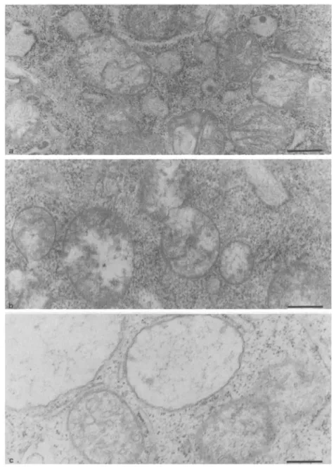
FIG. 3. Electron micrographs of control (a), TD (b), and TDA (c) cells. Abnormally swollen mitochondria can be seen in am-prolium-treated cells. The magnification bar represents 0.5 gym.
detectable under these conditions, suggesting that the cells contained G0.1 pmol TTP/mg of protein. From these results, we can assume that the critical amount of cell TDP required for survival is ~ 10 pmol/mg.
In parallel with thiamine derivatives, we also mea-sured the ATP and ADP contents of the cells ( Fig. 2 ) . Shortly after the onset of deficiency, we observed a transient decrease in ATP content followed by a recov-ery. This was concomitant with an increase in ADP content. After addition of amprolium, a further de-crease in ATP content was observed, followed by a partial recovery.
Table 1 gives a comparison of some biochemical parameters between cells grown in normal medium (10 ~M thiamine) , TD cells, and TD cells grown in the presence of 20 ~M amprolium ( TDA) . A slight depo-larization was observed in the latter condition, though
THIAMINE DEFICIENCY IN NEUROBLASTOMA CELLS amprolium was without effect on membrane potential
in thiamine-rich medium (not shown) . This might be due to a decrease in active cation transport as suggested by a decrease in ouabain-sensitive góRb + transport. In-tracellular volume as well as the kinetic parameters of high-affinity thiamine transport (Km and VmaX)
re-mained unchanged. Cell division decreased by ^-40% in TD cells as estimated by the decrease in [3H] -thymidine incorporation. In TDA cells lactate produc-tion was significantly increased and cell division was further slowed down. We thus compared the rates of Oz consumption in normal, TD, and TDA cells . The results are summarized in Table 2. As expected, control cells in thiamine-rich medium had a high rate of oxy-gen consumption; this was partially blocked by oligo-mycin and stimulated by uncouplers such as CCCP. Simple thiamine deficiency did not markedly reduce OZ consumption, though the uncoupled respiratory control (URC; Table 2) tended to decrease. In TDA cells, however, OZ consumption was markedly lower, even in the presence of the uncoupler and the URC was close to unity. This suggests severe impairment of mi-tochondrial function. The decrease in respiratory rate was expected as oxidative decarboxylation of pyruvate and a-ketoglutarate are blocked in the absence of TDP; our data show that respiratory control tends to be lost as well.
Electron microscopic examination revealed the pres-ence, in amprolium-treated cells, of abnormally large mitochondria ( Fig. 3 ) . The interior of these mitochon-dria was essentially electron translucent and no intact cristae were seen. In TD cells, no significant increase in mitochondrial size was seen, though the matrix ap-peared to be more disorganized than in nondeficient cells. In TDA cells, however, the main section of mito-chondria was 1 .2 ± 0.5 ~Cm (mean ± SD; n = 38) compared with 0.93 ± 0.26 (n = 30) and 0.96 -!- 0.27 ~Cm ( n = 37 ) for TD and normal cells, respectively (p = 0.0011, ANOVA test) . No significant changes were detected in the number of mitochondria per cell as a function of thiamine status.
In addition to changes in mitochondrial morphology, the density of ribosomes strongly decreased with the
FIG. 4. Binding of [3H] PK 11195 (1 nM) to neuroblastoma cells vs . incubation time. Binding experiments were performed at 37°C as described in Materials and Methods. Error bars represent mean ± SD values for three experiments. (O), total binding; ("), nonspecific binding (measured in the presence of 1 ~M unlabeled PK 11195) ; (O), specific binding.
201 7
FIG. 5. Displacement of [3H]PK 11195 binding (1 nM) by vari-ous ligands. Each point, expressed as a percentage of control, is the mean for three to six experiments. (O), PK 11195; ("), Ro 5-4864; (+), rotenone ; ( " ), clonazepam ; (D), Ro 15-1788.
severity of thiamine deficiency. As we have no evi-dence that TD or even TDA cells are appreciably swol-len, compared with normal ones (see Table 1), the decrease in apparent density of ribosomes most proba-bly reflects a decrease in the number of ribosomes per cell.
[~H] PK 11195, an isoquinoline carboxamide chemi-cally unrelated to benzodiazepines, is known to be a specific ligand of PBR (Le Fur et al., 1983) . Figure 4 shows the binding of [3H]PK 11195 to the cells as a function of time in the absence or presence of 1 ~M unlabeled PK 11195. Specific binding was calculated from the difference between total and nonspecific bind-ing. Equilibrium was reached after 15-30 min. In con-trast to most studies published in the literature, binding experiments were performed at 37°C instead of 4°C, as at the latter temperature equilibrium was not yet obtained after 90 min (not shown) . This result sug-gests an intracellar binding of PK 11195 in agreement with the known location of PBRs in the outer mito-chondrial membrane. The binding characteristics (Kp and Bm,x) should not be markedly temperature depen-dent as binding of PK 11195 is essentially entropy driven (Le Fur et al., 1983 ), whereas diffusion through membranes (which depends on membrane fluidity) may have a higher temperature coefficient.
Figure 5 shows the displacement of [ 3H]PK 11195 binding by various unlabeled ligads. As expected, PK 11195 and Ro 5-4864 (a benzodiazepine specific for PBRs; Marangos et al., 1982) were most effective. Ro 15-1788 and clonazepam, which are specific central benzodiazepine receptor ligands (Anholt et al., 1986) were without effect up to 1 ~M. Rotenone, an inhibitor of mitochondrial complex I, also inhibited [3H] PK 11195 binding in agreement with the results obtained by Hirsch et al. (1988b) on isolated rat kidney mito-chondria.
[3H] Flunitrazepam, which binds to PBRs as well as central-type receptors (Rohde and Harris, 1982; Schoemaker et al., 1983), was not displaced by the central ligand Ro 15-1788. Likewise, no specific bind-ing of [3H]Ro 15-4513 (another specific central li-gand; Sieghart et al., 1987 ) was observed (not shown ) . These results indicate that neuroblastoma cells contain
2018
FIG. 6. Scatchard plots for [3H]PK 11195 specific binding on
normal (O) and TD neuroblastoma cells (") . Nonspecific binding was estimated in the presence of 1 pM unlabeled PK 11195 (see Materials and Methods) and subtracted .
only the peripheral benzodiazepine receptors and are devoid of the central-type (GAGA ) receptor.
Figure 6 shows Scatchard plots of [3H]PK 11195
binding to cells cultured in normal and TD medium. A linear relationship was obtained for [3H]PK 11195
concentrations varying between 0.25 and 10 nM . Table 3 gives the KD and Bmax values for [3H]PK 11195
binding for cells grown in normal medium, TD cells, and TD cells returned to thiamine-rich medium for 2 weeks; the apparent affinity for [3H]PK 11195 binding
decreased in TD cells compared with normal cells whereas BmaX increased. These changes were fully re-versible after 2 weeks of culture in thiamine-rich me-dium.
Several authors have reported the existence of a low-affinity (i.e., micromolar) binding site for PBR ligands (Bowling and DeLorenzo, 1982; File et al., 1984) . We also observed a low-affinity binding in our cells. Figure 7 shows the displacement of 1 ~M [3H]PK 11195
binding by various ligands. The order of efficiency of these ligands was PK 11195 > Ro 5-4864 > diazepam > clonazepam > phenytoin > Ro 15-1788 . Like the high-affinity binding site, the low-affinity binding site showed a specificity for peripheral ligands versus cen-tral ligands, though clonazepam was significantly more efficient than Ro 15-1788 .
No displacement of [3H]PK 11195 binding from
TALE 3. Effect of thiamine deficiency on K and B ,~ for high-affinity binding of [3H]PK 11195
Kp and Bmax for [3H]PK 11195 in normal, TD, and
thiamine-deficient cells returned to thiamine-rich medium for 2 weeks as estimated from Scatchard plots. The significance of the differences was determined by the nonparametric test of Krnskal-Wallis : fur
Ko,p = 0.0024; forBm~x, p =0.039. The Mann-Whitney U test
for post hoc comparison between groups showed that both Ko and
Bm~xfor TD cells are significantly different from the same parameters
estimated in normal and TD recovered cells (mean ± SD values for n independent experiments) .
J. Neurochem., Vol. 64, No. 5, 1995
L. BETTENDORFF ET AL.
FIG. 7. Displacement of low-affinity [3H]PK 11195 binding by
various ligands. Binding was estimated in the presence of 1 nM [3H]PK 11195 and 1 pM PK 11195. The results are expressed
as percentage of control ± SD values for three experiments. (O), PK 11195; ("), Ro 5-4864 ; ( "), diazepam ; ("), clona-zepam; (~), phenytoin; (~), Ro 15-1788.
the high-affinity or the low-affinity binding site was observed with thiamine or its analogues pyrithiamine and oxythiamine up to 1 mM . It is interesting, how-ever, that we found thiamine transport was inhibited by different benzodiazepines and other PBR ligands at micromolar concentrations . Figure 8 shows the inhibi-tion of thiamine transport by various ligands. The order of efficiency was PK 11195 = Ro 5-4864 > diazepam > clonazepam > Ro 15-1788 = phenytoin, i.e., the same as the one obtained for the displacement of [ ~H ] PK 11195 from the low-affinity binding site. Re-cently, Patrini et al. (1993) suggested, on the basis of in vivo kinetic measurements, that phenytoin might interfere with thiamine uptake into the brain. Our re-sults suggest that such an effect is not likely to be mediated by the high-affinity thiamine transporter, as phenytoin is without effect up to 0.1 mM. No inhibi-tion of thiamine transport by GABA or picrotoxin was observed ( not shown ) . These results suggest that the inhibition of thiamine transport by benzodiazepines is not mediated through GABA,, receptors. This is in agreement with the observation that no increase in
~6C1 - uptake by GABA could be demonstrated in these
cells and that no specific binding of ['H ] muscimol (a specific GABAA receptor agonist; Harris and Allan, 1985 ) could be detected in Neuro 2a cells ( not shown ) . PBR ligands have been shown to affect mitochon-dria] respiratory control ( Vorobjev and Zorov, 1983;
FIG. 8. Effect of various compounds on high-affinity thiamine transport in neuroblastoma cells. Thiamine uptake (percentage of control) was determined as described in Materials and Meth-ods. Each point is the mean for three to nine experiments. (O), PK 11195; ("), Ro 5-4864 ; ("), diazepam ; ( "), clonazepam ; (4), phenytoin; (D), Ro 15-1788. Kp (~ BmaX(Pmol/mg) n Normal 1.4 ±0.2 1.7 ~ 0.2 6 TD 2.6±0.6 2.2±0.3 10 TD recovered 1.6 ± 0.7 1.6 ~ 0.6 5
THIAMINE DEFICIENCY IN NEUROBLASTOMA CELLS
TABLE 4. Effect ofRo 5-4864 on the distribution of intracellular thiamine derivatives and ATP content
T TMP TDP TTP ATP
Control 32±4 7±I 130±8 2.0±0.7 10.3±0 .6 Ros-4864(lOpA"1) 36-2 8±1 146±7 2.1±0.3 10 .8±0 .6 Ro s-4864 (100 pM) 40 . 3° 6 . 1 129 . 5 1 .7 - 0.5 8 - 1°
The cells were preincubated for 1 h with Ro 5-4864, and thiamine and ATP concenüations were determined as described in Materials and Methods. Results are expressed as picomoles per milligram for thiamine derivatives and as nanomoles per milligram for ATP (mean ± SD values of four experi-ments). TMP, thiamine monophosphate.
°p < 0.05, for comparison with the controls (ANOVA followed by the Dunnett test for post hoc comparison).
Hirsch et al., 1988a ) . As thiamine transport is depen-dent on intracellular ATP (Bettendorff and Wins, 1994) , it might be thought that these compounds act by decreasing intracellular ATP content, leading to an inhibition of thiamine transport.
However, after a 60-min preincubation in the ab-sence of 10 ~cM Ro 5-4864, no effect on the distribution of thiamine derivatives and ATP content of the cells was observed (Table 4) . The slight (25%) decrease in ATP, observed at 100 ~,M Ro 5-4864, may explain pan of the inhibition of thiamine transport, but this can be excluded at lower concentrations of the inhibitor. Furthermore, Ro 5-4864 inhibited thiamine transport even without preincubation ( not shown) , suggesting a more direct effect.
DISCUSSION
Reduction of extracellular thiamine concentration from 10 ~M to 6 nM leads to a 16-fold decrease in intracellular total thiamine content within 8 days. All four thiamine derivatives were decreased, including TTP, which has been previously reported to be particu-larly conserved in animal models (Pincus and Grove, 1970; Thornber et al., 1980) . Under our conditions, the cells remain perfectly viable, though the growth rate decreased by ^-40%. Uncoupled respiration was decreased but not the respiration in the presence of oxidative phosphorylation, suggesting that substrate availability is becoming a rate-limiting factor. This suggests that oxidative metabolism remains active even at very low external thiamine concentrations . Pre-sumably, pyruvate oxidation still occurs under mild thiamine deficiency; otherwise, pyruvate or lactate would accumulate and 02 consumption of the cells would decrease strongly, which is not the case (Tables
1 and 2) .
In TDA cells, total intracellular thiamine was further reduced. ATP levels also decreased, whereas lactate production increased, ouabain-dependent active$6Rb+
decreased, and the cells were slightly depolarized. At this stage, the rate of respiration was seriously im-paired. It should be pointed out that one of the earliest morphological changes observed in experimental
thia-2019 mine deficiency in brain involves glial edema, and this is observed before any symptomatic change (Robert-son et al., 1968 ) . This has been explained by decreased ATP levels and, thus, impairment of Na,K-ATPase ac-tivity. However, during thiamine deficiency, ATP lev-els rather tend to increase in cultured glioma cells (Schwanz and McCandless, 1976 ) as well as in rat brain (McCandless, 1982) . Furthermore, inhibition of Na,K-ATPase activity by ouabain in cultured astrocytes ( Kimelberg, 1981) and neuroblastoma cells (L. Bettendorff, unpublished results) leads to cell shrinkage rather than swelling. Increased ATP levels in TD cells are most probably the result of decreased ATP use rather than increased synthesis, in agreement with decreased [3H ] thymidine incorporation and
de-creased density of ribosomes (Fig. 3 ) .
About 25% of the mitochondria in amprolium-treated cells were abnormally large and translucent. No cristae were seen. That, on the same micrographs, normally sized mitochondria are seen close to large and empty ones suggests that we are not dealing with an artifact due to, e.g., the fixation procedures. Robert-son et al. (1968) reported the existence of such mito-chondria in TD rat brain but also in control material. Pawlik et al. (1977) found disorganized mitochondria in the plantar nerves of TD rats. Our results suggest that impairment of oxidative metabolism induces mito-chondrial abnormalities leading to the cell mortality in amprolium-treated cells. No clearcut modifications were observed in nuclei. It is known that the early observable event in apoptosis is chromatin condensa-tion (Willie, 1980) . This was not seen in our thiamine-deprived cells. It should be emphasized that only extreme thiamine deficiency as induced by thiamine antimetabolites leads to significantly increased cell mortality.
PBR, a 17-18-kDa protein ( Sprengel et al., 1989 ) , is located mainly in the outer mitochondrial membrane and has been claimed to be associated with the voltage-dependent anion channel ( porin ) and the adenine nu-cleotide carrier protein (McEnery et al., 1992) . It could be located at contact sites of the outer and inner mitochondrial membranes and regulate the activity of inner mitochondrial channels responsible for the up-take of ions and other metabolites (Kinnally et al., 1993) .
Leong et al . (1994 ) reported an increase of [3H ] PK
11195 binding sites in the brains of animals with pyri-thiamine-induced thiamine deficiency, whereas Bena-vides et al. (1987 ) reported an increase in PBRs in rat brain after neurotoxic lesions and suggested glial proliferation accompanying neuronal degeneration. Other studies have shown a higher density of PBRs in glioma versus neuroblastoma cells (Syapin and Skol-nick, 1979 ) and glial versus neuronal cell ( Sher and Machen, 1984), but PBRs are by no means absent in cells of neuronal origin (for review, see Le Fur et al., 1988 ) . Our results suggest an alternative and maybe complementary explanation; i .e., biochemical and
2020
structural modifications in mitochondria after thiamine deficiency would lead to an increase in PBR.
In this respect, it is interesting that [3H] Ro 5-4864
binding is increased in the temporal cortex of patients with Alzheimer's disease (Owen et al., 1983), whereas other studies report a decrease in a-ketoglutarate dehy-drogenase activity in Alzheimer's disease (Mastrogia-como et al., 1993 ) . Furthermore, added TDP induced a significantly higher stimulation of this enzyme in Alzheimer patients compared with controls, suggesting a premortem reduction of TDP levels (Mastrogiacomo et al., 1993) . Gibson et al. (1988) also reported a 75% decrease in a-ketoglutarate dehydrogenase and at least a 45% decrease in transketolase activities in the brains of patients with Alzheimer's disease.
In addition to high-affinity peripheral binding sites, we observed a low-affinity (micromolar) binding site for [3H] PK 11195. This binding also showed higher
apparent affinity for peripheral benzodiazepines, but that does not mean that those low-affinity "receptors" are of the peripheral type; low-affinity binding sites were ---1,000 times more abundant than PBRs (not shown ) . Low-affinity binding sites for benzodiaze-pines have been reported by several authors ( see Bowl-ing and DeLorenzo, 1982 ) . In this study, we also show that benzodiazepines and PK 11195 in a concentration range of 1-100 ~,M inhibit thiamine transport, the most effective compound being PK 11195 a "peripheral-type" ligand. No competition with thiamine or its anti-metabolites pyrithiamine and oxythiamine was ob-served and the central benzodiazepines were practi-cally ineffective.
At present it cannot be determined whether there is only one category of binding sites having multiple ac-tions and among them inhibition of thiamine transport or if we are dealing with numerous binding sites in-cluding the thiamine transporter. We favor the second hypothesis for the following reasons : We were unable to obtain reproducible Scatchard plots and tentative extrapolations yielded a Bm~ of -2,000-5,000 pmol/ mg, an extremely high value (compare with the 1 .7-2.2 pmol/mg for the high-affinity binding site in Table 3 ) . Such high values have also been reported by other investigators (Bowling and DeLorenzo, 1982) . The hydrophobic character of benzodiazepines would favor multiple binding to membrane components. The reason peripheral ligands are more effective than central ones is not clear. Altogether, our results suggest that, at least in Neuro 2a cells, the mortality observed under extreme thiamine deficiency (as induced by thiamine antime-tabolites ) is the consequence of impaired mitochon-drial function rather than the result of excitotoxicity or apoptosis. Indeed, even if the cells excrete gluta-mate, its extracellular concentration is unlikely to reach "neurotoxic" levels in the culture medium. In TD mammalian brain, other factors such as decreased neu-rotransmitter synthesis (for review, see Haas, 1988 ) , excitotoxic phenomena (Hazell et al., 1993; Langlais and Zhang, 1993 ) , or effects on membrane chloride
J. Neurochem., Vol. 64, No. 5, 1995
L. BETTENDORFF ET AL.
permeability (Bettendorff et al., 1993 ) should also be considered, as they might interfere with normal brain function and performance and might result in neuronal death.
In the absence of more precise knowledge about the function of PBRs, it is not possible to know the physiological significance of an increase in high-affin-ity peripheral binding sites in thiamine deficiency ex-cept that this might be some sort of compensation for impaired mitochondrial function.
Acknowledgment: We thank the National Funds for Sci-entific Research (Belgium) for a grant to L.B. M.B . is a Senior Research Assistant and P.W. is a Research Associate at the National Funds for Scientific Research.
REFERENCES
Anholt R. R. H., Pedersen P. L., De Souza E. B., and Snyder S. H. (1986) The peripheral-type benzodiazepine receptor. Localiza-tion to the mitochondrial outer membrane. J. Biol. Chem. 261, 576-583.
Benavides J., Fage D., Carter C., and Scatton B. (1987) Peripheral type benzodiazepine binding sites are a sensitive indirect index of neuronal damage. Brain Res. 421, 167-172 .
Bettendorff L. (1994) The compartmentation of phosyphorylated thiamine derivatives in cultured neuroblastoma cells. Biochim. Biophys . Acta 1222, 7-14.
Bettendorff L. and Wins P. (1994) Mechanism of thiamine transport in neuroblastoma cells: inhibition of a high affinity carrier by sodium channel activators and dependence of thiamine uptake on membrane potential and intracellular ATP. J. Biol. Chem. 269, 14379-14385 .
Bettendorff L., Peeters M., Jouan C. Wins P., and Schoffeniels E. (1991) Determination of thiamin and its phosphate esters in cultured neurons and astrocytes using an ion-pair reversed phase high-performance liquid chromatographic method. Anal. Bio-chem. 198, 52-59.
Bettendorff L., Kolb H.-A., and Schoffeniels E. (1993) Thiamine triphosphate activates an anion channel of large unit conduc-tance in neuroblastoma cells. J. Membr. Biol. 136, 281-288 . Bowling A. C. and DeLorenzo R. J. (1982) Micromolar affinity
benzodiazepine receptors: identification and characterization in central nervous system. Science 216, 1247-1249 .
Butterworth R. F. and Héroux M. (1989 ) Effect of pyrithiamine treatment and subsequent thiamine rehabilitation on regional cerebral amino acids and thiamine-dependent enzymes. J. Neu-rochem. 52, 1079-1084.
Catterall W. A., Ray R., and Morrow C. S. (1976) Membrane poten-tial dependent binding of scorpion toxin to action potenpoten-tial Na+ ionophore. Proc. Natl. Acad. Sci. USA 73, 2682-2686 . File S. E., Green A. R., Nutt D. J., and Vincent N. D. (1984) On
the convulsant action of Ro 5-4864 and the existence of a micromolar benzodiazepine binding site in rat brain. Psycho-pharmacology 82, 199-202 .
Gibson G. E., Sheu K. R., Blass J. P., Baker A., Carlson K. C., Harding B., and Perrino P. (1988) Reduced activities of thia-mine-dependent enzymes in the brains and peripheral tissues of patients with Alzheimer's disease. Arch. Neurol. 45, 836-840 . Haas R. H. (1988) Thiamine and the brain. Annu. Rev. Nutr. 8,
483-515.
Harris R. A. and Allan A. M. (1985 ) Functional coupling of y-aminobutyric acid receptors to chloride channels in brain mem-branes. Science 228, 1108-1110.
Hazell A. S., Butterworth R. F., and Hakim A. M. (1993) Cerebral vulnerability is associated with selective increase in extracellu-larglutamate concentration in experimental thiamine deficiency.
THIAMINE DEFICIENCY IN NEUROBLASTOMA CELLS
Hill M., Dupaix A., Nhiri M., Guyen L., and Arrio B. (1988) ATP:AMP phosphoüansferase activity, a new characteristic of Cantharanthus roseus tonoplasts . FEBS Lett. 230, 47-50. Hirsch J. D., Beyer C. F., Malkowitz L., Beer B., and Blame A. J.
(1988a) Mitochondrial benzodiazepine receptors mediate inhi-bition of mitochondrial respiratory conáol . Mol. Pharmacol. 34, 157-163 .
Hirsch J. D., Beyer C. F., Malkowitz L., Loullis C. C., and Blame A. J. (19886 ) Characterization of ligand binding to mitochon-dria) benzodiazepine receptors. Mol. Pharmacol. 34, 163-172. Kimelberg H. K. (1981) Active accumulation and exchange üans-port of chloride in astroglial cells in culture . Biochem. Biophys . Acta 646, 179-184 .
Kinnally K. W., Zorov D. B., Antonenko Y. N., Snyder S. H., McEnery M. W., and Tedeschi H. (1993 ) Mitochondria) benzo-diazepine receptor linked to inner membrane ion channels by nanomolar actions of ligands. Proc. Natl. Acad. Sci. USA 90,
1374-1378 .
Langlais P. J. and Zhang S. X. (1993 ) Exüacellular glutamate is increased in thalamus during thiamine deficiency-induced le-sions and is blocked by MK-801 . J. Neurochem. 61,
2175-2182.
Le Fur G., Vaucher N., Perrier M. L., Flamier A., Benavides J., Renault C., Dubroeucq M. C., Guérémy C., and Uzan A. (1983) Differentiation between two ligands for peripheral benzodiaze pine binding sites, [3H]Ro5-4864 and [3H]PK 11195, by ther-modynamic studies . Life. Sci. 33, 449-457.
Le Fur G., Gueremy C., Benavides J., Mestre M., Mizoule J., Doble A., and Uzan A. (1988 ) "Peripheral" benzodiazepine binding sites, in GABA and Receptors, Vol. 2 (Squires R. F., ed), pp.
15-34. CRC Press, Boca Raton, Florida.
Leong D. K., Lê O., Oliva L., and Butterworth R. F. (1994) In-creased densities of binding sites for the "peripheral-type" ben-zodiazepine receptor ligand ['H]PK 11195 in vulnerable re gions of the rat brain in thiamine deficiency encephalopathy . J. Cereb. Blood Flow Metab. 14, 100-105 .
Marangos P. J., Patel J., Boulanger J.-P., and Clark-Rosenberg R. (1982) Characterization of peripheral-type benzodiazepine binding sites in brain using [3H]Ro 5-4864 . Mol. Pharmacol. 22, 26-32.
Masüogiacomo F., Bergeron C., and Kish S. J. (1993 ) Brain a-ketoglutarate dehydrogenase complex activity in Alzheimer's disease. J. Neurochem. 61, 2007-2014 .
McCandless D. W. (1982) Energy metabolism in the lateral vestibu-lar nucleus in pyrithiamine-induced thiamine deficiency. Ann. NY Acad. Sci. 378, 355-364.
McEnery M. W., Snowman A. M., Trifiletti R. R., and Snyder S. H. (1992) Isolation of the mitochondria) benzodiazepine receptor: association with the voltage-dependent anion channel and the adenine nucleotide carrier. Proc. Natl. Acad. Sci. USA 89, 3170-3174.
Noll F. (1974) L-(+)-Lactate . Determination with LDH, GPT and NAD, in Methods of Enzymatic Analysis, Vol. 3, 2nd edit. (Bergmeyer H. U., ed), pp. 1475-1479. Academic Press, New York.
Owen F., Poulter M., Waddington J. L., Mashal R. D., and Crow T. J. (1983 ) ['H]Ro5-4864 and [3H] fluniüazepam binding in kainate-lesioned rat striatum and in temporal cortex of brains from patients with senile dementia of the Alzheimer type. Brain Res. 278, 373-375 .
Page M. G., Artkoma-Sey V., Coulson W. F., and Bender D. A. (1989) Brain glutamate and y-aminobutyrate (GABA) metabo-lism in thiamine-deficient rats. Br. J. Nutr. 62, 245-253. Patrini C., Perucca E., Reggiani C., and Rindi G. (1993) Effects of
phenytoin on the in vivo kinetics of thiamine and its phosphate esters in rat nervous tissues. Brain Res. 628, 179-186.
2021
Pawlik F., Bischoff A., and Bitsch I. (1977) Peripheral nerve changes in thiamine deficiency and starvation. An electron mi-croscopic study. Acta Neuropathol. (Berl.) 39, 211-218 . Peterson G. L. (1977) A simplification of the protein assay method
of Lowry et al. which is more generally applicable . Anal. Bio-chem. 83, 346-356.
Pincus J. H. and Grove I. (1970) Distribution of thiamine phosphate esters in normal and thiamine-deficient brain. Exp. Neurol. 28, 477-483.
Robertson D. M., Wasan S. M., and Skinner D. B. (1968 ) Ulüastruc-tural features of early brain stem lesions of thiamine-deficient rats. Am. J. Pathol. 52, 1081-1087 .
Rogers E. F. (1970) Thiamine antagonists, in Methods in Enzymol-ogy, Vol. 18A (McCormick D. B . and Wright L. D., eds), pp. 245-258. Academic Press, New York.
Rogister B ., Leprince P., Delree P., Van Damme J., Billiau A., and Moonen G. (1990) Enhanced release of plasminogen activator inhibitors) but not of plasminogen activators by cultured rat glial cells üeated with interleukin-1 . Glia 3, 252-257 . Rohde B. H. and Harris R. A. (1982) GABA and flunitrazepam
binding to neuroblastoma cell membranes-effects of growth conditions, ethanol and pentobarbital . Brain Res. 253, 133-141 .
Schoemaker H., Boles R. G., Horst W. D., and Yamamura H. I. (1983 ) Specific high-affinity binding sites for ['H]Ro-4864 in rat brain and kidney . J. Pharmacol. Exp. Ther. 225, 61-69. Schwartz J. P. and McCandless D. W. (1976) Glycolytic metabolism
in cultured cells of the nervous system. IV. The effects of thiamine deficiency on thiamine levels, metabolites and thia-mine-dependent enzymes of the C-6 glioma and C-1300 neuro-blastoma cell lines. Mol. Cell. Biochem. 13, 49-53.
Schwanz J. P., Lust W. D., Shirazawa R., and Passoneau J. V. (1975) Glycolytic metabolism in cultured cells of the nervous system. III. The effects of thiamine deficiency and pyrithiamine on the C-6 glioma and C-1300 neuroblastoma cell lines. Mol. Cell. Biochem. 9, 73-78.
Sher P. K. and Machen V. L. (1984 ) Properties of ['H]diazepam binding sites in cultured marine glia and neurons. Dev. Brain Res. 14, 1-6.
Sieghan W., Eichinger A., Richards J. G., and Möhler H. (1987 ) Photoaffinity labeling of benzodiazepine receptor proteins with the partial inverse agonist ['H]Ro 15-4513 : a biochemical and autoradiographic study. J. Neurochem. 48, 46-52.
Sprengel R., Werner P., Seeburg P. H., Mukhin A. G., Santi M. R., Grayson D. R., Guidotti A., and Krneger K. E. (1989) Molecu-lar cloning and expression of cDNA encoding a peripheral-type benzodiazepine receptor. J. Biol. Chem. 264, 20415-20421 . Starosta-Rubinstein S., Ciliax B . J., Penney J. B., McKeever P., and
Young A. B. (1987) Imaging of a glioma using peripheral benzodiazepine receptor ligands . Proc. Natl. Acad. Sci. USA 84, 891-895 .
Syapin P. J. and Skolnick P. (1979) Characterization ofbenzodiaze-pine binding sites in cultured cells of neural origin. J. Neuro-chem. 32, 1047-1051 .
Thornber E. J., Dunlop R. H., and Gawthorne J. M. (1980) Thiamin deficiency in the lamb: changes in thiamin phosphate esters in the brain. J. Neurochem. 35, 713-717.
Vayssière J.-L., Larcher J.-C., Berthelot F., Benlot C., Gros F., and Croizat B. (1986) Effects on mitochondria) metabolism of CCA, one inducer of neuroblastoma differentiation . Biochem. Biophys. Res. Commun . 140, 789-796.
Vorobjev I. A. and Zorov D. B. (1983) Diazepam inhibits cell respiration and induces fragmentation of mitochondria) reticu-lum. FEBS Lett. 163, 311-314.
Willie A. H. (1980) Glucocorticoid-induced thymocyte apoptosis is associated with endogenous endonuclease activation. Nature 284, 555-556.

![FIG. 4. Binding of [ 3 H] PK 11195 (1 nM) to neuroblastoma cells vs . incubation time](https://thumb-eu.123doks.com/thumbv2/123doknet/6353174.167626/5.892.529.744.151.300/fig-binding-h-pk-neuroblastoma-cells-incubation-time.webp)

