Université de Montréal
H3K27M/I mutations promote context-dependent
transformation in acute myeloid leukemia with RUNX1 alterations
par Yu Wei Zhang
Programme de Biologie Moléculaire Faculté de Médicine
Thèse présentée
en vue de l’obtention du grade de Maitrise en Biologie Moléculaire
option Générale
Résumé
Les mutations néomorphiques faux-sens affectant certaines lysines critiques des histones H3 jouaient un rôle important dans le développement d’une grande variété de tumeurs solides. Malgré la forte prévalence des mutations H3K27M dans les glioblastomes pédiatriques, se traduisant par une perte globale de H3K27me2/3, ces mutations n’ont pas été étudiées dans les cancers hématologiques humains. En utilisant les transgènes H3.3K27M/I, nous avons étudié les effets d’une perte de H3K27me2/3 sur les cellules hématopoïétiques normales et transformées.
Nous avons identifié des mutations d’histone H3K27M/I dans des leucémies myéloïdes aigües (LMA) accompagnées d’une diminution significative de H3K27me2/3. Les profils mutationnels des patients H3K27M/I a révélé que ces lésions génétiques survenaient invariablement de manière concomitante avec des mutations affectant le gène RUNX1. L’expression ectopique de H3K27M/I dans un modèle murin de leucémie induite par AML-ETO9a a accéléré la prolifération cellulaire in vitro et le développement leucémique in vivo. Cependant l’expression de H3.3K27M dans des cellules hématopoïétiques humaines CD34+ de sang de cordon a mené à une expansion sélective d’une population primitive de progéniteurs mais n’a pas été suffisante pour induire seule le développement leucémique.
Ces résultats démontrent l’existence d’une collaboration entre RUNX1 et la perte de H3K27me2/3. Dans un contexte de LMA avec mutation RUNX1, l’hypo-H3K27me3 accélère la progression de la maladie. Nous concluons que H3K27me2/3 possède une activité suppressive des LMA mutées dans RUNX1. Nos résultats ont le potentiel de guider le développement d’une approche thérapeutique ciblée pour le traitement des LMA avec mutations RUNX1.
Mots-clés: les cellules hématopoïétiques, leucémies myéloïdes aigües, H3.3K27M/I , oncohistones, RUNX1, Leucegene
Abstract
Neomorphic missense mutations affecting crucial lysine residues in histone H3 genes have been shown to be initiating mutation in a variety of solid tumors. Despite the high prevalence of H3K27M mutations in pediatric glioblastoma proceeded by the loss of global
H3K27me2/3, these mutations have not been studied in human hematological cancers. Using the H3.3K27M/I transgenes we studied the effects of the loss of H3K27me2/3 on normal and transformed hematopoietic cells.
We identified oncogenic histone H3K27M/I mutations in human acute myeloid leukemia (AML) that led to a significant reduction of H3K27me2/3. The mutation profiles of H3K27M/I patients revealed that these lesions invariably co-occurred with RUNX1 alterations. Expression of H3.3K27M/I in an AML1-ETO9a driven mouse model resulted in accelerated in vitro proliferation and in vivo disease latency. The expression of H3.3K27M in human CD34+ umbilical cord blood cells led to a selective expansion of a primitive hematopoietic progenitor population, but was insufficient to drive leukemogenesis on its own.
This demonstrates a collaboration between RUNX1 and loss of H3K27me2/3. In the context of RUNX1 alterations in AML, hypo-H3K27me3 accelerates disease progression. We conclude that H3K27me2/3 has a leukemia suppressive function in RUNX1 mutant and translocated AML. Our findings have the potential to inform the development of a targeted therapeutic strategy for the treatment of RUNX1mut AML patients.
Keywords: Hematopoietic Stem Cells. Acute Myeloid Leukemia, H3.3 K27M/I, oncohistones, RUNX1, Leucegene
Table des matières
Résumé ... i
Abstract ... ii
Table des matières ... iii
Liste des figures ... vi
Liste des tableaux ... vii
Liste des sigles ... viii
Liste des abréviations ... x
Remerciements ... xi
CHAPTER 1 – INTRODUCTION ... 1
Chromatin Biology ... 1
1.1.1 Chromatin Structure ... 1
1.1.2 Chromatin Regulation by Histone Post-Translational Modifications ... 3
1.1.3 Histone Modification: Methylation ... 4
Stem Cell Biology ... 6
1.1.4 Epigenetic Regulation of Normal Hematopoiesis ... 8
1.1.5 Hematopoietic Malignancies ... 9
1.1.6 Acute Myeloid Leukemia ... 10
1.1.7 EZH2 in Disease Hematopoiesis ... 11
1.1.8 Inhibition of PRC2 Activity through H3K27 Mutation ... 12
Gene Expression Regulation by Transcriptional Factors ... 15
1.1.9 Structure Characterization of RUNX ... 15
1.1.10 RUNX1 Alterations in Leukemia ... 15
General Rationale and Hypothesis ... 18
CHAPTER 2 – RESULTS ... 19
2.1 H3K27M/I mutations in Acute Myeloid Leukemia ... 19
2.1.2 Identification of H3K27M/I Mutation in AML Patients ... 22
2.1.3 Collaboration between H3K27M/I with AML1-ETO9a Accelerates Disease Progression ... 25
2.1.4 H3K27me2/3 in RUNX1alteredAML Patients ... 28
2.2 Functional Investigation of H3K27M in Normal Hematopoiesis ... 30
2.2.1 Reduction of H3K27 Methylation Selectively Expands Human Progenitor Population ... 30
2.2.2 shRUNX1 phenocopies immunophenotype of H3.3K27M ... 33
2.2.3 H3.3K27M Expression Enhances the Activity of Committed Myeloid Progenitors and Results in Mild Loss of LT-HSC Self-renewal ... 34
2.3 Investigation of Targeted Therapy for RUNX1 altered AML Patients ... 40
2.3.1 t(8;21) and RUNX1 mutant AMLs are sensitive to H3K27 demethylase inhibition.40 2.3.2 Genetic validation of the sensitive of GSK-J4 in OCI-AML5 ... 41
CHAPTER 3 – DISCUSSION ... 46
Identification of H3K27 mutation in AML ... 46
Genetic context of H3K27 mutation in AML ... 47
Characterization and Genetic Validation of GSK-J4 activity ... 47
EZH inhibitors in the treatment of RUNX1 altered AML patient ... 50
H3.3KM system as a replacement for traditional genetic manipulation of methyltransferase activity ... 51
Uncovering the mechanism behind the genetic synergy of hypo-H3K27me3 and RUNX1 alterations ... 52
CHAPTER 4 – METHODS ... 54
4.1 Primary AML Specimens ... 54
4.2 Next-generation RNA sequencing and mutation validation. ... 54
3.3 Human cord blood cell collection and processing. ... 54
4.4 CD34+ cell culture. ... 54
4.5 Flow cytometric quantification of H3K27me2/3. ... 55
4.8 Virus Production and UCB Cell Infection. ... 56
4.9 Western blot analysis. ... 57
4.10 Transplantation and monitoring of H3.3-infected and EZH1/2 treated UCB cells. ... 58
4.11 Estimation LT-HSC numbers by limiting dilution analysis. ... 58
4.12 Bone marrow cell transduction and generation of mouse AML models. ... 58
4.13 RNAi and CRISPR-Cas9 ... 59
Contributions ... 60
Bibliographie ... i
Appendix ... vii
Lentivirus Production and Transduction of CD34+ cord blood cells ... vii
Liste des figures
Chapter 1
Figure 1. Illustration of Chromatin Compaction ... 2
Figure 2. Illustration of Histone Modifications. ... 4
Figure 3. The Polycomb Repressive Complex 2 in coordinating epigenetic silencing activity 6 Figure 4. Hematopoietic stem cell hierarchy ... 7
Figure 5. H3K27M inhibits PRC2 methyltransferase activity. ... 14
Figure 6. Organization of cooperating lesion in RUNX1mut or t(8;21) AML ... 17
Chapter 2 Figure 7. Establishment of a FACS-based assay to quantify global levels of H3K27me2/3 . . 20
Figure 8. Flow cytometry is an effective tool to quantify H3K27me2/3 ... 21
Figure 9. Oncogenic histones H3K27M and H3K27I identified in AML patients to occur at low frequencies ... 25
Figure 10. Collaboration between H3.3K27I/M and AML1-ETO9a (AE9a) accelerated disease development ... 27
Figure 11. Quantification of global H3K27me2/3 in AML patient specimens ... 29
Figure 12. Low expression of core PRC2 members in t(8;21) AML ... 30
Figure 13. Reduction of H3K27me2/3 expands a selective human progenitor population. ... 32
Figure 14. shRUNX1 mimics H3.3K27M in the expansion of CD34+/CD45RA-/CD90+/CD133-... 34
Figure 15. H3.3K27M expression enhances the activity of committed myeloid progenitors. ... 35
Figure 16. Reduction of H3K27me2/3 has a mild impact on mice LT-HSC ... 36
Figure 17. Reduction of H3K27me2/3 has a mild impact on human LT-HSC ... 38
Figure 18. Limiting Dilution Assay assessing HSC frequency. ... 39
Figure 19. Selective sensitivity of t(8;21) and RUNX1mut AML cell lines towards H3K27 demethylase inhibitor GSK-J4.. ... 41
Figure 20. shUTX/JMJD3 validation of pharmacological specificity of GSK-J4 in OCI-AML5. ... 43
Figure 21. sgUTX/JMJD3 validation of pharmacological specificity of GSK-J4 in OCI-AML5. ... 44
Liste des tableaux
Table 1. Overview of histone modifications and their associated functions ... 4 Table 2. EZH2 inhibitors and their status in clinical development ... 14
Liste des sigles
Acute lymphocytic leukemia (ALL) Acute myeloid leukemia (AML)
Bone marrow (BM)
ChIP-sequencing (ChIP-seq) Chronic myeloid leukemia (CML)
Clustered regularly interspaced short palindromic repeats (CRISPR) Core-binding factor (CBF)
Colony forming unit (CFU)
Common myeloid progenitor (CMP) Common lymphoid progenitor (CLP)
Diffuse intrinsic pontine glioma (DIPG) Diffuse large B-cell lymphoma (DLBCL) DNA methyltransferase (DNMT)
Embryonic ectoderm development (EED) Embryonic stem cell (ESC)
Encyclopedia of DNA Elements (ENCODE) Enhancer of Zeste 1 (EZH1)
Enhancer of Zeste 2 (EZH2)
Glioblastoma multiforme (GBM)
Granulo/erythroid/monocytic/megakaryocytic (GEMM) Granulocyte-macrophage progenitor (GMP)
Histone deacetylase (HDAC) Histone demethylase (HDM) Histone methyltransferase (HMT)
Leukemia stem cell (LSC) Limiting dilution assay (LDA)
Long-term hematopoietic stem cell (LT-HSC) Lysine-to-methionine (KM)
Mix lineage leukemia (MLL) Multiplicity-of-infection (MOI) Multipotent progenitors (MPP) Myelodysplastic syndrome (MDS) Myeloproliferative neoplasm (MPN)
Polycomb repressive complex 1 (PRC1) Polycomb repressive complex 2 (PRC2) Post-translational modification (PMT)
RNA interference (RNAi)
Runt-related transcription factor (RUNX)
S-adenosyl-methionine (SAM)
Short-term hematopoietic stem cell (ST-HSC) Suppressor of zeste 12 (SUZ12)
The Cancer Genome Atlas (TGCA) Transcriptional factor (TF)
Variant allele frequency (VAF)
Liste des abréviations
For example (e.g.) That is (i.e.)
Remerciements
In writing this memoire, I have had the opportunity to reflect on the last two years of my Master’s and realized that I have been extremely fortunate to be surrounded by so many talented and generous people who made this work possible. The experiences and knowledge that I will take from IRIC will continue to shape my PhD and for years beyond that.
First and foremost, I would like to thank my research director, Dr. Guy Sauvageau for the opportunity to join his team. It is rare to find a supervisor that has exceed in so many areas of science. The incredible experiences that I have had at IRIC would not have been possible without the members of the team that he has assembled.
A special thanks to Bernhard Lehnertz for being a patient and thoughtful supervisor. I am extremely fortunate to have a found a mentor that who has allowed me to grow scientifically. Thank you to Tara MacRae, Jalila Chagraoui, Elisa Tomellini, Richard Bisaillon, Irene Baccelli and Laura Simon for your guidance and the many hours of discussions. Finally, to Julie Lessard and Alain Verreault, for being a part of my Master’s committee.
In closing, I believe the years that I have spent in the Sauvageau Lab was a period of remarkable growth for me as a scientist. This was thanks to the collaborative effort of everyone at the IRIC to provide opportunities for young scientists and build a collaborative research environment. Finally, this work would not have been possible without a community that values science and the generous support of FRQS.
CHAPTER 1 – INTRODUCTION
Chromatin Biology
The primary function of the chromatin is to efficiently package the mass quantity of DNA to fit into the nucleus. The chromatin consists of complexes of small proteins known as histones that are wrapped around by DNA. The tight or loose wrapping of the DNA around the histone regulates DNA replication, DNA damage, recombination, transcription and most importantly controls whether the genes can be expressed to form their encoded product. Chromatin packaging is highly dynamic and is governed by different chromatin modifying activities such as DNA methylation, histone modification, and ATP-dependent remodeling. The term “epigenetic” was coined to describe these changes that modified gene expression without alterations in DNA sequence.
1.1.1 Chromatin Structure
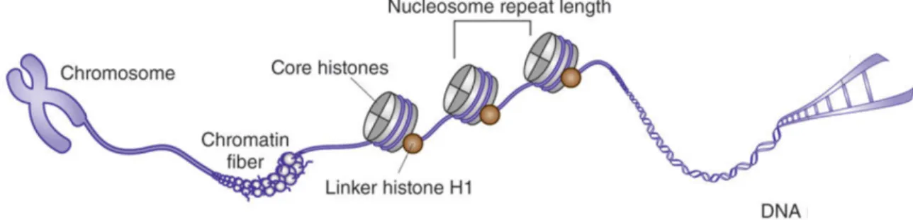
At its most fundamental level, chromatin structure is comprised of the nucleosome, a structure containing an octamer of four core histone proteins which is wrapped around by 147 base pairs (bps) in 1.7 turns around a protein octamer. The nucleosome core particle is composed of a hetero-octamer of histones comprising a tetramer of (H3-H4)2 flanked by two dimers of H2A-H2B. Each histone is composed of a globular domain and an unstructured tail domain. Two consecutive nucleosomes can be bound by linker histone H1. Together, the repeating structure of nucleosomes result in the formation of a structure that resembles “beads on a string”
(Figure 1). Nucleosomes coil and stack together forming chromatin, which is condensed into
higher-order structures and compacted into the chromosomes [1, 2]. Components of chromatin such as the histone protein and DNA are chemically or structurally modified to decrease or increase accessibility to regulatory proteins. For instance, a tight compaction restricts the regulatory elements of the gene to be inaccessible to the regulatory proteins, thereby blocking gene expression [3].
Multiple histone variants exist for the core histone H3, H2A, H2B and for the linker histone H1. Often, the histone variants only differ by a few amino acids or domains. Each variant localizes to specific chromatin domains and may be differentially expressed in various tissues. The substitution of the core histones with different variants will affect post-translational modifications (PMTs), protein interaction and higher order chromatin structure [4].
Figure 1. Illustration of Chromatin Compaction. Double helix DNA wraps around a
histone octamer that contains two copies of the major types of histones (H2A, H2B, H3 and H4) and connect through the linker histone H1 to form the nucleosomes. The nucleosomes further condense into chromatin fibers to be package into the nucleus. Modified from [5].
Chromatin is traditionally distinguished in two forms: euchromatin and heterochromatin. These terms were coined by Emil Heitz, who had observed heterochromatin as chromosomal segments that appeared extremely condensed and dark in the nucleus during interphase. In contrast, euchromatin appeared relatively light in color [6]. Since then, we have uncovered that euchromatin is less condensed and can be transcribed, whereas heterochromatin is highly condensed and cannot be transcribed. Heterochromatin can be further categorized into facultative and constitutive heterochromatin. Constitutive heterochromatin occupies the same regions (centromeric and telomeric chromosome) in every cell and in regions that are gene-poor. In contrast, facultative heterochromatin is associated with the silencing of genes due to developmental/environmental cues and scatters throughout the genome. These differing chromatin regions within the genome are marked by a variety of PTMs that give the regions their identity [7, 8].
1.1.2 Chromatin Regulation by Histone Post-Translational Modifications
The observation that histone modifications regulate specific and distinct functional outputs of the eukaryotic genome has led to the histone code hypothesis. Due to the structural protruding of the histone tail, the amino acids on the tail are easily accessible targets to enzymatic activities that form PTMs. Histone modifications occur on serine, lysine and arginine residues of the N-terminal tail of the four core histones. These residues can undergo methylation, acetylation, phosphorylation, ubiquitination or sumoylation (Figure 2) [9]. The post-translational enzymes are referred as “erasers” and “writers” referring to their ability to deposit or remove PTMs from the histone. PTMs are dynamically regulated, with many enzymes dedicated to their addition and removal. The numerous post-translational modifiers each target specific residues and can deposit/remove a specific modification. The interpretation of the modification depends on the “reader/effector” that will bind to a specific combination of histone modification and translate the code into a meaningful biological consequence [3, 10].
Figure 2. Illustration of Histone Modifications. The N terminal tail of the histones protrude
from the core of the nucleosome and are subject to a variety of modifications that influence chromatin structure. PTMs establish a signaling platform to recruit reader modules. The readers determine the functional outcome of the PTMs. The different post-translational modifications that can occur on each histone are illustrated in this figure. Adapted from [11].
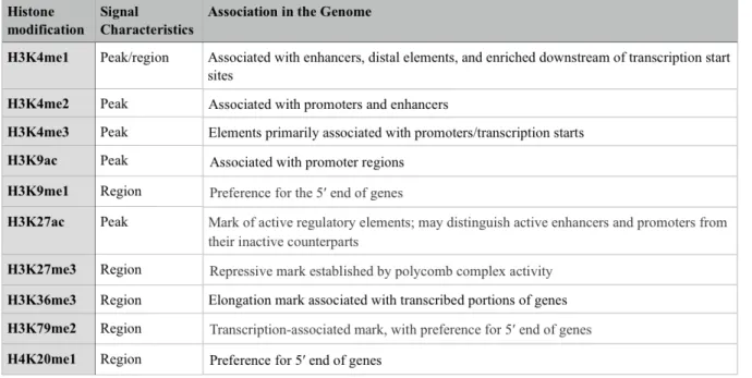
Genome wide pattern analyses of PTMs at regulatory regions (i.e. promoter and enhancer) have shown that PTMs associate at functionally distinct genomic regions and are successfully used to predict the presence of regulatory elements. For instance, H3K9ac and H3K4me2 tend to correlate with active promoters and H3K4me1 is present at distal enhancer regions (Table 1) [12].
Table 1. Overview of histone modifications and their associated genomic region. The
systemic mapping of transcription and transcription factors association through the Encyclopedia of DNA Elements (ENCODE) project identify that different histone
modifications can be used systematically to assign functional attributes to genomic regions. Table modified from [12].
1.1.3 Histone Modification: Methylation
Lysine methylation has a diverse number of roles that include heterochromatin formation, X-chromosome inactivation, and transcriptional regulation. Unlike acetylation which generally
correlates with transcriptional activation, histone lysine methylation can signal either activation or repression depending on the site of methylation [13]. The lysine methylation sites that have been associated with transcriptional activation include K4, K36 and K79 on histone H3. In contrast, di- and trimethylation of K9 and K27 on histone H3 are associated with transcriptional repression [14]. The degree of histone lysine methylation is determined by the antagonizing activities of site-specific histone methyltransferases (HMTs) and histone demethylases (HDMs). Almost all histone lysine methyltransferases rely on the catalytic activity of the SET domain for the addition of methyl-groups on the lysine residue [15] (DOT1/DOT1L is the only lysine methyltransferase that lacks a SET domain [16]). The two most conserved motifs within the SET domain (i) is a tyrosine which is essential for interacting with the lysine, and (ii) a knot like structure that is the S-adenosyl-methionine (SAM) binding pocket. The co-factor SAM serves as the methyl group donor for the covalent modification [17]. The removal of the methyl mark is achieved through the catalytic activity of histone lysine demethylases, amino oxidase or the JMJC family[18, 19].
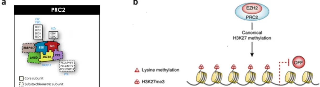
At the core of the H3K27 silencing system is Polycomb Repressive Complex 2 (PRC2) which is composed of either methyltransferase, Enhancer of Zeste 2 (EZH2) or its paralog Enhancer of Zeste 1 (EZH1). EZH2 functions with the assembly of other PRC2 proteins at the N terminal domain that include Embryonic Ectoderm Development (EED) and Suppressor of Zeste 12 (SUZ12). The catalytic SET domain of EZH1/2 deposits the repressive H3K27me2/3 mark which inhibits the accessibility of transcription factors through chromatin compaction. In addition, accessory subunits that influence PRC2 activity can be present in the complex including histone-binding protein RBBP4, zinc finger protein AEBP2 and PHF1
(Figure 3). Multiple factors influence recruitment of PRC2 to its genomic target including the
DNA sequence, transcription factors and pre-existing histone modifications. [20, 21].
Specific histone demethylases are involved in the removal of histone methylation marks. The lysine 27 methyl marks catalyzed by EZH1/2 are removed by the demethylase activity of HDMs, UTX and JMJD3. JMJD3/UTX are part of the KDM6 subfamily that contains a well-conserved JMJC domain in their C terminus which catalyzes the transition of H3K27me3 to
[22]. In summary, it is the dynamics between HDM and HMT activity that modulates gene expression.
Figure 3. The Polycomb Repressive Complex 2 in coordinating epigenetic silencing activity. (a) The mammalian PRC2 is composed of three essential core subunits: the catalytic
subunit Enhancer of Zeste 2 (EZH2) or Enhancer of Zeste 1 (EZH1), Embryonic Ectoderm Development (EED) and Suppressor of Zeste 12 (SUZ12). The accessory proteins of the PRC2 include PCL, RBBP4/7 and JARID that modulate PRC2 activity in different cellular contexts. (b) EZH2 mainly regulates transcriptional activity through the methylation of H3 on lysine 27 (H3K27) that is associated with gene silencing. Adapted from [21] and [23].
Stem Cell Biology
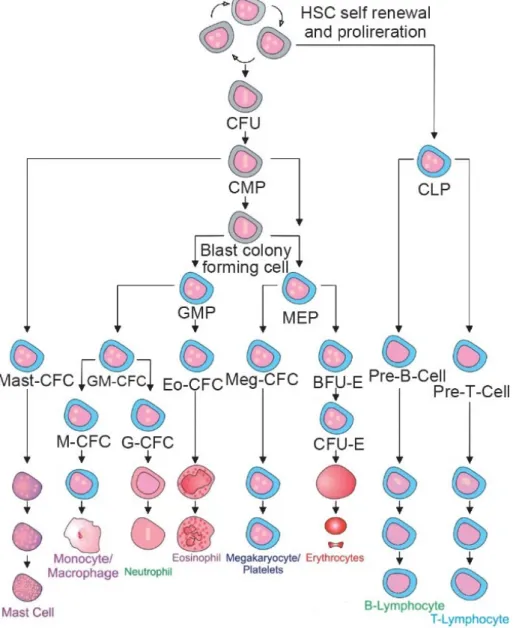
Throughout the life time of an individual, hematopoietic stem cells (HSCs) are responsible for replenishing the blood and immune cells. HSC differentiation is achieved through stepwise restriction of lineage potential. HSCs remain quiescent for most of their lifespan, while the more committed progenitors have very high proliferative capacities. For instance, HSCs give rise to a series of multipotent progenitors (MPPs) that have decreased self-renewal potential. MPPs differentiate towards more committed progenitors and eventually produce the mature cells that constitute the blood and the immune system (Figure 4). The balance between self-renewal, differentiation and proliferation in the hematopoietic lineage is critical for maintaining homeostasis and enables the rapid and robust response to physiological stresses, such as blood loss, infections and traumatic injuries [24].
It remains to be elucidated exactly how a stem cell determines whether to maintain its multi-potency or to differentiate. However, we can speculate that this process involves a fine
orchestration between cell cycle regulation and cell fate decision. During the transition from HSC to committed progenitor, there is a down-regulation of many HSC-associated genes which inversely correlate with the up-regulation of lineage-specific genes. The silencing or activation of gene expression and transcriptional programs is controlled by a highly regulated epigenetic process of DNA methylation and histone PTMs [24].
Figure 4. Hematopoietic stem cell hierarchy. Hematopoietic stem cells give rise to several
oligopotent progenitors (i.e. granulocyte-macrophage progenitors (GMP) and megakaryocyte/ erythrocyte progenitors (MEP)) and eventually fully differentiated cells (i.e.monocyte/macrophages/granulocytes and megakaryocytes/ platelets/ erythrocytes) [25].
1.1.4 Epigenetic Regulation of Normal Hematopoiesis
All cells of the hematopoietic lineage share the same genome but differ in their epigenome. During cellular division, dynamic changes occur in the epigenetic landscape allowing them to acquire their cellular identity.
A phenomenon that was originally demonstrated in embryonic stem cells (ESCs) describing key developmental genes that were simultaneously occupied by counteracting marks has also been reported in HSCs. These bivalent domains constitute the co-existence of repressive H3K27me3 and activating H3K4me3 marks. This mechanism ensures silencing while enabling quick activation [26]. Genome wide association mapping has identified an association of developmental genes with bivalent chromatin marks in HSCs and MPP cells. During lineage commitment, the bivalent marks will dissolve taking on the identity of being either repressive or activating. Interestingly, regions that become activating after lineage commitment associated with H3K4me1 and H3K27me1 in the gene regulatory regions. Overall this would suggest, the commitment to a specific mark during differentiation is already predetermined in HSCs and MPP cells [27].
PRC2 plays a major role in normal hematopoiesis by promoting pluripotency maintenance and self-renewal of hematopoietic stem cells. Interestingly, PRC2 regulates normal hematopoietic stem cell function in a developmental-stage-specific manner. For instance, the deletion of Eed exhausts adult HSCs, whereas neonatal HSCs are produced in normal numbers but are defective in maintenance/differentiation [28]. Furthermore, loss-of-function mutation of Ezh2 or Suz12 enhanced the activity of HSC and progenitor cells. By silencing pro-differentiation genes in LT-HSCs, Ezh2 maintains long-term self-renewal potential. The ectopic expression of Ezh2 causes a significant increase in HSCs and myeloid lineage cells [29]. In addition, the over-expression of Ezh2 in HSCs prevents exhaustion and maintains stem cell
potential over a series of transplantations [30]. This was in line with the observation that Ezh2 is abundantly expressed in highly purified HSCs and is rapidly down-regulated on differentiation. The ability of PRC2 to maintain HSCs may rely on its repression of Cdkn2a. Normally, Cdkn2a induces cycle arrest in G1 and G2 but is repressed by H3K27me3. The deletion of Cdkn2a partially rescues HSC deficiency in Eed(-/-) mice. Ezh2 also represses genes associated with cell cycling, HSC differentiation and pro-apoptosis (e.g. NOXA, p21 and WIG1) [28, 31]
During lymphopoiesis, Ezh2 expression is up regulated in proliferating cells such as human germinal center B cells, cycling T and B lymphocytes and plasmablasts. Then during B-cell differentiation and maturation, Ezh2 becomes down-regulated. This suggests that EZH2 acts as a checkpoint mechanism that regulates pro-B to pre-B cell transition. In support of this EZH2 was shown to regulate pre-B cell receptor in pro-B cell, a receptor implicated in both cycle exit and Igκ recombination expression [32]. In addition, Ezh2 can also inhibit the maturation of pro- to pre- B cells through Stat5 mediated recruitment of Ezh2 to repress Igκ transcription thereby inhibiting recombination [33].
1.1.5 Hematopoietic Malignancies
In many respects, leukemia cells share many common characteristics of a normal hematopoietic stem cell. Pathways classically associated with cancer also regulate normal stem cell function such as proliferation, survival, self-renewal and differentiation. Mutations can occur at any point during stem cell division and give rise to a leukemia-initiating mutation. Evidence suggests that the first leukemogenic mutations occur in a self-renewing cell (i.e. HSC) or in a more differentiated cell (i.e. MPP) to re-establish the ability to self-renew. These initial mutations confer pre-leukemic cells that will go on to accumulate successive mutations, and lead to the loss of normal hematopoietic functions and subsequently the formation of blast cells. In many cases, only one or two additional cooperating mutations are needed to generate the malignant founding clones [34, 35].
1.1.6 Acute Myeloid Leukemia
Acute myeloid leukemia (AML) is characterized by a block in myeloid differentiation and the uncontrolled proliferation of transformed myeloid progenitors. Current paradigms suggest that AML can be a (1) a step-wise progressive malignancy. When myelodysplastic syndromes (MDS) or myeloproliferative neoplasms (MPN) are untreated, they can progress into chronic myeloid leukemia (CML) and ultimately evolve to become AML. Alternatively, AML can develop from (2) genotoxic therapies from unrelated malignancies, but most cases arise (3)
de novo. The increased effort to characterize mutations occurring in AML have revealed key
insights into mechanisms of leukemogenesis [36].
AML is a rapidly progressing disease, and treatment needs to commence almost immediately after diagnosis. Treatment for AML usually consists of high dose chemotherapy and/or stem cell transplantation. Chemotherapy is highly successful at inducing remission of AML patients, but there is a high probability of relapse [37]. Newly developed technologies in next generation sequencing (NGS)/RNA sequencing (RNA-seq) have revealed large molecular heterogeneity of the disease and allowed for better characterization. These advancements have driven the development of precision medicine that is directed at targeting specific mutations. The translation of these advancements into improved therapy is just beginning. Overall, AML is a clinically and genetically heterogeneous disease that is marked by a multitude of chromosomal abnormalities and/or genetic alterations which translate to differences in drug response and survival.
There is a close connection between stem cell differentiation and wide-spread epigenetic remodeling. Epigenetic alterations are observed in several hematological malignancies, consistent with data from clinical patients describing epigenetic regulator mutations. The Cancer Genome Atlas(TCGA) performed whole exon sequencing of 200 AML patients, and revealed that 30% of mutations are within chromatin modifiers (e.g. ASXL, EZH2, MLL-PTD), 44% in DNA modification genes (e.g. DNMT3A, TET2, and IDH1/2) and myeloid transcription factors (e.g. CEBPA, RUNX1) [38]. The prevalence of mutations in epigenetic modifiers make them an attractive drug target for treatment.
1.1.7 EZH2 in Disease Hematopoiesis
Genetic and epigenetic profiles can influence disease progression thereby understanding these aberrations can provide insight into the mechanism of tumorigenesis. The first indication of EZH2 being a driver mutation came from the observation that EZH2 over-expression was associated with poor prognosis in prostate cancer patients [39]. Since then, high EZH2 expression in tumour cells has been documented in breast, bladder and endometrial cancer correlating with aggressive and advanced disease stages [40].
The PRC group of chromatin regulators are critical for HSC lineage specification, but often become deregulated during leukemogenesis. For instance, somatic heterozygous point mutation at tyrosine 641 (Y641) in the SET domain of EZH2 increases global H3K27me3 and is found in 22% of diffuse large B-cell lymphomas (DLBCL) and in 7% to 12% of follicular lymphomas [41]. In line with this, Yap et al. (2011) proposed that the inactivation of UTX results in increased H3K27me3 leading to the formation of blast cells, overall suggesting that hyper-H3K27me3 is an oncogenic mark [41]. These genetic studies have led to an intense effort to develop EZH2 inhibitors, that prevent methylation through inhibiting the SAM-binding pocket in the SET-domain of EZH2(e.g. GSK126 and UNC1999. Currently, there are eight EZH2 inhibitors and their therapeutic status varies between preclinical and phase I clinical trial [42, 43] (Table 2). Preclinical trials for GSK126 was performed in DLBCL and has progressed onto phase I clinical trial [42]. Similarly, UNC1999 which has been hailed as the inhibitor for both EZH1 and EZH2, has been shown to selectively kills DLBCL cells [44].
Although, gain-of-function mutation of EZH2 in hematopoietic malignancies would suggest its role as a proto-oncogene, EZH2 has also been characterized to be a tumour suppressor. Inactivating mutation of EZH2 are often observed in T-ALLs, MDS and MPN [45, 46]. Overexpression of JMJD3 was found in cases of Hodgkin's lymphoma and is associated with the loss of H3K27me3 at derepressed target genes [47]. It clearly has been demonstrated the fluctuation of H3K27 methylation due to EZH2 mutations has multi-faceted functions in hematological disorders that is highly context-dependent.
Table 2. EZH2 inhibitors and their status in clinical development. Modified from [23].
1.1.8 Inhibition of PRC2 Activity through H3K27 Mutation
Recently, lysine-to-methionine (KM) mutations affecting H3K27 have been reported in pediatric brain cancers, more specifically diffuse intrinsic pontine gliomas (DIPGs) and supratentorial glioblastoma multiforme (GBMs). Often, in glioma a missense mutation AGG encoding a lysine is mutated into ATG, and shifts the amino acid codon to a methionine on lysine 27 of histone H3. The identified missense mutations were found predominantly in the genes encoding H3.3, H3F3A and H3F3B, and to a lesser extent in the genes encoding H3.1, HIST1H3B and HIST1H3C. Non-canonical histone H3.3 is a replication independent histone, while canonical H3.1 and H3.2 are expressed for replication coupled deposition. These variants differ by four amino acids, but these residues have crucial roles in specialized functions and localization in the genome. For instance, terminal differentiate neurons are composed of 90% of H3.3 of total histone H3. Whereas in dividing cells, H3.3 comprises ~20% of total histone H3. Interestingly, the specific H3.1 or H3.3 mutations were found to differ by the tumour type, patient age and location. H3K27 mutations appear to be mutually exclusive with IDH1 mutation and EGFR amplification, and commonly associate with p54 overexpression and ATRX loss [48, 49].
H3K27M mutations were shown to have a potent dominant inhibitory effect on the EZH1/2 enzymes thereby causing a profound and specific reduction in H3K27me2/3 and a modest
increase in H3K27ac [50]. All other amino acid substitutions were surveyed for a reduction of methylation, at a lesser extend a histone lysine-to-isoleucine mutation(H3K27I) was also able to
achieve reduced H3K27me3. Similarly, transgene over expression of lysine 4, 9, or 36 to methionine on H3(to be referred as H3KM) subsequently resulted in the decrease of methylation on the corresponding lysine residue (Figure 5) [50].
A histone lysine-to-methionine mutations targets the catalytic SET domain of HMTs, sequesters the enzyme and prevents binding with the endogenous H3. As little as 1% contribution from the mutated histone H3.3K27M to total H3 was sufficient to cause a profound reduction in H3K27me2/3, with little changes to other histone marks. Reduction of histone methylation is achieved through a low expression level of the mutated histone due to the strong hydrophobic interaction occurring between the substituted methionine binding with the catalytic SET domain. Y641N mutation in the SET domain of EZH2 results in a less potent inhibition by H3K27M peptide. This suggests that the aromatic-hydrophobic interaction plays a role in the stabilization of EZH2 at the lysine mark [50]. Recently, the crystal structure of EZH2 and an H3K27M peptide revealed that the methionine residue rests in the pocket that normally accommodates the lysine residue in the SET domain. The H3.3K27M substitution has a stronger binding of EZH2 due to the (1) unbranched hydrophobic residue that leads to stronger binding; (2) high avidity due to SUZ12 and RbAp48 binding to H3. Overall, K27M mutant nucleosomes have a 22-fold higher affinity to EZH1/2 compared to the wild type lysine residue, thereby sequestering the enzyme and efficiently inhibiting H3K27me3 [51].
GSK-J4 was developed as a specific inhibitor for UTX/JMJD3 through inhibition of the catalytic JMJC domain, and leads to an increase in H3K27me2/3 [52]. In vitro treatment of patient samples revealed a dose-dependent inhibition of cellular viability using GSK-J4 that was specific to H3.3K27M mutated specimens [53]. Together, this provided evidence that histone
alterations can directly promote tumorigenesis and that these tumors could be sensitive to K27 demethylase inhibition.
Figure 5. H3K27M inhibits PRC2 methyltransferase activity. (a) K27M targets the catalytic
sites of EZH1/2 competing with its endogenous substrate binding and inhibiting its ability to deposit H3K27me2/3. (b) A survey of all amino acid substitutions at H3K27 demonstrate that K27M reduces H3K27me3 and to a lesser extent K27I. (c) Other SET-domain proteins are sensitive to lysine-to-methionine substitution. H3.3 transgenes of K4M, K9M, and K36M decreased overall amounts of H3K9me2/3, H3K36me2/3 and a small reduction in H3K4me2/3. Modified from [50].
Gene Expression Regulation by Transcriptional Factors
Transcriptional factors(TFs) are also key cellular components that control gene expression, just like epigenetic factors their activity can affect cellular identity. TFs are commonly deregulated in numerous disease and are a major class of cancer cell dependencies.
1.1.9 Structure Characterization of RUNX
RUNX proteins are part of a family of metazoan transcription factors that are essential for development, with multiple roles ranging from proliferation, differentiation, apoptosis and lineage differentiation. Within a mammalian cell, there are three RUNX genes: RUNX1, RUNX2 and RUNX3 each with diverse functions that depend on cell context [54].
Structurally, RUNX proteins have a highly conserved DNA-binding domain termed the Runt domain and a divergent C terminus that has both an inhibitory and activation domain. At the DNA binding domain, RUNX1(formally known as AML1) interacts with its co-factor, core-binding factor b (CBFb) to form the CBF complex and stabilizes the interaction between RUNX1 and the DNA motif 5ʹ-PuACCPuCA-3ʹ [55]. The functional diversity of RUNX is based on the multiple protein interactions taking place at the C terminus. The interacting partners of RUNX are the basis of their ability to function as either a transcriptional repressor or activator. For instance, RUNX1 tends to be a weak transcriptional regulator by itself but achieves its full level of activity when interacting with other proteins. Many chromatin regulators are interacting partners that are found at the C terminus including Polycomb Repressive Complex 1 (PRC1) (i.e. Bim1), Histone Deacetylase (HDAC) (i.e. HDAC1,3), H3K4 methyltransferase Mixed Lineage Leukemia (MLL) and p300 Acetyltransferase [56, 57].
1.1.10 RUNX1 Alterations in Leukemia
Approximately 5-10% of AML cases carry the RUNX1 mutation and frequently co-occur with complex gene mutation involving epigenetic modifiers (AXSL1, IDH2, KDMT2A, EZH2) and components of the spliceosome complex (SRSF2, SF3B1)[58]. Interestingly, EZH2 mutations are rare in AML but found in higher frequencies within RUNX1 mutant and RUNX1-RUNX1T1 AML (Figure 6) [59, 60]. In the context of MDS, the loss of Ezh2 promoted the
patients suffering from RUNX1mut AML highlights the need to better understand the genetics of
this disease and to develop more specific and efficient therapeutic strategies [56].
Approximately 10–15% of AML cases carry the t(8;21)(q21;q22) translocation, which involves the RUNX1 and CBFb gene [62]. At the molecular level, the t(8;21) translocation results in the creation of the novel fusion protein AML1-ETO (RUNX1-RUNX1T1). Specifically, the full length AML1- ETO has 752 amino acids, comprising of the N terminal portion of the Runt domain of RUNX1 with the four nervy homology regions (NHR1-4) of ETO. AML1-ET09a(AE9a) is a truncated version of AML1-ETO generated by alternative splicing during transcription and comprises of 575 amino acids [63]. The NHR2 within ETO is an essential interacting surface for oligomerization, leading to a stable transcription complex and interaction with transcriptional regulators that will promote early myeloid self-renewal and interferes proper hematopoietic differentiation [64]. Like RUNX1mut AML, we observed an
enrichment of cooperating lesions occurring in epigenetic modifier [60]. AML1-ETO expression acts as a negative dominant of RUNX1 activity and is associated with good prognosis core binding factor subgroup.
Figure 6. Organization of cooperating lesion in RUNX1mut or t(8;21) AML. (a) Top axis
represents mutation frequency of RUNX1mut AML cooperating lesions. Bottom axis represents mutation frequency of RUNX1wt AML cooperating lesions. The red/black asterisk marks represent significantly co-occurring cooperating lesions in either RUNX1mut or RUNX1wt AML. RUNX1mut AML were significantly associated with mutations of epigenetic modifiers (ASXL1, IDH2, KMT2A, EZH2), components of the spliceosome complex (SRSF2, SF3B1) and STAG2, PHF6, BCOR (b) Mutation data for CBFb-MYH11 and RUNX1-RUNX1T1 AML. Mutations in epigenetic modifiers (AXSL2, EZH2 and KDM6A) and cohesin (SCM1A, RAD21 and SMC3) were significantly enriched in RUNX1-RUNX1T1 AML represented in black bars. Both (a) and (b) showed that EZH2 mutations predominately occur in the context of a RUNX1 alteration. Adapted from [60] and [59].
General Rationale and Hypothesis
In a series of landmark papers beginning in 2012, recurrent mutations in histone H3K27M/I were reported as frequently occurring mutations in adult/paediatric high-grade and diffuse intrinsic pontine glioma [48, 49]. Within the context of hematopoietic diseases, we observed H3.3K27M mutations in paediatric T-ALL, but presence of these histone mutations have not been reported in myeloid cancers to date [65]. However, inactivating mutations of EZH2 have been identified in MDS, MPN and AML [45, 46]. Furthermore, EZH2 mutations are rare in de novo AML, but a relative enrichment for PRC2 mutations have been observed predominantly in t(8;21) [57], RUNX1 mutant AML while they are virtually absent in inv(16) and RUNX1 wild type AML [58]. Based on this rationale, I can present the two hypotheses below.
Part I: I hypothesize histone H3K27 mutations may occur in AML.
Part II: If H3K27 mutations are present in AML, I hypothesize H3K27 mutations may occur in a specific genetic context.
My project will utilize Leucegene, one of the largest genomic and RNA sequenced collection of AML specimens consisting of 415 diseased patient samples. The tools provided through Leucegene will enable the scavenge for H3K27 mutations in AML specimens, but may also allow the identification of cooperating lesions, and quantification of H3K27me3 levels in the affected primary patient samples.
CHAPTER 2 – RESULTS
2.1 H3
K27M/Imutations in Acute Myeloid Leukemia
2.1.1 Quantitative Analysis of H3K27me2/3 Through Flow Cytometry
To accurately quantify methylation, we needed to develop an easily applicable method to measure the level of H3K27me2/3 in H3.3K27M/I transgene expressing cells and in patient samples. Traditionally, western blot analyses have been used to measure H3K27me2/3. However, western blots have limited sensitivity and are unable to quantify methylation levels of individual cells. Flow cytometry provided a good alternative as it was widely used in clinical hematology and allowed us to examine heterogeneous cell populations at a single cell level in AML patient samples.
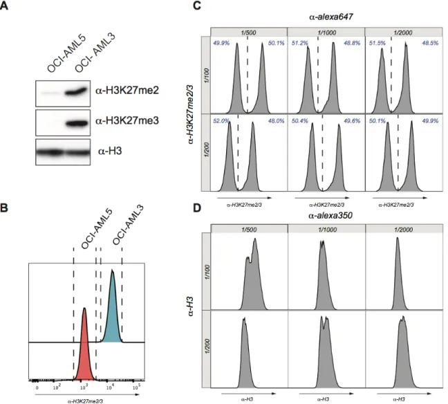
Development of this flow cytometry assay relied on the incidental discovery that OCI-AML5 had a significantly reduced level of H3K27me2/3 compared to OCI-AML3 (Figure 7A). This dramatic reduction of H3K27me2/3 was explained by a homozygous point mutation (R690H) in the catalytic core of the EZH2 SET domain. The difference of H3K27me2/3 between OCI-AML5 and OCI-AML3 was then validated using flow cytometry and showed excellent agreement compared to the western blot thereby validating our protocol (Figure 7B). Using a 1:1 mixture of OCI-AML3 and OCI-AML5 cells, we efficiently titer our monoclonal H3K27me2/3 antibody, and determined the optimal dilution to be at 1/100 and the secondary antibody at 1/500 (Figure 7C). Accurate measurement of H3K27me2/3 relied on normalization of the methylation signal to the total H3 content as H3K27me2/3 can increase dramatically in cases of hyperploid and variations in cycle cell. To normalize our H3K27me2/3 signal to the variation of nuclear DNA content, we titered an H3 antibody and determined the optimal dilution at 1/200, and the secondary antibodies at 1/500 (Figure 7D). Using this dual antibody staining, we normalized H3K27me2/3 to H3 signal in FlowJo allowing us to reliably measure H3K27me2/3 in patient AML samples.
Figure 7. Establishment of a FACS-based assay to quantify global levels of H3K27me2/3
(A) Western blot analysis from whole cell lysates of AML 3 and AML 5. OCI-AML3 and OCI- AML5 have sharply contrasting levels of H3K27me2/3. (B) Flow cytometry on AML3 and AML5 cells reflected results from western blot analysis. OCI-AML3 had a significantly higher H3K27me2/3 signal in comparison to OCI-AML5. (C and D) Titration of H3K27me2/3 and H3 antibody. Histogram represents the H3K27me2/3 or H3 signal. Vertical axis shows different dilutions of primary antibody. Horizontal axis indicates the different dilutions of secondary antibody. Percentage of low and high H3K27me2/3 are represented in blue.
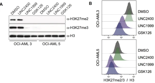
To validate the sensitivity of our flow cytometry assay, we cultured OCI-AML3 and OCI-AML5 cells in the presence of DMSO, UNC2400, UNC1999 or GSK126 for 72 hours. UNC1999 and GSK126 served as inhibitors of EZH1/2, while UNC2400 was the inactive molecule that was used as an additional control. Proceeding treatment, the cells were lysated for western blot analysis or fixed with ethanol for flow cytometry. Western blot analysis showed that OCI-AML3 treated with EZH inhibitors (UNC1999/GSK126) had significantly reduced H3K27me2/3 compared to the DMSO/UNC2400 controls. Similar, results were shown in the flow cytometry assay. By western blot, OCI-AML5 cells showed a complete loss of H3K27me2/3 regardless of the treatment condition. However, by flow cytometry OCI-AML5 treated with EZH inhibitors showed an even greater degree of reduction in H3K27me2/3. This level of sensitivity was previously undetectable in the western blot (Figure 8A and 8B). There was an excellent agreement between western blot and flow cytometry results, but flow cytometry proved to be a superior method as it (1) allows single cell analysis (2) much higher level of sensitivity (3) greater time efficiency. Going forward we decided that given the numerous advantage of flow cytometry, that we would use this assay to validate loss of H3K27me2/3 in cells expressing the K27M transgenes and AML patient samples.
drug vehicle (DMSO), inactive EZH inhibitor (UNC2400) or EZH inhibitors (GSK126 and UNC1999) for 72 hours (B) Alternatively, OCI-AML3 and OCI-AML5 cells were also fixed in cold ethanol and processed through flow cytometry. The H3K27me2/3 signal was then normalized to the H3 signal.
2.1.2 Identification of H3
K27M/IMutation in AML Patients
Of the 415 sequenced adult AML patients within the Leucegene cohort, we identified two patients that had a mutation on lysine 27 of histone H3. Patient 04H138 had a mutation on the
HIST1H3H gene where lysine 27 was mutated into a methionine (HIST1H3HK27M). Patient 12H183 had a mutation on the HIST1H3F gene where lysine 27 was mutated into isoleucine (HIST1H3FK27I). Next generation sequencing revealed that the H3.1K27M and H3.1K27I mutations
occurred at a variant allele frequency (VAF) of less than 50%, suggesting a minor hemizygous mutant sub-clone (Figure 9a). Gene expression profiles revealed that the mutated H3.1 gene contributed to 27.8% of total canonical histone H3 gene expression in patient 04H138 and 4.4% of total canonical H3 gene expression in patient 12H183. Together, this suggested that both variants are present in different sub-stoichiometric levels in the chromatin incorporate pool of histone H3 (Figure 9c). The somatic nature of the mutations was confirmed by sequencing of the matching DNA obtained from buccal swaps. While the HIST1H3FK27I mutation was also detected in the normal tissue sample, other AML mutations were also identified indicating the likelihood of a contamination in the swap (Figure 9b). Furthermore, in The Cancer Genome Atlas (TCGA) cohort of 200 AML patient samples, one patient was identified with a K27M mutation on HIST1H3D (HIST1H3DK27M) [38]. Overall the H3.1K27M/I mutated histone occurs at a frequency of 0.05% (1/200), indicating the rareness of this mutation in adult AML. Mutation profiles of H3.1K27M/I AML patients showed that the mutated histone was likely to co-occur with other epigenetic modifiers that were suggested to have an impact on H3K27me3 including
BCOR, ASXL1, AXSL2, and EZH2. Interestingly each of these H3.1K27M/I AML patients contained a unique mutation that altered RUNX1, suggesting a genetic synergy between mutated RUNX1 and reduced H3K27me3 (Figure 9d).
In glioma patients, H3K27M/I mutationswere proceeded by a reduction in H3K27me3 [50], to assess methylation levels in the heterogeneous AML patient samples we utilized our newly developed flow cytometry assay. In the HIST1H3HK27M patient specimen the reduction in
H3K27me2/3 was detectable in approximately 50% of the cells, which was consistent with the
HIST1H3HK27M VAF of 27.9%. In the HIST1H3FK27I AML patient specimen we did not observe a loss of H3K27me2/3 representative of the VAF of 41%, nonetheless a small population within the patient specimen exhibited a loss of H3K27me3 (Figure 9e). The discrepancy of the H3K27me2/3 signal between the two patients can be partially explained by (1) the mutated histone allele being expressed at 27.6% in patient 04H138 compared to 4.4% in patient 12H183
(Figure 9c) and (2) in vitro data suggesting that H3K27M was the more potent inhibitor of EZH1/2 (49). To validate, the presence of the mutated histone within the low H3K27me2/3 population, we performed cell sorts of the high and low H3K27me3 population, followed by Sanger sequencing. As anticipated, the histone mutation occurred exclusively within the population exhibiting reduced methylation (Figure 9f). Taken together, our data were the first to identify rare heterogeneous H3.1K27M/I mutations in adult AML that have a dominant-negative effect on H3K27me2/3.
Figure 9. Oncogenic histones H3.1K27M and H3.1K27I identified in AML patients to occur
at low frequencies (a) Within the Leucegene cohort containing 415 sequenced AML patients
with diverse cytogenetics, only two patients were identified to contain either the histone
H3.1K27M or H3.1K27I mutation. (b) Sanger sequencing validated somatic mutations in
H3.1K27M/I AML patients. Genomic DNA from AML cells (top) and buccal swaps (bottom). The missense mutation is highlighted in the red box. (c) Level of each histone H3 gene contributing to the total canonical histone H3 expression. In patient 04H138 the
HIST1H3HK27M variant is present on the highest expressed H3 (27.6% of all H3 genes,
highlighted in bold). In patient 12H183, expression of the HIST1H3F gene constitutes 4.4% of total canonical histone H3. (d) Mutation profile of H3.1K27M/I AML patients. Each patient harbored additional mutations effecting epigenetic modifiers and unique mutations altering RUNX1 (e) A subpopulation of diseased cells from 04H138 had 10-fold less H3K27me2/3. Reduction in H3K27me2/3 was less prominent in 12H183. (f) As indicated in the blue box, cells from H3.1K27M/I AML patients were sorted per low and high H3K27me3. Sorted population were processed through Sanger sequencing and revealed that the missense histone mutation occurred specifically in the sub-population expressing low H3K27me3.
2.1.3 Collaboration between H3
K27M/Iwith AML1-ETO9a Accelerates Disease
Progression
Closer examination of the molecular profile of AML patients harboring the H3K27M/I mutation revealed that all three patients had a RUNX1 gene alteration. The 04H138 (HIST1H3HK27M) patient had a heterozygous truncation of RUNX1 at L98-, 12H183 (HIST1H3FK27I) patient carried the RUNX1-RUNX1T1 translocation and TCGA K27M (HIST1H3DK27M) patient had biallelic point mutations on the RUNX1 gene (Figure 9d). Overall
this suggested that (1) H3K27 mutation associates with RUNX1mut AML (2) H3K27 mutation were likely to be a secondary mutation that arise from RUNX1mut AML. We hypothesized a leukemia-accelerating collaboration between RUNX1 alterations and reduced H3K27me2/3.
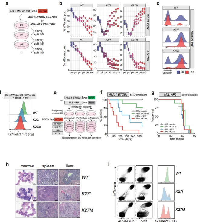
To investigate, the collaboration between RUNX1 and H3K27M/I mutations,
cells were infected with a panel of H3.3KM transgenes including: H3.3WT, H3.3K27I, and H3.3K27M
(Figure 10a). The proliferation kinetics were monitored over several passages in a competitive
assay. Initially, it appeared that there was no proliferative advantage in any of the conditions, however by passage 3 in H3.3K27M AE9a and passage 7 in H3.3K27I AE9a, the frequency of the tdTomato marked cells significantly accelerated, while simultaneously the H3.3WT tdTomato-marked cells continued to decline. More importantly, H3.3WT, H3.3K27I, and H3.3K27M all conferred a similar disadvantage in MLL-AF9 cultures (Figure 10b). By the end of passage 10 it was clear that AE9a expressing H3.3K27M/I that had originally occupied ~70% of the culture had proliferated to dominate the culture as shown by the strong transgene expression (Figure
10c). H3K27me3 levels were validated through flow cytometry and showed that H3.3K27I/M
infected AE9a cells exhibited significant reductions in H3K27me2/3 signal relative to H3.3WT.
(Figure 10d). Together, these results demonstrated that expression of H3.3K27I and H3.3K27M synergized with AE9a but not MLL-AF9 AML cells in vitro.
To further validate our hypothesis that H3.3K27I and H3.3K27M confer a selective proliferation advantage to AE9a but not MLL-AF9 AML, the engineered murine leukemia cells expressing H3.3WT, H3.3K27I or H3.3K27M were transplanted into mice recipients (Figure 10e). The first mice to reach disease end stage were AE9a driven AML expressing H3.3K27M, H3.3K27I and then followed by H3.3WT much later. This demonstrated a clear acceleration of disease development in the H3.3K27M/I mice compared to H3.3WT in AE9a AML (Figure 10f). Histological analysis from bone marrow isolated from the diseased leukemic AE9a mice showed an accumulation of characteristic AML blast cells. Furthermore, histological analysis of livers and spleens displayed a high infiltration of leukemic cells indicative of full-blown AML pathologies (Figure 10h). All diseased recipients contained a high fraction of c-Kit positive cells characteristic of AML blasts in this model. Isolated leukemic bone marrow cells contained readily discernible cell populations with largely decreased H3K27me2/3 levels specific to the H3.3K27I and H3.3K27M groups (Figure 10i). In contrast, MLL-AF9 expressing H3.3K27I/M did not experience changes in disease latencies compared to the control group (Figure 10g). This reiterated our previous conclusion that in the context of AML1-ETO9a-driven leukemia, H3K27me3 acts as a tumour suppressor. Notably, the more potent inhibitory effect of the
H3.3K27M mutant on PRC2 compared to H3.3K27I resulted in a stronger cooperation with AE9a demonstrating that varying degrees of PRC2 inhibition have quantitatively different effects on leukemia progression in t(8;21) AML.
assays to examine AML1-ETO9a or MLL-AF9 immortalized mouse bone marrow cells with MSCV H3.3KM mutants IRES tdTomato. Cellular proliferation was measured by monitoring percentage tdTomato over several passages using flow cytometry (b) Transformed AML1-ETO9a or MLL-AF9 cells expressing the H3.3KM mutants were passaged 10 times to reveal cell proliferation activity. (c) Histogram representation tdTomato FACS profiles at passages 0 and 10 indicate selection for high expression of H3.3K27I/M at the end of the culture period in AE9a cells. (d) Histogram representation of global H3K27me2/3 normalized to H3 signal. AML1-ETO9a cells expressing the H3.3K27M/I were sorted on tdTomato positivity and passaged 10 times. (e) Experimental strategy to assess AML progression upon H3.3KM expression in vivo. (f-g) Survival curves of mice transplanted with 5*105 AML1-ETO9a or 2*105 MLL-AF9 cells expressing the H3.3K27M/I mutants. (h) Pathological analysis of diseased AE9a cell recipients indicates typical AML end-stage characteristics. Bone marrow cells collected from disease mice showed a selection for cells expressing high level H3K27M marked by tdTomato and c-Kit. H3K27M/I mice exhibited a reduction of H3K27me2/3.
2.1.4 H3K27me2/3 in RUNX1
altered
AML Patients
EZH2 mutations occurred almost exclusively in the context of RUNX1 mutations in
AML (either RUNX1-RUNX1T1 or RUNX1mut). This suggested further epigenetic instability from EZH2 mutations may be required to complement RUNX1 mutated patients. Hence, we hypothesized that RUNX1 alteredAML may express lower levels of H3K27me3 compared to
RUNXWT AML. This prompted us to further investigate H3K27me2/3 in primary AML patients with RUNX1 alterations. To this end, we examined H3K27me2/3 in 29 AML patient samples, our panel included: 13 control patients, 10 patients with t(8;21)(q22;q22) translocation and 6 patients with mutations in the RUNX1 gene. 5,000 single cell events were examined from each of the described patients. Our result indicated that with the exception of the H3.1K27M/I patient no other patient sample exhibited any significant reduction in H3K27me3 that was detectable by flow cytometry (Figure 11). However, it should be mentioned that flow cytometry has limited detection compared to ChiP-sequencing (ChiP-seq).
Figure 11. Quantification of global H3K27me2/3 in AML patient specimens. 5,000 single
cell events representing the level of H3K27me2/3 in AML patients specimens were acquired through flow cytometry in RUNX1mut, t(8;21) and control patient specimens.
From transcriptome data of Leucegene patients, we examined differentially expressed genes in RUNX1mut, t(8;21) and RUNX1WT AML patients, the primary aim was to assess if there were any indications that H3K27me3 was hindered in patients harboring a defective RUNX1 gene. Comparative transcriptomic analysis revealed there was a decrease in expression of components of PRC2 including EED and SUZ12 in t(8;21) patients, that would suggest a decrease in H3K27me3 (Figure 12). In summary, there was no clear loss of H3K27me2/3 in RUNX1 altered patients other than the histone H3.1K27M/I patients previously described. However, t(8;21) AML patients have decreased expression of PRC2 components, that could suggest reduced levels of H3K27me3 that was not detectable through flow cytometry.
Figure 12. Low expression of core PRC2 members in t(8;21) AML. Transcriptome
analysis of core components of PRC2 in patients categorized based on RUNX1 mutation status into RUNX1mut, t(8;21) and other (i.e. RUNX1WT) group. Significant reduction in expression of EED and SUZ12, but not EZH1/2 was observed in t(8;21) AML.
2.2 Functional Investigation of H3
K27Min Normal Hematopoiesis
2.2.1 Reduction of H3K27 Methylation Selectively Expands Human
Progenitor Population
To investigate the influence of H3K27 methylation on differentiation, self-renewal and proliferation of human HSPCs, we infected cells with lentivirus expressing the H3.3KM mutants. The lentiviral constructs included the no insert (i.e. empty vector), H3.3WT, H3.3K9M and H3.3K27M mutant followed by an IRES GFP cassette. These cells were maintained in optimized
HSC expansion conditions that contained the small molecule UM171 [66]. By western blot analysis, we observed a global reduction of H3 methylation on the corresponding lysine marks
in lentiviral infected HSPCs. For instance, expression of H3.3K27M reduced global H3K27me2/3
(Figure 13a). At day 10, we examined the surface phenotype of the expanded cells and detected
striking surface phenotype changes on HSPC expressing H3.3K27M. Specifically, these cells maintained higher frequencies of primitive CD34+/CD45RA- cells compared to all other tested conditions (empty vector, H3.3WT and H3.3K9M). A more detailed analysis revealed that expression of H3.3K27M led to the eightfold expansion of a CD34+/CD45RA-/CD90+/CD133 -population and a mild expansion of a CD34+/CD45RA-/CD90+/CD133+ population. The expansion of the CD133- population indicated to us that H3.3K27M infected HSPCs had expanded a progenitor population and not an HSC population (Figure 13b and 13c).
We hypothesized that H3.3K27M mediated expansion of the described progenitor population was due to the loss of H3K27me2/3. Therefore, inhibiting the methyltransferase activity of EZH1/2 should mimic the H3.3K27M phenotype. As predicted, pharmacological inhibition of EZH1/2 not only ablates the presence of H3K27me2/3, but can expand the CD34+CD45RA -CD90+ population in a dose dependent manner. The treated cultures reached the absolute maximal expansion of CD34+/CD45RA-/CD90+ cells at 1µm. At this concentration, this was accompanied by an almost 50% reduction in total cell numbers in the same cultures, suggesting that although pharmacological inhibition of EZH1/2 augments the expansion of CD34+/CD45RA-/CD90+ cells, its impact on the remainder of the culture is detrimental (Figure
13d and 13e).
Given that H3.3K27M reduces methylation through the inhibition of EZH1/2 [50], we hypothesized that the expression of H3.3K27M and EZH1/2 inhibition likely expanded CD34+/CD45RA-/CD90+ cells through the same mechanism (the loss of H3K27me2/3). To validate this hypothesis, we exposed HSPCs infected with the H3.3KM lentiviral constructs to
either DMSO, GSK126, UNC2400 or UNC1999 at 1µM. The combination of H3.3KM expression with GSK126 or UNC1999 treatment led to a similar expansion of the CD34+/CD45RA-/CD90+/CD133- population in all lysine-to-methionine culture conditions (vector only, H3.3WT, H3.3K9M , H3.3K27M) (Figure 13f).
In summary, the data demonstrated that the expression of H3.3K27M caused a marked reduction of H3K27me2/3 and led to profound phenotypic changes in primitive human hematopoietic cells. Given the strikingly similar phenotypical effect of H3.3K27M expression and EZH1/2 inhibition on these cells, the effect of H3.3K27M on human HSPCs is likely prompted by its inhibition of EZH1/2.
Figure 13. Reduction of H3K27me2/3 expands a selective human progenitor population
(a) Modification of histone marks by H3.3KM mutant expression were analyzed by western blot using whole cell lysates. Efficient decrease in H3K27me2/3 or H3K9me3 from expression of H3.3K27M or H3.3K9M. (b) Phenotypic characterization of H3.3KM mutants expressed in human HSPC, and cultured for 10 days. Presentation of flow cytometry gating
strategy to analyze subpopulations. Gated on the CD34+CD45- subpopulation followed by gating into either CD90+/CD133- or CD90+/CD133+. Percentage of each gated subpopulation is represented. (c) Summary of population frequency of subpopulations. HSPCs expressing H3.3K27M displayed a selective expansion of the CD34+/CD45RA-/CD90+/CD133- population and a mild increase in the CD34+/CD45RA-/CD90+/CD133+ population. (d) Dose-dependent increase in the percentage of CD34+/CD45RA-/CD90+ cells (left panel) in EZH1/2 inhibitor treated HSPCs. Expansion of CD34+/CD45RA-/CD90+ cells in absolute cell numbers (middle panel). Absolute cell numbers in total culture (right panel) (e) Dose-dependent decrease of H3K27me2/3 in EZH1/2 inhibitor treated HSPCs. EZH1/2 inhibitor treated cultures from (d) were pooled and used for Western blot analysis using the indicated antibodies. (f) All H3.3KM HSPCs treated with EZH1/2 inhibitors(1µM) led to a similar fold-expansion of the CD34+/CD45RA-/CD90+/CD133- population. Cells were analyzed at d10 post-infection and EZH1/2 inhibitor treatment.
2.2.2 shRUNX1 phenocopies immunophenotype of H3.3
K27MBased on our previous observation that H3.3K27I or H3.3K27M exhibited a genetic synergy with AML1-ETO9a mouse model of t(8;21) AML, we hypothesized that hindering RUNX1 expression in human HSPCs could mimic the expansion of the CD34+/CD45RA -/CD90+/CD133- population previously seen in HSPCs expressing H3.3K27M.To validate this hypothesis, we designed shRNAs targeting RUNX1. Knockdown was validated using western blot analysis and showed a 50-80% decreased expression of the RUNX1 protein (Figure 14a). When surface phenotype was examined at d10, we observed a knockdown dependent expansion of CD34+/CD45RA-/CD90+/CD133- cells and to a much lesser extent a mild expansion of the CD34+/CD45RA-/CD90+/CD133+ cells in HSPCs (Figure 14b). These results mimicked the changes in the surface phenotype that we had observed in HSPCs expressing H3.3K27M. Overall, it highlighted the importance of further investigating the mechanisms governing the genetic synergy between the loss of RUNX1 and hypo-H3K27me2/3.
Figure 14. shRUNX1 mimics H3.3K27M in the expansion of CD34+/CD45RA-/CD90+/CD133- (a) Lentiviral expression of shRUNX1 induces a specific reduction in
RUNX1 expression in cultured human HSPCs as shown by western blot analysis. (b) Phenotypic characterization of HSPCs expressing shRUNX1. CD34+ cells derived from UCB were infected with lentivirus expressing shRUNX1 and cultured for 10 days. A similar gating strategy as previously described was applied, and revealed that knockdown of RUNX1 mimicked H3.3K27M in its ability to expand the progenitor CD34+/CD45RA-/CD90+/CD133- population.
2.2.3 H3.3
K27MExpression Enhances the Activity of Committed Myeloid
Progenitors and Results in Mild Loss of LT-HSC Self-renewal
To delineate the HSPC differentiation stages that were modulated by H3.3K27M, we first maintained empty vector control, H3.3WT, H3.3K9M or H3.3K27M infected human HSPC cultures ex vivo for 14 days and plated equal numbers of GFP+ cells into methylcellulose media to assess their clonogenic potential. Strikingly, we observed increased numbers of unipotent mature colonies that were granulocytic and granulo-monocytic but not multipotent immature colonies (granulo/erythroid/monocytic/megakaryocytic; GEMM) colony forming cells upon expression of H3.3K27M compared to control and H3.3K9M conditions (Figure 15).
![Table 2. EZH2 inhibitors and their status in clinical development. Modified from [23]](https://thumb-eu.123doks.com/thumbv2/123doknet/2043782.4919/25.918.122.799.120.406/table-ezh-inhibitors-status-clinical-development-modified.webp)