Acute administration of GPR40 receptor agonist potentiates
glucose-stimulated insulin secretion in vivo in the rat
Lalit S. Doshi, Manoja K. Brahma, Sufyan G. Sayyed, Amol V. Dixit, Prakash G. Chandak,
Venu Pamidiboina, Hashim F. Motiwala, Somesh D. Sharma, Kumar V.S. Nemmani
⁎
Piramal Life Sciences Limited (formerly Nicholas Piramal Research Centre), 1A, 1B, and 1C, Nirlon Complex, Near NSE Complex, Off Western Express Highway, Goregaon (E), Mumbai–400 063, India
Received 21 December 2007; accepted 20 October 2008
Abstract
Recently, several in vitro studies have shown that GPR40 receptor activation by free fatty acids (FFAs) results in glucose-dependent insulin secretion. However, whether GPR40 receptor activation results in glucose-dependent insulin secretion in vivo in rats is not known. Therefore, we evaluated the effect of synthetic GPR40 receptor agonist (compound 1) on glucose tolerance test (GTT) in fed, fasted, and insulin-resistant rats. In oral GTT, intraperitoneal GTT, and intravenous GTT, GPR40 receptor agonist improved glucose tolerance, which was associated with increase in plasma insulin level. Interestingly, in GTTs, the rise in insulin levels in agonist-treated group was directly proportional to the rate of rise and peak levels of glucose in control group. Although glibenclamide, a widely used insulin secretogogue, improved glucose tolerance in all GTTs, it did not display insulin release in intraperitoneal GTT or intravenous GTT. In the absence of glucose load, GPR40 receptor agonist did not significantly change the plasma insulin concentration, but did decrease the plasma glucose concentration. Fasted rats exhibited impaired glucose-stimulated insulin secretion (GSIS) as compared with fed rats. Compound 1 potentiated GSIS in fasted state but failed to do so in fed state. Suspecting differential pharmacokinetics, a detailed pharmacokinetic evaluation was performed, which revealed the low plasma concentration of compound 1 in fed state. Consequently, we examined the absorption profile of compound 1 at higher doses in fed state; and at a dose at which its absorption was comparable with that in fasted state, we observed significant potentiation of GSIS. Chronic high-fructose (60%) diet feeding resulted in impaired glucose tolerance, which was improved by GPR40 receptor agonist. Therefore, our results demonstrate for the first time that acute GPR40 receptor activation leads to potentiation of GSIS in vivo and improves glucose tolerance even in insulin-resistant condition in rats. Taken together, these results suggest that GPR40 receptor agonists could be potential therapeutic alternatives to sulfonylureas.
© 2009 Elsevier Inc. All rights reserved.
1. Introduction
Glucose-stimulated insulin secretion (GSIS) is compro-mised in conditions of starvation as well as in pathophysio-logic conditions like impaired glucose tolerance and type 2 diabetes mellitus[1-3]. Glucose-stimulated insulin secretion occurs through the classic glucose and free fatty acid (FFA)– mediated pathways. The classic glucose pathway is a KATP
-dependent Ca2+-mediated process. On the other hand, FFA-mediated insulin secretion results from intracellular utiliza-tion of fatty acids through the malonyl coenzyme A (CoA)/
long-chain CoA signaling network and triglyceride/FFA cycling pathway[4]. A recent study demonstrated that FFAs act as endogenous ligand for extracellularly located GPR40 receptor[5].
GPR40 belongs to class A (rhodopsin-like) of the superfamily of G-protein–coupled receptors and is preferen-tially expressed on pancreaticβ-cells[6]. In vitro activation of GPR40 receptor by FFAs has been shown to potentiate GSIS [7-9]. Studies in MIN6 and INS-1 cells indicate that saturated, monounsaturated, and polyunsaturated (medium and long chain) fatty acids augment GSIS through GPR40 receptor activation[5,10]. Furthermore, silencing of GPR40 receptor expression by small interfering RNA in rat islet[11]
and in INS-1 cells led to significant inhibition of FFA-induced insulin secretion[10]. In GPR40−/−knockout mice, intralipid infusion-induced insulin secretion was reduced
Metabolism Clinical and Experimental 58 (2009) 333–343
www.metabolismjournal.com
⁎ Corresponding author. Tel.: +91 22 3081 8404; fax: +91 22 3081 8411.
E-mail address:[email protected](K.V.S. Nemmani). 0026-0495/$– see front matter © 2009 Elsevier Inc. All rights reserved. doi:10.1016/j.metabol.2008.10.005
approximately by 50% as compared with that in wild-type C57BL/6J mice[12]. All these studies together underscore the role of GPR40 as an FFA receptor and its effect on GSIS. Moreover, a recent study has shown that GPR40 receptor activation using synthetic molecule causes glucose-depen-dent insulin secretion in MIN6 cells [13]. Thus, GPR40 receptor agonists could serve as potential therapeutic alternative to sulfonylureas, which cause insulin release independent of glucose stimulus causing a prolonged hypoglycemia that limits their clinical use.
Based on the findings that GPR40 receptor activation potentiates GSIS in vitro, we designed a study to determine the effect of GPR40 receptor activation on GSIS in vivo in rat using small molecule agonist compound 1 [14]. Compound 1 is a full GPR40 receptor agonist and binds to hGPR40 receptors cloned in Chinese hamster ovary cell lines with a pEC50of 6.03 ± 0.04μmol/L[14]. It is well known
that FFA-mediated GSIS is impaired in fasting conditions during which it is preferentially subjected to β-oxidation, thereby limiting its availability to stimulate insulin secretion
[1]. However, it is not known whether potentiation of GSIS occurring through GPR40 receptor–mediated mechanisms is altered in fasting states. Hence, our second objective was to compare the potentiation of GSIS by GPR40 receptor agonist in fasted and fed states. It is also well known that FFAs exhibit dual effect on GSIS. Acute exposure of β-cells to fatty acids potentiates GSIS [15], whereas long-term exposure inhibits the process [16,17]. Indeed, prolonged elevation of FFA by chronic feeding of high-fructose diet to rats results in reduced GSIS [18]. Therefore, our third objective was to study whether GPR40 receptor agonist potentiates GSIS in rats chronically fed with 60% high-fructose diet. When we submitted our work, no study examining the effect of small molecule GPR40 receptor agonist in vivo was reported. While this article was under review, Tan and associates[19]reported that small molecule GPR40 receptor agonist enhances glucose-dependent insulin secretion in isolated islets and improves glucose tolerance in mice. In contrast to in vitro studies on glucose-dependent insulin secretion by Tan and associates[19], this study has extensively investigated GSIS in vivo in rats.
2. Materials and methods 2.1. Animals
Male Sprague-Dawley rats aged 7 to 9 weeks (180-200 g) were procured from the central animal facility, Piramal Life Sciences, Mumbai. Animals were housed in individually ventilated cages at a room temperature of 22°C ± 2°C, humidity 55% ± 5%, with a 12-hour/12-hour light/dark cycle and had access to water and standard chow (Amrut Laboratory Animal Feed, Sangli, India; protein, 22.12%; fat, 4.13%; and carbohydrate, 55.43%) or 60% fructose-enriched diet (D 0011301; Research Diets, New Brunswick, NJ) ad libitum. The guidelines of the
Commit-tee for the Purpose of Control and Supervision on Experiments on Animals, Government of India, were followed; and all experimental procedures were approved by the Animal Ethics Committee.
2.2. Chemicals and reagents
GPR40 agonist compound 1 (3-[4-{4-chlorobenzyla-mino}phenyl] propanoic acid) was synthesized at the Department of Medicinal Chemistry, Piramal Life Sciences, Mumbai. Heparin was purchased from Biological E, Hyderabad, India. All other chemicals were procured from Sigma-Aldrich, St Louis, MO.
2.3. Oral glucose tolerance test
Glucose load (2 g/kg) was given orally to rats in this study. Rats were mildly anesthetized for 1 to 2 minutes using isoflurane anesthesia (3.5% vol/vol isoflurane in O2); and
about 80μL of blood was collected from retroorbital plexus into heparinized (20 U/mL) microcentrifuge tubes at different time points, viz, −60, 0, 2, 5, 15, 30, 60, and 120 minutes. The plasma was separated by centrifugation at 6000g for 7 minutes at 4°C. Plasma was used immediately for glucose estimation; an aliquot was stored at−20°C for insulin estimation later.
2.4. Intraperitoneal glucose tolerance test
Incretin hormones are known to cause insulin secretion in response to oral glucose[20]. To eliminate the incretin effect on insulin secretion, glucose tolerance tests (GTTs) were also performed by administering glucose through parenteral route (intraperitoneal and intravenous). The same procedure was followed as described for oral GTT (OGTT), except that glucose load was administered by intraperitoneal route. 2.5. Intravenous glucose tolerance test
The same procedure was followed as described for OGTT, except that a glucose load of 1.5 g/kg was administered intravenously through rat-tail vein (volume of injection, 1 mL/kg).
2.6. Treatment regimen
All the experiments were performed at 9:00 AM in the morning on overnight-fasted animals unless otherwise stated in protocol. Compound 1 and glibenclamide were prepared as a suspension in 0.5% carboxymethylcellulose (CMC) with 25μL Tween 80 and administered orally (1 mL/kg). 2.6.1. Effect of GPR40 receptor agonist on GSIS
To assess dose-dependent effect of compound 1 on GSIS, overnight-fasted rats were randomly divided into 4 groups and were subjected to an intraperitoneal GTT (IPGTT) as described above. One of the following treatments was assigned to each of the 4 groups: 0.5% CMC (1 mL/kg) or 50, 100, and 200 mg/kg of compound 1 given 1 hour before intraperitoneal glucose load.
2.6.2. Effect of GPR40 receptor agonist on plasma glucose and insulin in absence of glucose load
Overnight-fasted rats were randomly divided into 4 groups: 0.5% CMC (1 mL/kg) and compound 1 (50, 100, and 200 mg/kg). The animals were bled before treatment at 0 minute and after treatment at 0.5, 1, 2, 4, 6, and 8 hours; and plasma glucose and insulin were assessed as described below. 2.6.3. Effect of GPR40 receptor agonist on GSIS in GTTs
Overnight-fasted rats were randomly divided into 3 groups and were assigned treatments as follows: 0.5% CMC (1 mL/kg), glibenclamide (30 mg/kg), and compound 1 (100 mg/kg); and the animals were subjected to OGTT, IPGTT and intravenous GTT (IVGTT) as per the protocol described above.
2.6.4. Effect of GPR40 receptor agonist on GSIS in fasted and fed rats
Rats were randomly divided into 2 groups: 1 group was fasted overnight, whereas the other group was fed with the standard chow. Fed and fasted groups were further randomly divided into 3 groups: 0.5% CMC (1 mL/kg), glibenclamide (30 mg/kg), and compound 1 (100 mg/kg); and IPGTT was performed. In addition, the study was repeated using compound 1 at a dose of 180 mg/kg in fed rats.
2.6.5. Effect of GPR40 receptor agonist on GSIS in rats fed a high-fructose diet
Insulin resistance was induced by chronic feeding of rats for 8 weeks with 60% fructose-enriched diet. A group fed normal pellet diet was also used that served as a control. Rats fed a high-fructose diet were divided into 2 groups: 1 group served as control (0.5% CMC, 1 mL/kg), whereas the other group was treated with compound 1 (100 mg/kg). The animals fed normal pellet diet and 60% high-fructose diet were subjected to IPGTT as described above.
2.7. Biochemical estimation
Plasma glucose concentration was estimated immedi-ately after sample collection using the glucose determina-tion kits (DiaSys, Holzheim, Germany; glucose oxidase/ peroxidase [GOD/POD] enzymatic assay method) with a Hitachi 902 biochemical autoanalyzer (Hitachi Science Systems, Ibaraki, Japan). Plasma insulin was estimated using an enzyme-linked immunosorbent assay kit as per the manufacturer's instructions (Linco Research, St Charles, MO). Insulinogenic index was estimated as per the previously described method [21]. It was calculated by taking the sum of the difference in plasma insulin at each time point with respect to basal levels (up to 30 minutes) and dividing it by the sum of the difference in plasma glucose at each time point (up to 30 minutes) with respect to basal levels. 0 R30DI 0 R30DG 2.8. Pharmacokinetic studies
Rats were randomized into 2 groups (n = 4 per group): 1 group was fasted overnight (∼16 hours), whereas the other group was fed with normal diet. The fasted rats received an oral dose of 100 mg/kg, whereas fed animals received 100, 140, 180, and 250 mg/kg of compound 1. Plasma concentra-tions were determined by a high-performance liquid chroma-tography method developed in our laboratory. Briefly, blood was collected from retroorbital plexus under isoflurane anesthesia in heparinized (20 U/mL) microcentrifuge tubes. Plasma was obtained by centrifugation at 6000g for 7 minutes at 4°C in a Sigma refrigerated centrifuge (model no. 3K30, rotor no. 12153). Plasma samples were processed by transferring a 150-μL quantity into a microcentrifuge tube followed by equal volume of acetonitrile. The sample was vortex mixed for 2 minutes and centrifuged at 9000g for 5 minutes. The supernatant was collected and subjected to high-performance liquid chromatography analysis. The chromatographic system consisted of a Waters 2695 Separa-tions Module with 2996 Photodiode Array Detector (Waters, Milford, MA). Separation was carried out at room temperature on a Waters X-Terra C18 column of 100 × 3 mm internal diameter and a particle size of 5μmol/L. Elution was carried out with a gradient of 100% acetonitrile and 0.1% trifluoroacetic acid at a flow rate of 1 mL/min. Ultraviolet absorbance was monitored at photodiode array 3D Max plot wavelength. The retention time for compound 1 was 5.8 minutes. Calibration curve was obtained by dissolving a known quantity of compound 1 in rat plasma to obtain concentrations ranging from 1 to 100 μg/mL. A linear relationship (r2= 0.9998) was obtained when peak areas were plotted against plasma concentration. Coefficients of variation were lower than 10%, whereas accuracy ranged from 90% to 110%. The detection limit of the method was 0.5μg/mL. 2.9. Statistical analysis
All the results are expressed as mean ± SEM. Unpaired Student t test (2-tailed) was performed for statistical analysis of differences between mean values when comparisons were made between 2 groups. When comparisons were to be made between more than 2 groups, 1-way analysis of variance followed by Tukey post hoc analysis was used. Furthermore, for multiple comparisons of plasma glucose and insulin values at individual time points between 3 groups, we performed 1-way analysis of variance with repeated measures with “time” as a “within factor” and “treatment groups” as a “between factor.”
3. Results
3.1. Effect of GPR40 receptor agonist on GSIS
The effect of compound 1 on plasma glucose, insulin, and insulinogenic index in IPGTT is shown inFig. 1(A, B, and C). Compound 1 reduced plasma glucose at 30 minutes at a
dose of 50 (Pb .05), 100, and 200 mg/kg (P b .01). Increase in insulin levels was observed at a dose of 100 (Pb .05) and 200 mg/kg (Pb .01) of compound 1. Insulinogenic index of compound 1–treated animals at 100 and 200 mg/kg was higher than that of control group (Fig. 1C). The increase in insulinogenic index of compound 1 at both 100 and 200 mg/kg over the control group was similar; and hence, the dose of 100 mg/kg was selected for further studies.
3.2. Effect of GPR40 receptor agonist on plasma glucose and insulin in fasted rats
In the absence of glucose load, compound 1 per se caused significant glucose reduction at the dose of 100 and 200 mg/kg at 2 (Pb.01) and 4 hours (100 mg/kg, P b.05; 200 mg/kg P b .01;Fig. 2A). However, no significant change in insulin level was observed at any of the doses tested (Fig. 2B).
3.3. Effect of GPR40 receptor agonist on GSIS in GTTs 3.3.1. Oral glucose tolerance test
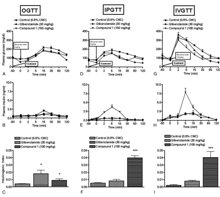
Fig. 3A illustrates that compound 1 displayed better glucose tolerance with significant (Pb.05) glucose reduction at 60 minutes as compared with control animals. In addition, compound 1 treatment resulted in increase in plasma insulin levels, with significant increase observed at 30 minutes (Pb .05) as compared with control group (Fig. 3B). Treatment of compound 1 and glibenclamide potentiated GSIS as demon-strated by the increase in insulinogenic index (Fig. 3C). 3.3.2. Intraperitoneal glucose tolerance test
In IPGTT, increase in plasma insulin levels was observed at earlier time points (2 and 5 minutes, P b .01), whereas
Fig. 1. Effect of GPR40 receptor agonist (50, 100, and 200 mg/kg) on plasma glucose (A) and insulin release (B) in IPGTT in overnight-fasted rats. Compound 1 was administered orally 1 hour before glucose load (2 g/kg, intraperitoneal). Fig. 1 C illustrates insulinogenic index. Data represent mean ± SEM of 6 rats. *Pb .05 vs control group.
Fig. 2. Effect of GPR40 receptor agonist on blood glucose and insulin release in overnight-fasted rats in the absence of glucose load. Effect of different doses of compound 1 (50, 100, and 200 mg/kg) on plasma glucose (A) and insulin release (B). Data represent mean ± SEM of 6 rats. *Pb.05 vs control group.
decrease in plasma glucose was observed at later time points (5 minutes onward, P b .01) (Fig. 3D and E). This rise in insulin secretion in terms of insulinogenic index was greater in the compound 1–treated group when compared with that of control group (Fig. 3F). In contrast, glibenclamide improved glucose tolerance but did not exhibit insulin release (Fig. 3D and E).
3.3.3. Intravenous glucose tolerance test
The intravenous glucose load of 1.5 g/kg caused the plasma glucose to shoot up within 2 minutes to a high level of 350 to 400 mg/dL (Fig. 3G) in the control group. Compound 1 improved glucose tolerance. Plasma insulin levels showed a peak at 5 minutes (P b .01), whereas
reduction in plasma glucose was observed 15 minutes onward (P b .01), suggesting that the released insulin resulted in glucose reduction. As in IPGTT, glibenclamide did not cause any significant rise in insulinogenic index when compared with that of control group (Fig. 3I). Compound 1 treatment resulted in higher insulinogenic index than that of control group.
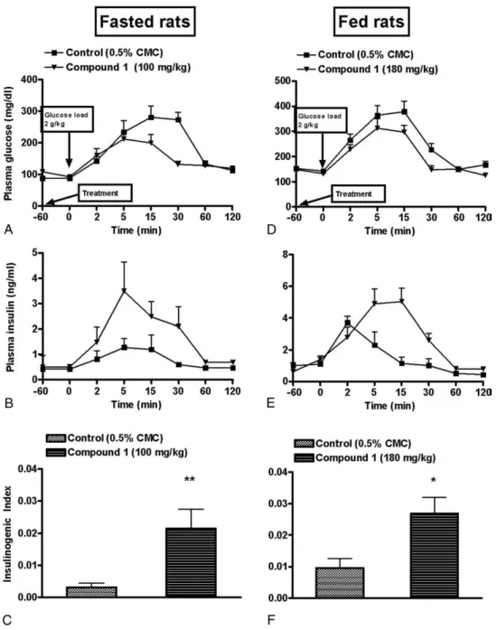
3.4. GPR40 receptor agonist potentiated GSIS in fasted and fed rats
The basal levels of plasma glucose (fed vs fasted rats: 127.2 ± 3.9 vs 78.1 ± 3.5) and insulin (fed vs fasted rats: 0.85 ± 0.2 vs 0.40 ± 0.02) were higher in fed group (Fig. 4D
Fig. 3. Effect of GPR40 receptor agonist on GSIS in overnight-fasted rats. Compound 1 (100 mg/kg) was administered orally 1 hour before glucose load (2 g/kg, oral; or 2 g/kg, intraperitoneal; or 1.5 g/kg, intravenous). Glibenclamide (30 mg/kg, 1 hour prior) was used as a reference compound. Changes in blood glucose level, corresponding insulin release, and insulinogenic index in OGTT (A-C), IPGTT (D-F), and IVGTT (G-I) studies. Data represent mean ± SEM of 6 rats. *Pb .05 and ***Pb .001 vs respective control group.
and E) as compared with fasted group (Fig. 4A and B). Fasting-induced impairment of GSIS was evident from the insulinogenic index when compared with that of fed state (Fig. 4C and F). Glibenclamide-treated animals displayed reduction in plasma glucose and increased insulinogenic index when compared with controls in fed group but not in fasted group (Fig. 4C and F).On the other hand, compound 1 potentiated GSIS in fasted state but failed to do so in fed state as is evident from insulinogenic index (Fig.4Cand F). However, a careful assessment revealed that the rise in
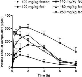
plasma insulin levels in fed animals in the compound 1– treated group at 2 minutes was significantly higher (Pb .05) when compared with that in the controls (Fig. 4D and E). Suspecting differential pharmacokinetics in fed and fasted condition, a detailed pharmacokinetic evaluation was performed, which revealed the low plasma concentration of compound 1 in fed state (Fig. 5). As expected, increasing dose of compound 1 improved its pharmacokinetic profile in a dose-dependent manner. In fed state, compound 1 at 180 mg/kg exhibited a Cmaxof 250.3μg/mL and AUC0-last
Fig. 4. Effect of GPR40 receptor agonist on GSIS in fasted or fed rats. Compound 1 (100 mg/kg) was administered orally 1 hour before glucose load (2 g/kg, intraperitoneal) in fed or fasted rats. Glibenclamide (30 mg/kg, 1 hour prior) was used as a reference compound. Changes in blood glucose levels and corresponding insulin release in fasted (A, B) and fed (D, E) rats. Insulinogenic index in fasted (C) and fed (F) states. Data represent mean ± SEM of 6 rats. *Pb .05 and **Pb .01 vs respective control group.
of 1068.8 μg*h/mL, which were similar to the Cmax
(256.9 μg/mL) and AUC0-last (1022.2 μg*h/mL) of
compound 1 at 100 mg/kg in fasted state (Table 1). Accordingly, we studied the effect of compound 1 on GSIS at 180 mg/kg in fed state vs 100 mg/kg in fasted state. Compound 1 (180 mg/kg) showed a significant decrease in plasma glucose (Pb .05) and an increase in insulin (P b .05) levels as compared with controls in fed state (Fig. 6D and E). Compound 1 also showed an increase in insulinogenic index as compared with control group in both fed and fasted state (Fig. 6C and F).
3.5. Improvement of GSIS by GPR40 receptor agonist in rats fed a high-fructose diet
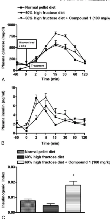
Feeding of animals with 60% fructose diet resulted in significant glucose intolerance (P b .01 at 15, 30, and 60 minutes vs normal pellet diet control; Fig. 7A). Furthermore, the group fed 60% high-fructose diet exhibited reduced insulinogenic index (Fig. 7C), which was not significant. Treatment with compound 1 (100 mg/kg) potentiated GSIS as indicated by the rise in the insulinogenic index (Fig. 7B and C).
4. Discussion
Recently, the GPR40 receptor involvement in glucose homeostasis has generated much interest because of the discovery of FFAs as its endogenous ligands[22]. Several in vitro and ex vivo studies show that acute stimulation of GPR40 receptor with fatty acids (ie, palmitate, intralipid) potentiates GSIS[7,9,10]. However, potentiation of GSIS by FFA is compromised in chronically FFA elevated states such as insulin resistance induced by feeding of a high-fructose diet to rats[18]. Recently, small molecule GPR40 receptor agonists were shown to potentiate glucose-dependent insulin
secretion in MIN6 cell line[13]and in pancreatic islets, and improved glucose tolerance in mice[19]. However, whether GPR40 receptor activation results in potentiation of GSIS in vivo in rats is not known. Therefore, by using small molecule agonist, we assessed the effect of GPR40 receptor activation on GSIS in normal and pathophysiologic conditions in rats. We found that GPR40 receptor activation results in potentiation of GSIS in fasted, in fed, and, most importantly, in insulin-resistant rats.
In OGTT, compound 1 showed better glucose tolerance and significantly improved plasma insulin levels, suggesting potentiation of GSIS. Potentiation of GSIS was also observed in IPGTT and IVGTT. In these studies, increase in insulin level in compound 1–treated group precedes the glucose reduction, which implies that potentiation of GSIS is responsible for improved glucose tolerance. Interestingly, in GTTs, the insulin level in agonist-treated groups was directly proportional to the rate of rise and peak levels of glucose in control group (ie, intravenous or intraperitoneal insulin release compared with oral). Tan and associates [19] have also observed increase in plasma insulin level at early time point and improved glucose tolerance in mice. Glibenclami-de's insulinotropic action is compromised in fasted animals in IPGTT and IVGTT. There is evidence in literature suggesting that fasting compromises the insulin secretory response of glibenclamide and tolbutamide [23]. However, in OGTT, insulin release in glibenclamide-treated animals is higher as compared with that in controls. The plausible explanation to this observation is that, in OGTT, the observed insulin release is a synergistic action of both glibenclamide and incretin hormones released upon oral glucose load[24]. Despite the lack of insulin release, glibenclamide improved glucose tolerance in fasted animals, which might be through extrapancreatic mechanisms like nonoxidative glucose dis-posal and increased glucose transport[25-27].
In the absence of glucose load, compound 1 dose dependently lowered glucose, but without associated change in insulin levels. Immunohistochemical and messenger RNA studies have demonstrated that GPR40 receptors are expressed specifically in the rat pancreatic tissue and rodent pancreatic β-cell lines (MIN6, βTC-3, HIT-T15, and RINm5F) [5,7]. Therefore, the observed glucose reduction might be an extrapancreatic effect, unrelated to GPR40
Fig. 5. Pharmacokinetic profile of GPR40 receptor agonist in fasted or fed rats. The oral absorption profile of compound 1 in fed (100, 140, 180, and 250 mgkg) and fasted (100 mg/kg) rats. Data represent mean ± SEM of 4 rats.
Table 1
Pharmacokinetic parameters after oral administration of compound 1 in fed (100, 140, 180, and 250 mg/kg) and fasted (100 mg/kg) state in rats
Compound 1 (mg/kg)
Parameters Fasted state Fed state
100 100 140 180 250 Cmax(μg/mL) 256.93 96.75 175.26 250.29 307.04 Tmax(h) 0.50 1.00 2.00 2.00 2.00 AUC0-last(μg*h/mL) 1022.22 388.21 880.40 1068.80 1863.15 AUC0-∞(μg*h/mL) 1067.17 443.19 957.14 1156.81 2521.45 Half-life (h) 1.54 2.50 1.72 1.87 3.76 Data represent mean values of 4 rats.
receptor stimulation. Altogether, our results suggest that GPR40 receptor agonist potentiates insulin release in a glucose-dependent manner. In contrast, Flodgren and co-workers[28] have reported that GPR40 is present on α-cells and causes glucagon release. In the same study, linolenic acid (a endogenous agonist of GPR40) shows a dose-dependent glucagon release at both low and high concentra-tions of glucose. It is thereby expected that released glucagon, after GPR40 receptor agonist treatment, should result in increase in glucose levels. However, our studies showed a dose-dependent reduction in glucose preceded by insulin
release in the GTT (Fig. 1A-C). It may be likely that theβ-cell effect might be overriding a subtle effect mediated byα-cells because, in normal physiology, theβ-cell population in islets is higher than that of α-cells. Nevertheless, further studies are needed for better understanding of this observation.
It is well known that fasting causes impairment of GSIS. The impairment of GSIS was shown to be due to increased β-oxidation and reduced esterification of FFA in fasted state, thus making them unavailable for insulin exocytosis through formation of complex lipids[1,29]. In our study, we found that there was a marked impairment of GSIS in fasting state
Fig. 6. Effect of GPR40 receptor agonist on GSIS in fed and fasted rats at doses resulting in similar plasma exposure. Compound 1 was administered orally 1 hour before glucose load (2 g/kg, intraperitoneal). Fasted rats were administered a dose of 100 mg/kg and fed animals with a dose of 180 mg/kg, resulting in a comparable pharmacokinetic profile. Changes in blood glucose levels, corresponding insulin release, and insulinogenic index for fasted (A-C) and fed (D-F) rats. Data represent mean ± SEM of 6 rats. *Pb .05, **P b .01 vs respective control group.
as compared with fed state. Earlier, Stein and coworkers[30]
reported that circulating FFA is essential for GSIS in fasting state in rats.
Although it is very clear that intracellular actions of FFA get compromised in fasting state, the role of extracellular GPR40 receptor–mediated actions in GSIS in fasting state is not yet known. Hence, in the present study, we examined the role of GPR40 receptor stimulation by compound 1 on GSIS in both fasting and fed state. Compound 1 treatment resulted
in potentiation of GSIS in fasted state and, upon increasing the dose, in fed state as well. In contrast, glibenclamide potentiation of GSIS is compromised in fasting state. This observation is in line with previous studies where intrave-nous glibenclamide showed reduced insulin release in fasted rats[23]. Glibenclamide's insulin secretory action involves intracellular mechanisms because 90% of its binding sites are located inside the β-cell[31]. Moreover, both glucose and glibenclamide stimulate insulin release by inhibiting carni-tine palmitoyltransferase 1 activity, which switches fatty acid metabolism fromβ-oxidation to protein kinase C–dependent insulin exocytosis. Recent studies have shown that the ability of glibenclamide and malonyl-CoA to inhibit carnitine palmitoyltransferase 1 gets impaired during fasting[32]. In view of our results with GPR40 receptor agonist in fed and fasted state and the fact that glibenclamide and glucose (which act mainly by intracellular pathway) fail to potentiate/ mediate GSIS in fasted state, we infer that GSIS impairment induced by fasting involves alteration in intracellular FFA action rather than modulation in GPR40 receptor signaling. The acute stimulating effect of FFA on GSIS has been well described both in vitro [33,34] and in vivo [35]. In contrast, several in vitro studies inβ-cell lines and in rodent and human islets have confirmed that insulin secretion at high glucose concentration is impaired in a time-dependent manner by chronic exposure to FFA. Islets from prediabetic Zucker diabetic fatty rats and from fructose-fed insulin-resistant rats appear to be more susceptible to this FFA-mediated desensitization of GSIS[36,18]. Although deleter-ious effects of long-term exposure of FFA on glucose tolerance have been well studied, the role of GPR40 receptor in these conditions is still not clear. There are contradicting reports regarding involvement of GPR40 receptor in impaired glucose tolerance associated with insulin-resistant condition. According to Steneberg and coworkers [37], GPR40−/− deletion confers protection against obesity-induced hyperinsulinemia, hyperglycemia, and glucose intolerance, whereas Latour and associates [12] demon-strated that deleterious effects of FFA on GSIS were similar in both GPR40−/−and wild-type mice islets, indicating that GPR40 receptor does not mediate the long-term deleterious effects of FFA. In our study, we observed that chronic feeding with 60% fructose diet to rats resulted in impaired glucose tolerance. This implies that rats chronically fed a diet containing 60% fructose were insulin resistant. Treatment with GPR40 receptor agonist enhanced insulin release, resulting in improved glucose tolerance. Improvement in glucose tolerance in mice fed a high-fat diet using GPR40 receptor agonist was reported by Tan and associates [19]. These findings indicate that GPR40 receptor agonist potentiates GSIS even in insulin-resistant condition.
In conclusion, our study for the first time shows that acute administration of a small molecule GPR40 receptor agonist results in potentiation of GSIS in vivo in rats fed a normal and a chronic high-fructose diet. The potentiation of GSIS in high fructose diet–induced insulin-resistant rats suggests that
Fig. 7. Effect of GPR40 receptor agonist on GSIS in rats fed with 60% high-fructose diet for 8 weeks. The rats were subjected to IPGTT after overnight fasting. Compound 1 (100 mg/kg) was administered orally 1 hour before glucose load (2 g/kg, intraperitoneal). Changes in blood glucose levels (A), corresponding insulin release (B), and insulinogenic index (C) in rats fed 60% fructose diet. Data represent mean ± SEM of 6 rats. *Pb .05 vs group fed 60% high-fructose diet.
the actions of GPR40 receptor are intact even in states of chronically elevated FFA. Unlike sulfonylureas, GPR40 receptor agonist potentiated GSIS; and therefore, it may not cause severe hypoglycemia. Exposure to isoflurane is known to cause glucose intolerance [38]. However, prolonged exposure causes acute hyperglycemia in fed but not in fasted rats [39]. In this study, except in 1 experiment (Fig. 6), animals were fasted overnight and all the rats were exposed to isoflurane anesthesia for a brief period. Therefore, the effect of isoflurane on glucose tolerance is expected to be minimal and, if any, will be same across all the groups. Further studies are in progress to determine whether chronic administration of GPR40 receptor agonist improves fasting glucose and diabetic complications in animal models of diabetes. Nevertheless, these findings together suggest that GPR40 receptor agonist may offer a novel treatment option to control postprandial hyperglycemia.
Acknowledgment
We express sincere thanks to Dr Jeffery S Mogil, Professor, McGill University, Canada, for his kind assistance in statistical analysis; Mr Asif Pathan for his assistance in writing the manuscript; and Mr Nitin Deshmukh and Ms Pooja Bhatt for technical assistance. We are highly grateful to Dr AK Gangopadhyay for providing compound 1 and to Dr Smita Tankhiwale and Dr Sandeep Bhigawade for providing the experimental animals.
References
[1] Tamarit-Rodriguez J, Vara E, Tamarit J. Starvation-induced changes of palmitate metabolism and insulin secretion in isolated rat islets stimulated by glucose. Biochem J 1984;221:317-24.
[2] Jones CN, Abbasi F, Carantoni M, et al. Roles of insulin resistance and obesity in regulation of plasma insulin concentrations. Am J Physiol Endocrinol Metab 2000;278:E501-8.
[3] Kosaka K, Kuzuya T, Hagura R. Insulin secretory response in Japanese type 2 (non–insulin-dependent) diabetic patients. Diabetes Res Clin Pract 1994;24(Suppl):S101-10.
[4] Nolan CJ, Madiraju MS, Delghingaro-Augusto V, et al. Fatty acid signaling in the {beta}-cell and insulin secretion. Diabetes 2006;55 (Suppl 2):S16-23.
[5] Itoh Y, Hinuma S. GPR40, a free fatty acid receptor on pancreatic beta cells, regulates insulin secretion. Hepatol Res 2005;33:171-3. [6] Vassilatis DK, Hohmann JG, Zeng H, et al. The G protein–coupled
receptor repertoires of human and mouse. Proc Natl Acad Sci U S A 2003;100:4903-8.
[7] Itoh Y, Kawamata Y, Harada M, et al. Free fatty acids regulate insulin secretion from pancreatic beta cells through GPR40. Nature 2003;422: 173-6.
[8] Kotarsky K, Nilsson NE, Olde B, et al. Progress in methodology. Improved reporter gene assays used to identify ligands acting on orphan seven-transmembrane receptors. Pharmacol Toxicol 2003;93: 249-58.
[9] Shapiro H, Shachar S, Sekler I, et al. Role of GPR40 in fatty acid action on the beta cell line INS-1E. Biochem Biophys Res Commun 2005; 335:97-104.
[10] Schnell S, Schaefer M, Schofl C. Free fatty acids increase cytosolic free calcium and stimulate insulin secretion from beta-cells through activation of GPR40. Mol Cell Endocrinol 2007;263:173-80.
[11] Fujiwara K, Maekawa F, Yada T. Oleic acid interacts with GPR40 to induce Ca2+signaling in rat isletβ-cells: mediation by PLC and L-type Ca2+channel and link to insulin release. Am J Physiol Endocrinol Metab 2005;289:E670-7.
[12] Latour MG, Alquier T, Oseid E, et al. GPR40 is necessary but not sufficient for fatty acid stimulation of insulin secretion in vivo. Diabetes 2007;56:1087-94.
[13] Briscoe CP, Peat AJ, McKeown SC, et al. Pharmacological regulation of insulin secretion in MIN6 cells through the fatty acid receptor GPR40: identification of agonist and antagonist small molecules. Br J Pharmacol 2006;148:619-28.
[14] Garrido DM, Corbett DF, Dwornik KA, et al. Synthesis and activity of small molecule GPR40 agonists. Bioorg Med Chem Lett 2006;16: 1840-5.
[15] Dobbins RL, Chester MW, Stevenson BE, et al. A fatty acid-dependent step is critically important for both glucose and non –glucose-stimulated insulin secretion. J Clin Invest 1998;101:2370-6. [16] Kashyap S, Belfort R, Gastaldelli A, et al. A sustained increase in
plasma free fatty acids impairs insulin secretion in nondiabetic subjects genetically predisposed to develop type 2 diabetes. Diabetes 2003;52: 2461-74.
[17] Mason TM, Tracy G, Tchipashvili V, et al. Prolonged elevation of plasma free fatty acids desensitizes the insulin secretory response to glucose in vivo in rats. Diabetes 1999;48:524-30.
[18] Chen NG, Reaven GM. Fatty acid inhibition of glucose-stimulated insulin secretion is enhanced in pancreatic islets from insulin-resistant rats. Metabolism 1999;48:1314-7.
[19] Tan CP, Feng Y, Zhou YP, Eiermann GJ, et al. Selective small-molecule agonists of G protein–coupled receptor 40 promote glucose-dependent insulin secretion and reduce blood glucose in mice. Diabetes 2008;57:2211-9.
[20] Gautier JF, Fetita S, Sobngwi E, et al. Biological actions of the incretins GIP and GLP-1 and therapeutic perspectives in patients with type 2 diabetes. Diabetes Metab 2005;31:233-42.
[21] Hanson RL, Pratley RE, Bogardus C, et al. Evaluation of simple indices of insulin sensitivity and insulin secretion for use in epidemiologic studies. Am J Epidemiol 2000;151:190-8.
[22] Milligan G, Stoddart LA, Brown AJ. G protein–coupled receptors for free fatty acids. Cell Signal 2006;18:1360-5.
[23] Bosboom RS, Zweens J, Bouman PR. Effects of feeding and fasting on the insulin secretory response to glucose and sulfonylureas in intact rats and isolated perfused rat pancreas. Diabetologia 1973;9:243-50. [24] Gutniak MK, Juntti-Berggren L, Hellstrom PM. Glucagon-like peptide
I enhances the insulinotropic effect of glibenclamide in NIDDM patients and in the perfused rat pancreas. Diabetes care 1996;19:857-63. [25] Muller G, Satoh Y, Geisen K. Extrapancreatic effect of sulphonylureas —a comparison between glimepiride and conventional sulphonylur-eas. Diabetes Res Clin Pract 1995;28(Suppl 1):S115-37.
[26] William PS, Anderson DK, Scott J. Selective potentiation of insulin mediated glucose disposal in normal dogs by the sulphonylurea Glipizide. J Clin Invest 1981;37:1016-23.
[27] Kaku K, Inou Y, Kaneko T. Extrapancreatic effects of sulphonylurea drugs. Diabetes Res Clin Pract 1995;28(Suppl 1):S105-8.
[28] Flodgren E, Olde B, Meidute-Abaraviciene S, et al. GPR40 is expressed in glucagon producing cells and affects glucagon secretion. Biochem Biophys Res Commun 2007;354:240-5.
[29] Fink G, Gutman RA, Cresto JC, et al. Glucose-induced insulin release patterns: effect of starvation. Diabetologia 1974;10:421-5.
[30] Stein DT, Esser V, Stevenson BE, et al. Essentiality of circulating fatty acids for glucose-stimulated insulin secretion in the fasted rat. J Clin Invest 1996;97:2728-35.
[31] Ozanne SE, Guest P, Hutton JC, et al. Intracellular localization and molecular heterogeneity of the sulphonylurea receptor in insulin-secreting cells. Diabetologia 1995;38:277-82.
[32] Cook G. The hypoglycemic sulfonylureas glyburide and tolbutamide inhibit fatty acid oxidation by inhibiting carnitine palmitoyltransferase. J Biol Chem 1987;262:4968-72.
[33] Crespin SR, Greenough WB, Steinberg D. Stimulation of insulin secretion by long-chain free fatty acids. A direct pancreatic effect. J Clin Invest 1973;52:1979-84.
[34] Warnotte C, Gilon P, Nenquin M. Mechanisms of the stimulation of insulin release by saturated fatty acids. A study of palmitate effects in mouse beta-cells. Diabetes 1994;43:703-11.
[35] Carpentier A, Mittelman SD, Lamarche B, et al. Acute enhancement of insulin secretion by in humans is lost with prolonged elevation. Am J Physiol Endocrinol Metab 1999;276:E1055-66.
[36] Hirose H, Lee YH, Inman LR, et al. Defective fatty acid–mediated beta-cell compensation in Zucker diabetic fatty rats. Pathogenic
implications for obesity-dependent diabetes. J Biol Chem 1996;271: 5633-7.
[37] Steneberg P, Rubins N, Bartoov-Shifman R, et al. The receptor GPR40 links hyperinsulinemia, hepatic steatosis, and impaired glucose homeostasis in mouse. Cell Metab 2005;1:245-58.
[38] Laber-Laird K, Smith A, Swindle MM, et al. Effects of isoflurane anesthesia on glucose tolerance and insulin secretion in Yucatan minipigs. Lab Anim Sci 1992;42:579-81.
[39] Saha JK, Xia J, Grondin JM, et al. Acute hyperglycemia induced by ketamine/xylazine anesthesia in rats: mechanisms and implications for preclinical models. Exp Biol Med 2005;230:777-84.