GUEST EDITORIAL
Valvuloarterial impedance in aortic stenosis: look
at the load, but do not forget the flow
Patrizio Lancellotti
*
and Julien Magne
University of Lie`ge, Heart Valve Clinic, Department of Cardiology, University Hospital Sart Tilman, B-4000 Lie`ge, Belgium
This editorial refers to ‘Valvuloarterial impedance does not improve risk stratification in low-ejection fraction, low-gradient aortic stenosis: results from a multicentre study’ by F. Levy et al., on page 358.
The vast majority of patients with severe calcified aortic stenosis (AS) have normal left ventricular (LV) ejection fraction, even in the
presence of symptoms.1Nevertheless, ≈20% of patients with AS
and undergoing valve surgery were found with reduced LV ejection
fraction (,50%)1in the last Euro Heart Survey. This characteristic is
often the result of a concomitant coronary artery disease. In some patients, however, LV hypertrophy, due to the chronic pressure overload, is inadequate to normalize systolic wall stress, resulting
in an afterload mismatch and a decrease in LV ejection fraction.2
Reduced LV function may lead to flow state and thus to low-gradient, despite the presence of severe AS. In fact, three main types of patients with severe AS, according to LV function and flow, are generally observed: (i) normal LV ejection fraction and normal flow, (ii) reduced LV ejection fraction and reduced flow, and (iii) normal LV ejection fraction and reduced flow.
Low-ejection fraction/low-gradient severe AS represents a chal-lenging clinical entity. The classification of patient in the so-called low-flow/low-gradient (LF/LG) severe AS subset may considerably vary from different studies and is generally based on the presence of the three following haemodynamic criteria: (i) an aortic valve
area (AVA) ,1 cm2, (ii) a LV ejection fraction ,30– 45%, and (iii) a
mean transvalvular pressure gradient (MPG) ,30–40 mmHg.3–12
LF/LG severe AS is associated with a poor outcome under
conser-vative management13and a high operative mortality risk.14
More-over, even after aortic valve replacement (AVR), the prognosis of such patients is worse than those with preserved LV function, and the improvement of symptoms remains limited.
True-severe or pseudo-severe
aortic stenosis
The reduced survival reported in LF/LG severe AS, when com-pared with ‘classic’ severe AS, is obviously related to both the LV disease and the inappropriate timing of surgery generally
reported in such patients. Indeed, due to the low-flow state, the apparent discrepancy between AVA and MPG may be considered as an artefact or a measurement error, which, in turn, could under-estimate the severity of symptom and delay intervention. In this regard, the cornerstone of the evaluation of LF/LG AS is the dis-tinction between true- and pseudo-severe AS. In the former, the aortic valve is really severely stenotic, the afterload mismatch is the main cause of LV dysfunction, the symptoms are essentially valve-related, and AVR is recommended and beneficial. In the latter, the aortic valve could be only mildly or moderately stenotic and the small reported AVA is due to an inability of the impaired LV to generate enough forces to open the calcified aortic cups. In this context, low-dose dobutamine stress echocardiography (DSE) is strongly recommended for the assessment of such
patients. The DSE is crucial in the management of LF/LG AS15
and allows (i) as to distinguish true-severe from pseudo-severe
AS3,12and (ii) to evaluate the presence of LV contractile reserve,
which is a marker of better peri-operative outcome.6,7 During
dobutamine infusion, a pseudo-severe AS may increase its AVA and only exhibits small changes in MPG. In contrast, true-severe AS had no or minimal augmentation in AVA during DSE and had a marked increase in MPG.
Nevertheless, in the presence of an excessive LV afterload, with
no or few LV contractile reserve (≈one-third of patients), the
nor-malization of flow rate is not possible and, unmasking pseudo-severe AS is challenging. In this regard, the calculation of the projected AVA
at a normal transvalvular flow rate (Qmean. 250 mL/s) may be very
useful and more accurate than the traditional echocardiographic indices (e.g. valvular resistance, dobutamine-induced increase in
AVA, or MPG) to differentiate true from pseudo-severe AS.5
Global left ventricular afterload
Concomitantly to the progressive aging of the general population, AS is nowadays becoming a part of the general atherosclerotic disease process, which progressively decreases the compliance of the vascular bed downstream the aortic valve. Besides the LV dys-function, LF/LG AS is often associated with concomitant systemic
hypertension.5,16,17 This may induce a low-flow state despite
The opinions expressed in this article are not necessarily those of the Editors of the EJECHO or of the European Society of Cardiology.
*Corresponding author. Tel:+32 4 366 71 94, Fax: +32 4 366 71 95, Email: [email protected]
Published on behalf of the European Society of Cardiology. All rights reserved.&The Author 2011. For permissions please email: [email protected]
European Journal of Echocardiography (2011) 12, 354–357 doi:10.1093/ejechocard/jer044
at Bibliotheque Fac de Medecine on January 19, 2012
http://ehjcimaging.oxfordjournals.org/
normal LV ejection fraction. As a result, the LV faces a double afterload: (i) a valvular load, due to the AS and (ii) an arterial load, as a consequence of reduced arterial compliance.
The valvuloarterial impedance (Zva) is a new index proposed by
the group of Pibarot and coworkers,18 assessing the global LV
haemodynamic load (i.e. total load) that can be measured by Doppler echocardiography. The Zva is defined as the ratio of the estimated LV systolic pressure [i.e. the sum of systolic arterial pressure (SAP) and MPG] to the stroke volume indexed (SVi) for
body surface area: Zva= (SAP + MPG)/SVi. This index in fact
represents the valvular and arterial factors that oppose ventricular ejection by absorbing the mechanical energy (transformed in heat) developed by the LV.
The Zva is associated with LV myocardial dysfunction,19 and
with longitudinal, radial, and circumferential LV deformation
impairment,20 especially in low-flow patients. Of note, the LV of
patients with moderate AS and concomitant hypertension may face a global haemodynamic load equivalent, or even superior, than patients with severe AS but no hypertension. In this regard, patients may probably develop myocardial dysfunction and symp-toms because of the combination of moderate AS and some degrees of hypertension.
The concept of ‘global afterload’ emerges as appealing and, in a clinical standpoint, may be very useful to reconcile the apparent discordance between moderate AS and the symptomatic status. In the case of a low Zva, the symptoms may be related to another concomitant disease. On the contrary, in high-Zva patients, the symptoms could be the result of the additive effects of a moderate AS and reduced arterial compliance and/or increased vascular resistance.
Furthermore, as expected, high Zva is associated with a poorer
outcome.21 Retrospectively, Hachicha et al.22 found a graded
relationship between increased Zva and reduced overall survival.
In addition, we recently found that high Zva (≥5 mmHg/mL m2)
was a powerful predictor of reduced cardiac event-free survival
in asymptomatic patients with moderate to severe AS.21
Impact of valvuloarterial
impedance in low-ejection
fraction/low-gradient severe aortic
stenosis
The study by Levy et al.23was aimed to evaluate the prognostics
value of Zva in patients with low-ejection fraction, low-gradient severe AS. From 1995 to 2005, consecutive symptomatic patients with severe LF/LG AS who underwent DSE were included in this French multicentre registry. The Zva was retrospectively calculated in 184 patients (71 + 10 years, 75% of male) and confronted to other demographic or echocardiographic parameters in predicting the outcome. A total of 88 patients (48%) had a high Zva
(≥5.5 mmHg/mL/m2). Compared with the low-Zva group, these
patients had more severe AS, significant lower ejection fraction and LV end-diastolic diameter, and had more frequently a
contrac-tile reserve. Based on a DSE-induced increase in AVA ≥0.3 cm2
associated with a peak DSE AVA≥1 cm2, pseudo-severe AS was
found in 12% of the cohort. Interestingly, the Zva value was stat-istically similar between true and pseudo-severe AS and therefore, was not helping to discriminate these patients.
As previously published,19–21the authors reported a significant
association between reduced LV function and increased Zva. They elegantly found that this relationship seems to be more pro-nounced in patients with very low LV ejection fraction (,20%).
In the whole cohort, a high Zva was not associated with 5-year reduced survival. In addition, in the subset of patients who were operated on, Zva was not predictive of both operative and 5-year post-operative mortality.
The authors concluded that, by opposition to LV contractile reserve, Zva had no prognostic value and seems to be useless for predicting mid-term survival and both operative and 5-year post-AVR mortality.
However, the lack of relationship between Zva and outcome in LF/LG severe AS might be explained, in part, by fluid mechanics.
Valvuloarterial impedance: a
flow-dependent parameter
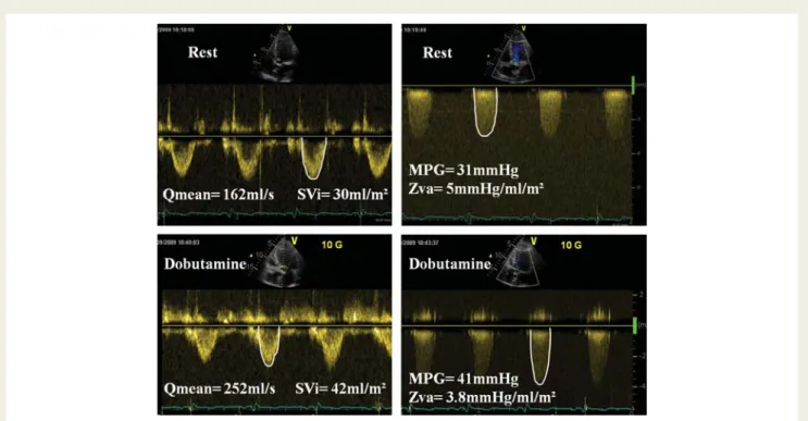
By nature, Zva is flow-dependent and may considerably vary in a same patient over time and during an echocardiographic examin-ation, more specifically in the presence of low-flow state. More-over, two patients with similar AVA and degree of hypertension (i.e. similar LV global afterload), may have different Zva values. The MPG (included in the numerator of the equation for the cal-culation of Zva) is highly flow-dependent and had a square relationship with the Qmean. Because the SVi is the only par-ameter included in the denominator, the impact of flow on the variability of Zva is more important in low-flow state than in a normal or high-flow situation. Subtle changes in SVi and in heart rate may result in high variation in Zva in LF/LG patients
(Figure1). In addition, the impact of minor error in the
measure-ment of SVi on the calculation of Zva may be stronger in low-flow patients. The weak correlation reported by the authors between Zva and MPG confirms that these two parameters are subject to broad variability.
Under dobutamine, a small increase in Qmean may rapidly and noticeably decrease Zva. In patients with low-ejection fraction and low-flow, the Qmean may also markedly vary over patients, from very low (,100 mL/s) to quite normal (.250 mL/s). As
empha-sized in Figure2, the Zva calculated for a patient with severe AS
(AVA ¼ 0.7 cm2) considerably varies according to the flow.
Inter-estingly, this simulation is obtained with a constant SAP (120 mmHg), as it is often the case in low-flow patients due to the adaptation of vascular resistance, suggesting that the variability of Zva is not only related to the changes in SAP. Furthermore, the extent of the flow dependency is higher in low-flow state (SV ,60 – 50 mL). While Zva rises by only 10% between 120 and 60 mL of SV, the increase in low-flow state is very high (.45%). This observation suggests that the calculation of Zva in LF/LG AS is less accurate for estimating the LV global haemodynamic load. Zva was previously found to be associated with a poor outcome in a large series of patients with AS and preserved LV
function.21,22Hence, as highlighted by the results of Levy et al,23
Guest Editorial
355
at Bibliotheque Fac de Medecine on January 19, 2012
http://ehjcimaging.oxfordjournals.org/
it might be argued that the presence of poor LV function rep-resents the main determinant of outcome in LF/LG severe AS. The outcome of such patients thus seems to relate more to the
intrinsic LV myocardial dysfunction than to the global LV haemo-dynamic burden.
The authors reported no statistical differences in Zva between
pseudo and true-severe AS. Figure2revealed that the Zva is
mark-edly lower in pseudo than in true-severe AS patients when the SV is .60 mL. On the other hand, for a SV ,50 mL, Zva are very similar in both groups. This may also explain why Zva is not accu-rate to distinguish pseudo-severe from true-severe AS. This obser-vation also strengthens the idea that Zva at rest might not be a good parameter to evaluate the global LV haemodynamic burden in LF/LG AS. Thus, the use of peak DSE or DSE-induced changes in Zva might be of more interest.
Conclusion
To improve the risk stratification and the management of AS, which remains challenging in numerous cases, the comprehensive evaluation of the valvular and arterial load is mandatory. In this regard, the calculation of Zva appears as particularly useful. However, in LF/LG AS, the Zva seems to be less precise in the assessment of the LV global afterload, essentially due to its high-flow dependency in this specific subset of patient.
Conflict of interest: none declared.
Funding
J.M. is a research associate from the F.R.S-FNRS, Brussels, Belgium, and received a grant from the Fonds Le´on Fredericq, Lie`ge, Belgium. Figure 1 Doppler echocardiographic measurements in a patient with low-flow/low-gradient severe aortic stenosis. Resting indexed aortic valve area was 0.35 cm2/m2and increased up to 0.5 cm2/m2under 10mm/kg/min of dobutamine infusion. Systolic arterial pressure remained
unchanged during the test. The normalization of transvalvular flow rate (Qmean) during dobutamine stress echocardiography resulted in a significant decrease in valvuloarterial impedance (Zva). SVi indicates indexed stroke volume and MPG, mean pressure gradient.
Figure 2 Simulation of the relationship between valvuloarterial impedance and left ventricular stroke volume in patients with true-severe (aortic valve area ,0.7 cm2) and pseudo-severe aortic stenosis. In low-flow state (stroke volume ,50 mL), (i) the Zva markedly increased in response to small changes in stroke volume, and (ii) true- and pseudo-severe aortic stenosis exhibited similar Zva. Body surface area was assumed at 1.8 m2, left ventricular end-diastolic volume at 120 mL, heart rate at 65 b.p.m., and systolic arterial pressure at 120 mmHg.
Guest Editorial
356
at Bibliotheque Fac de Medecine on January 19, 2012
http://ehjcimaging.oxfordjournals.org/
References
1. Iung B, Baron G, Butchart EG, Delahaye F, Gohlke-Barwolf C, Levang OW et al. A prospective survey of patients with valvular heart disease in Europe: the Euro Heart Survey on Valvular heart Disease. Eur Heart J 2003;24:1231 – 43. 2. Krayenbuehl HP, Hess OM, Monrad ES, Schneider J, Mall G, Turina M. Left
ven-tricular myocardial structure in aortic valve disease before, intermediate, and late after aortic valve replacement. Circulation 1989;79:744 – 55.
3. Nishimura RA, Grantham JA, Connolly HM, Schaff HV, Higano ST, Holmes DR Jr, Low-output, low-gradient aortic stenosis in patients with depressed left ventricu-lar systolic function: the clinical utility of the dobutamine challenge in the cathe-terization laboratory. Circulation 2002;106:809 – 13.
4. Blitz LR, Herrmann HC. Hemodynamic assessment of patients with low-flow, low-gradient valvular aortic stenosis. Am J Cardiol 1996;78:657 – 61.
5. Blais C, Burwash IG, Mundigler G, Dumesnil JG, Loho N, Rader F et al. The pro-jected valve area at normal flow rate improves the assessment of stenosis severity in patients with low flow aortic stenosis: the multicenter TOPAS (Truly or Pseudo Severe Aortic Stenosis) study. Circulation 2006;113:711 – 21.
6. Monin JL, Monchi M, Gest V, Duval-Moulin AM, Dubois-Range JL, Gueret P. Aortic stenosis with severe left ventricular dysfunction and low transvalvular pressure gradients. J Am Coll Cardiol 2001;37:2101 – 7.
7. Monin JL, Quere JP, Monchi M, Petit H, Baleynaud S, Chauvel C et al. Low-gradient aortic stenosis: operative risk stratification and predictors for long-term outcome: a multicenter study using dobutamine stress hemodynamics. Circulation 2003;108: 319 – 24.
8. Quere JP, Monin JL, Levy F, Petit H, Baleynaud S, Chauvel C et al. Influence of pre-operative left ventricular contractile reserve on postpre-operative ejection fraction in low-gradient aortic stenosis. Circulation 2006;113:1738 – 44.
9. Pereira JJ, Lauer MS, Bashir M, Afridi I, Blackstone EH, Stewart WJ et al. Survival after aortic valve replacement for severe aortic stenosis with low transvalvular gradients and severe left ventricular dysfunction. J Am Coll Cardiol 2002;39: 1356 – 63.
10. Schwammenthal E, Vered Z, Moshkowitz Y, Rabinowitz B, Ziskind Z, Smolinski AK et al. Dobutamine echocardiography in patients with aortic stenosis and left ventricular dysfunction: predicting outcome as a function of management strategy. Chest 2001;119:1766 – 77.
11. Connolly HM, Oh JK, Schaff HV, Roger VL, Osborn SL, Hodge DO et al. Severe aortic stenosis with low transvalvular gradient and severe left ventricular dysfunc-tion. Result of aortic valve replacement in 52 patients. Circulation 2000;101: 1940 – 46.
12. deFilippi CR, Willett DL, Brickner E, Appleton CP, Yancy CW, Eichhorn EJ et al. Usefulness of dobutamine echocardiography in distinguishing severe from nonse-vere valvular aortic stenosis in patients with depressed left ventricular function and low transvalvular gradients. Am J Cardiol 1995;75:191 – 94.
13. Horstkotte D, Loogen F. The natural history of aortic valve stenosis. Eur Heart J 1988;9(Suppl. E):57 – 64.
14. Levy F, Laurent M, Monin JL, Maillet JM, Pasquet A, Le Tourneau T et al. Aortic valve replacement for low-flow/low-gradient aortic stenosis: operative risk strati-fication and long-term outcome: a European multicenter study. J Am Coll Cardiol 2008;51:1466 – 72.
15. O’Connor K, Lancellotti P, Pierard LA. Stress Doppler echocardiography in valv-ular heart diseases: utility and assessment. Future Cardiol 2010;6:611 – 25. 16. Clavel MA, Fuchs C, Burwash IG, Mundigler G, Dumesnil JG, Baumgartner H et al.
Predictors of outcomes in low-flow, low-gradient aortic stenosis: results of the multicenter TOPAS Study. Circulation 2008;118:S234 – 42.
17. Tribouilloy C, Levy F, Rusinaru D, Gueret P, Petit-Eisenmann H, Baleynaud S et al. Outcome after aortic valve replacement for low-flow/low-gradient aortic stenosis without contractile reserve on dobutamine stress echocardiography. J Am Coll Cardiol 2009;53:1865 – 73.
18. Briand M, Dumesnil JG, Kadem L, Tongue AG, Rieu R, Garcia D et al. Reduced systemic arterial compliance impacts significantly on left ventricular afterload and function in aortic stenosis: implications for diagnosis and treatment. J Am Coll Cardiol 2005;46:291 – 98.
19. Cramariuc D, Cioffi G, Rieck AE, Devereux RB, Staal EM, Ray S et al. Low-flow aortic stenosis in asymptomatic patients: valvular-arterial impedance and systolic function from the SEAS Substudy. JACC Cardiovasc Imaging 2009;2:390 – 99. 20. Lancellotti P, Donal E, Magne J, O’Connor K, Moonen ML, Cosyns B et al. Impact
of global left ventricular afterload on left ventricular function in asymptomatic severe aortic stenosis: a two-dimensional speckle-tracking study. Eur J Echocardiogr 2010;11:537 – 43.
21. Lancellotti P, Donal E, Magne J, Moonen M, O’Connor K, Daubert JC et al. Risk stratification in asymptomatic moderate to severe aortic stenosis: the importance of the valvular, arterial and ventricular interplay. Heart 2010;96:1364 – 71. 22. Hachicha Z, Dumesnil JG, Pibarot P. Usefulness of the valvuloarterial impedance
to predict adverse outcome in asymptomatic aortic stenosis. J Am Coll Cardiol 2009;54:1003 – 11.
23. Levy F, Monin JL, Rusinaru D, Petit-Eisenmann H, Lelguen C, Chauvel C et al. Does valvuloarterial impedance improve risk stratification in low-ejection fraction, low-gradient aortic stenosis? Results from a multicenter study. Eur J Echocardiogr 2011;12:358 – 63.
Guest Editorial
357
at Bibliotheque Fac de Medecine on January 19, 2012
http://ehjcimaging.oxfordjournals.org/