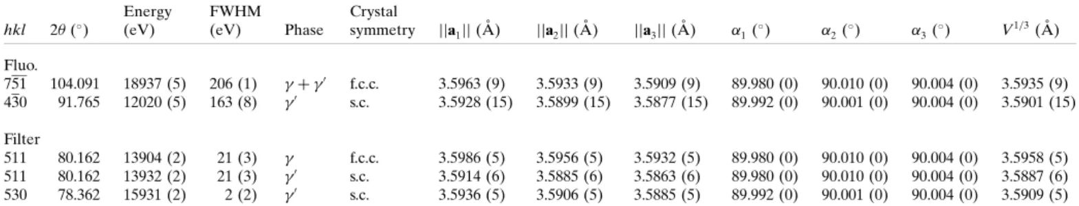
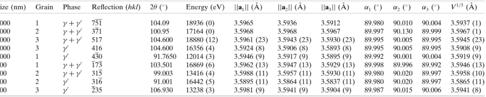
Full elastic strain tensor determination at the phase scale in a powder metallurgy nickel-based superalloy using X-ray Laue microdiffraction
13
0
0
Texte intégral
Figure
Documents relatifs