1
Ameliorative effect of Aristolochia longa and Aquilariamalaccensison
lead acetate inducednephrotoxicityin female rats
KHELEF Yahia , 1 , GHARBI Safa 1 , ZEGHIB Khaoula 1 DEROUICHE Samir
DEROUICHE Samir ¹*, ZEGHIB Khaoula1, GHARBI Safa1, KHELEF Yahia 1Department of Cellular and Molecular Biology, Faculty of Natural Sciences and Life, University of Echahid Hamma Lakhdar El Oued, El Oued 39000, (Algeria).
P
Received 2 September 2018/Accepted 27 November 2018/ Published online 01 December 2018 International Journal of Biological and Agricultral Reasearch
( IJBAR)
Journal home page: www http://www.univ-eloued.dz/ijbar/ ISSN: 2661-7056
* DEROUICHE Samir Department of Cellular and Molecular Biology, Faculty of Natural Sciences and Life, University of Echahid Hamma Lakhdar El Oued (Algeria)
E-mail: dersamebio@gmail.com
Abstract
Background:Aquilariamalaccensis And Aristolochia longa are plants used extensively in traditional medicine for the treatment of cancer in several regions in Algeria. We investigate the ameliorative effect of Aristolochia longa and Aquilariamalaccensison kidney toxicity induced by lead in female albino rats. Methods:Twenty five (25) apparently healthy female Wistar rats were randomly divided into five groups of five rats in each: control, Pb, Pb + A. longa (Ar), Pb+ A. malaccensis (Aq), and Pb+Ar+Aq. lead (100 mg/kg b.w) as Pb(C2H3O2)2 added in their drinking water for 75 days. A. longa (rhizome powder at a dose 1% w/w of diet) and A. malaccensis (heartwood powder at a dose 1% w/w of diet) were added to the feed during the last 15 days of lead exposed in the animals. Results:Obtained results revealed that lead treatment caused a significant increase in blood glucose level, serum urea, creatinine and uric acidlevel and in kidneyof MDA level and CAT activity. In contrast, it led to an decrease in the kidneyGST activityand GSH level in rats .Also, the results clearly showed that lead causes alterations of glomerular and tubular structure in comparison with controls.Our results showed that treatment with A. malaccensis and A. longa a partial correction of the previous parameters.The histological observations confirmed the nephroprotection results by the biochemical parameters. Conclusion:Results demonstrated beneficial effects of A. longa and A. malaccensis treatment againacetate lead-induced toxicity and kidney damage in rats.
2 Introduction
Lead is a chemical element that exists in nature. It is considered a major pollutant in the environment[1]. It is recognized as one of the most toxic and harmful heavy metals, even in small amounts [2].As one major heavy metal pollutants, lead is normally generated from industry and continuously present in soil, drinking water and aquatic environments[3].The non-biodegradable nature of lead is the prime reason for its prolonged persistence in the environment [4].Lead can transfer into the trophic chain by causing deleterious effects for humans and other living organisms. It is a toxic metals in the environment and target most organs of the human body[5]. The kidneys play an essential role in the elimination of lead from the body and the bioaccumulation of lead is mainly in the kidneys and then in the liver and brain in animals exposed by lead [6]. This metal induces a wide range of physiological, biochemical and behavioral dysfunctions in laboratory animals and humans, including respiratory, neurological, hepatic and male and female reproductive systems [7]. Renal toxicity of lead is characterized by alteration in proximal tubular, glomerular sclerosis and in interstitial fibrosis. Renal dysfunction in humans exposed to the excessive amount of lead includes enzymeuria, low- and high-molecular weight proteinuria, transport of organic anions and glycemic filtration rate. In addition, at the histopathological state, renal damage has been shown in humans, including intranuclear inclusion bodies and cellular necrosis in the proximal and interstitial tubule fibrosis [8]. Lead can cause oxidative stress in the blood and other tissues which is considered as one of the possible mechanisms of the harmful and toxic effects induced by this metal in the organism [9]. It has been postulated that lead causes oxidative stress by generating the release of reactive oxygen species (ROS) such as superoxide radicals, hydrogen peroxide and hydroxyl radicals and lipid peroxides [10], leading to the development of cancer, diabetes, neurodegenerative diseases and cardiovascular diseases [11].Aquilariamalaccensisand Aristolochia longa Are plants used extensively in traditional medicine in the treatment of cancer in several regions in Algeria. Aquilariamalaccensis has numerous biological activities, including activity, microbial, anti-tumor, anti-allergic, anti-oxidant [12].Aristolochia longa also has several therapeutic uses as against ovarian insufficiencies, healing, diuretic [13] , analgesic, inflammatory, anti-hyperglycemic [14]. The aim of present study was designed to investigate the effect of Aquilariamalaccensis and Aristolochia longa on nephrotoxicity, stress oxidative and kidney tissue alteration induced by lead in rats.
3 Materials and methods
Chemicals
Lead acetate and all other chemicals used in the study were obtained from Sigma-Aldarich Corporation (St. Louis, Missouri, USA).
Plant materials
Aristolochia longa roots and Aquilariamalaccensisheartwood were collected in herbalists shops from a local market of El-Oued.(voucherspecimen no: FSNV/DB/consult/2016/78-15-02 for Aristolochia longa and voucher specimen no: FSNV/DB/consult/2016/81-12-06 for
Aquilariamalaccansis) The vegetal materials were washed with water, and then dried at room temperaturefor 48 to 92 h. grounded into powder and stored at room temperature until use. Animals and Experiments
Female albino rats aged 8 weeks old weighting approximately 220g, were obtained from the animal house of Pasteur institute, Algeria. Animals had free access to standard diet and were provided distilled water. After the adaptation period, rats were divided into five groups of 5 rats in each and kept in animal’s house of Molecular and cellular biology Department, University of El Oued, Algeria. Standard rat diet and water were available ad libitum for the duration of the experiments unless otherwise noted. Animals were acclimated for two weeks under the same laboratory conditions of photoperiod (12h light/12 h dark) with a relative humidity 65.3 % and room temperature of 23 ± 2 C°. The experimental procedures were carried out according to the National Institute of Health Guide-lines forAnimal Care and approved by the Ethics Committee of our institution.The experiment was conducted over a period of 70 days. After a period of adaptation, the animals were divided into five experimental groups of 5 animals each:
Groupe I (control group): animals served as normal control.
Groupe II (lead-treated group): animals received acetate lead (100 mg/kg b.w) as Pb(C2H3O2)2 added in drinking water for 70 days in animals.
Groupe III (lead+ Aq-treated group): animals received a powder diet containing powder of heartwood of AquilariaMalaccensis (1% w/w in feed) for 15 days.
Groupe IV (lead+Ar-treated group): animals received a powder diet containing powder of rhizome of Aristolochialongua (1% w/w in feed) for 15 days.
4
Groupe V (lead+Aq+Ar-treated group): animals received a powder diet containing powder of both heartwood of Aquilariamalaccensis and rhizome of Aristolochialonguafor 15 days.
Blood was collected, some of which was centrifuged at 3,000 rpm for 10 min to obtain the plasma which was kept frozen at -20°C until used for biochemical analysis of urea, creatinine, uric acid andtotal protein level.
2.4. Tissue preparation
1g of kidney tissue of each rat of different groups was used. After milling and homogenizing the tissues in TBS (50 mMTris, 150 mMNaCl, pH 7.4), and centrifuged at 10 000 × g for 15 min at 4°C, then the supernatant obtained was stored at - 20 ° C while waiting for the determination of oxidative stress parameters.
Plasma Biochemical parameters
Biochemical markers were assayed according to standard methods. The levels of serum urea, creatinine, uric acid and total protein were estimated using commercial kits from Spinreact laboratories, Spain (urea, ref. 1001332; creatinine, ref. 1001113; uric acid,ref. 1001013 and total protein, ref. 1001291).
Tissue oxidative stress parameters Estimation of lipid peroxidation levels
The principle of this assay is based on the condensation of MDA in acidic and hot medium with thiobarbituric acid according to Yagi et al.[15]. The reaction results in the formation of a pink complex between two molecules of thiobarbituric acid which can therefore be measured by Absorption spectrophotometry at 532 nm and the level of MDA in liver was expressed as nmol/mg protein.
Determination of reduced glutathione (GSH) level
GSH concentration was performed with the method described by Ellman[16].The complex formed between GSH and 5,5'dithiodis-2-nitrobenzoic acid (DTNB) releases thionitrobenzoic acid (TNB) which has an absorbance at 412 nm.Total GSH content was expressed as nmol GSH/mg prot.
5
Determination of Glutathione-S-transferase (GST) activity
The activity of GSTs was measured spectrophotometrically according to the method of Habig et al. [17], based on the formation kinetics of a complex between a GST substrate: 1-chloro-2-4-dinitrobenzene (CDNB ), And glutathione (GSH). The complex formed can be visualized by increasing the optical density at a 340 nm. The enzyme activity was expressed as nmol CDNB conjugate/min/mg protein.
Determination of Catalase activity
The catalase activity consists in measuring the disappearance of catalase-induced H2O2 contained in the sample by measuring the absorbance of H2O2 at 560 nm using a spectrophotometer according to the method of Aebi, 1984 [18].
2.7.Histopathological Analysis
The kidney from eachrat was removed immediately and conserved in a sample flaskcontaining 10% formalin solution. Liver was processed bythe paraffin technique. Sections of 5 μm thickness were cut andstained by hematoxylin and eosin for histological examination.
Statistics Analysis
Mean data values are presented, with their standard deviations (mean ± SD). All statistical comparisons were made by Student’s t-test using Minitab 13software, and statistical significance was defined as P < 0.05.
Results
body weight, body weight gain and relative kidney weight
The results obtained (Table 1) show that Pb(C2H3O2)2 treatment at a dose (100 mg/kg b.w) caused a significant decrease (p<0.001) in body weight and asignificant increase (p<0.01) in Relative kidney weight compared to the control rats. But After the treatment time, the animals that received the powder of rhizome A. longa and heartwood A. malaccensisalone or combined showed reversion partial of this this change .
6 Table 1
Mean initial Body weight (g), final Body weight (g) and relative kidney weight of control and experimental groups
parameters Control (n=5) Lead (n=5) Lead + Aq (n=5) Lead + Ar (n=5) Lead +Aq+Ar (n=5) Initial Body weight 225±5.06 225.2±5.69 226.60±23.71 221±8.76 225.2±3.51 Final Body weight 291±3.07 198±2.32*** 196±5.00*** 215±1.15**a 231±1.61*a Relative kidney weight 0.56±0.01 0.73±0.04** 0.65±0.02* a 0.67±0.01*** b 0.59±0.01 c
* p<0.05, ** p<0.01, ***p<0.001: significantly different from control group. a p<0.05, c p<0.001: significantly different from Pb group.
Values are mean ± SEM, n=number of observations.
3.2. Biochemical parameters
The results obtained in Table 2 show that a significant increase(p < 0.05) in blood glucose, serum urea, creatinine and uric acidlevel and no significant difference in serum total proteinconcentration in the group exposed to acetate lead compared to the control group. Treatment with powder of heartwood Aq or of rhizome Artotally restored the level of blood glucose, serum urea, creatinine and uric acid Compared to lead group.
Table 2
Changes in biochemical parameters of control and experimental groups parameters Control (n=5) Lead (n=5) Lead + Aq (n=5) Lead + Ar (n=5) Lead +Aq+Ar (n=5) Blood glucose (g/l) 0.752±0.051 0.905±0.107* 0.80±0.053b 0.81±0.067 a 0.775±0.027c Urea (g/l) 0.64±0.067 0.81±0.085* 0.60±0.037 c 0.67±0.030 c 0.67±0.033b Creatinine (mg/l) 4.40±0.40 4.55±0.320* 4.40±0.245a 3.80±0.200* a 4.00±0.00a Uric acid (mg/l) 17.60±1.94 18±2.00* 18.8±1.32* 17.6±1.60a 18.6±1.36* Totale protein (g/l) 50.6±1.40 48.33±1.76 47.60±2.60 48.2±1.12 47.4±1.53
* p<0.05, ** p<0.01 : significantly different from control group. a p<0.05, b p<0.01: significantly different from Pb group. Values are mean ± SEM, n=number of observations.
Stress oxidative parameters
Our results illustrated in Table 3 show a significant increase (p <0.01 and p <0.001)in lipid peroxidation level and catalase activity respectively and a significant decrease (p <0.001 )in GSH concentration and GST activity inkidney of rats of the leadgroup compared to the control. In contrast our results also show a significant decrease in kidney MDA level (p <0.001) and catalase (p <0.01) activity in lead+Aq, lead+Ar and lead+Ar + Aq treated groups and a significant increase in kidney GSH concentration(p <0.01) and GST activity (p <0.05)in only lead +Ar group Compared to lead group.
7 Table 3.
MDA, GSH levels and antioxidant enzyme activities in kidney tissue of control and experimental groups parameters Control (n=5) Lead (n=5) Lead + Aq (n=5) Lead + Ar (n=5) Lead +Aq+Ar (n=5) MDA (µmol/mg pro) 3.16±0.41 4.37± 0.46** 2.12±0.32** c 2.11±0.13*** c 1.717 ±0.09*** c GSH (nmol/mg pro) 0.48±0.09 0.248±0.01*** 0.212±0.03*** 0.38±0.040* b 0.227±0.01*** GST (nmol/min/mg pro) 3.75±0.78 2.48±0.02*** 2.44±0.17** 3.01±0.14** a 2.62±0.53** CAT (UI/mg pro) 8.61±0.82 18.75±0.22*** 14.21±0.88*** b 11.78±0.77** c 11.51±0.18b
* p<0.05, ** p<0.01, *** p<0.001 : significantly different from control group. a p<0.05, b p<0.01: significantly different from Pb group.
Values are mean ± SEM, n=number of observations.
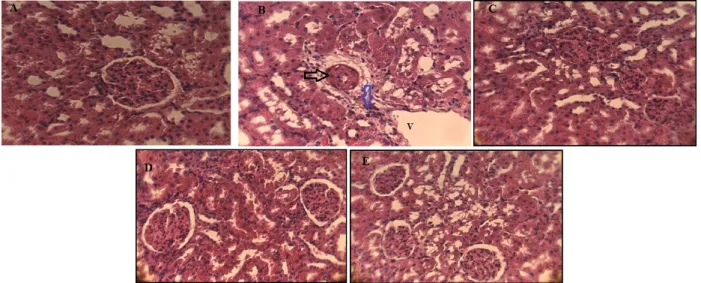
Histopathological studies
Microscopic observation of histological sections of kidney from the control rat showed normal renal parenchyma with glomeruli, tubules and normal cortical and medullar areas (Fig. 1A).However, histological sections of the lead-exposed rat kidney revealed Degeneration represented by necrosis and glomerular atrophy apparent to their architecture with tubular necrosis, dilatation and tabular vacuolation. In addition, haemorrhage, foci of inflammation and large space of Bowman, was observed in the kidney sections of this group (Figure 1B). Treatment with Aquilariamalacenssis and / or Aristolochia longa in diets in lead-contaminated rats for 15 days provided protection against tubular and glomerular histological damage induced by this metal. These plants specially Aristolochia longainduce a remarkable regeneration of kidney tissue. However, Aquilariamalacenssis partially decreased severe damage to kidney tissue (Figure 1C, 1D, 1E).
8
Figure 1. The histological examination of Kidney from a control rat showing normal architecture (Figure 1A, ×400). Acetate lead treated rats kidney showing Focus of inflammation (blue arrow), Glomerular necrosis (black arrow) and vacuolation (V ) (Figure 1B, ×400). Powder rhizome of A. longa -treated rat kidney showing normal appearance of tubule and glomerulus (Figure 1D, ×400). kidney sections of the animals administered powder of heartwood A. Malaccensis alone (Figure 1C, ×400) or combined with rhizome of A. longa (Figure 1E, ×400) of showed moderate degree of kidney damage, necrosis and inflammatory cell, protection from tubule and glomerulus degradation.
Discussion
The present study investigated the effects of treatment with powder of rhizome A. longa and heartworm A. malaccensisonbiochemical markers of kidney function, oxidative stress and kidney histology outcome of acetate lead-induced kidney damage in rats. In the present study, the reduction in body weight is used as an indicator of the deterioration of the overall health of the rat. Exposure of lead acetate to doses (100 mg / kg) in rats resulted in a decrease in body weight and increase in relative liver weight. This result is consistent with several studies [19, 20] who suggested that reduction in body weight is due to reduced food consumption by lead effects on nerve centers responsible for the regulation of satiety and hunger. Also the loss of body weight may be due the reduced in mass of muscle and cachexia due to the oxidative stress induced by lead [21].Increase in relative kidney weight may be due to lead-induced necrosis and apoptosis on this organ [22].After the treatment time, the animals that received A. longa and A. malaccensis showed reversion partial of this less body weight, due probably the improvement of behavior and biochemical parameters.Improvement of relative kidney weightwas probably due to the inhibitory effect of these plants against the accumulation of lead in kidney which reduces their injurious effect on kidney and therefore decreases their relative weight. Also, the protective effect of these plants could be due to the presence of anti-inflammatory compounds such as
9
flavonoids and phenolic acids [23].In the present study, lead exposure results in a significantincrease in blood glucose level. Our results in agreement with study ofIbrahim et al. (2012)[24].Lead enters into competition with calcium in metabolic processes exactly on the insulin excretion by several actions: inhibitory action on voltage-dependent calcium channels, decreased adenylatecyclase activity [25], Reduces the release of certain neurotransmitters such as serotonin [26]. Moreover, lead causes adverse effects on the pancreas exactly on the excretion of insulin by islets of Langerhans which increases blood glucose level [27]. Our results also demonstrated a significant decrease in the blood glucose level in rats treated with A. malaccensis and A. Longa alone or in combination compared with the lead group, the polyphenolic and flavonoid compounds of plants can influence glucose metabolism by several mechanisms such as inhibition of carbohydrate digestion and absorption of glucose in the intestine, Stimulation of insulin producing cells, modulation of glucose release from the liver, activation of glucose uptake in insulin-sensitive tissues, and modulation of hepatic glucose production [28,29].In our study, exposure of lead acetate to rats resulted in nephrotoxicity characterized by a significant increase in the serum urea, creatinine and uric acid level. These results are similar to those published in previous studies [30, 31, 32]. The results clearly show that this metal affects the excretory function of nephrons, the structural and functional unit of the kidneys.Treatment of rats with A. Malaccensis and / or A. Longa restored renal function. The protective effect of A. Malaccensis and A. Longa was reflected by decreased serum urea and creatinine level in rats treated with these plants compared to rats exposed to lead.In our study, the results show a significant decrease in GSH level and GST activity and a significant increase in MDA level and CAT activity in the kidney of rats exposed with lead acetate.These results are in agreement with several studies[33,34, 35]. There is a complex interaction between antioxidants and oxidants such as reactive oxygen species, which modulates the generation of oxidative stress [36]. Thesechanges are explained by the increase in the of free radicals, which can be due to the accumulation of 5-aminolevulinic acid (ALA) in kidney under the effect of lead [37].These also shows that lead is able to promote the generation of ROS, which results in lipid peroxidation in renal tissues, suggesting their deleterious effects in this tissue. Thus, another mechanism, lead can have a direct peroxidation activity that has a high binding affinity on the cell membrane [38]. Since oxidative stress is the first response to the environmental pollutants, liver cells may stimulate antioxidant and detoxification responses to counter heavy metal damages. The involvement of anti-oxidative enzymes such as GST play a considerable mission in protecting cells from oxidative stress [39].Increased CAT activity can be explained as a tissue defense
10
mechanism against increased H2O2 fluxes during Pb-induced oxidative stress [40].Our results show that treatment with A. malaccensis and / or A. longa could prevent lead-induced alteration, increasing the antioxidant activity of GSH and GST and decreasing lipid peroxidation. Suggest that both plants possess antioxidant activity based on the elimination of free radicals and the restoration of the oxidizing / antioxidant balance during toxicity induced by this metal. The presence of polyphenols and flavonoids in A. Malaccensis and A. Longa may be responsible for antioxidant activity and are considered good chelating agents for metal ions. In addition, the activity of phenolic acids depends on the number and position of the group (-OH) and (-OCH3) groups [41], and the flavonoids inactivate and stabilize the free radicals by their group Hydroxyl (C3-OH). They are also capable of chelating metal ions (released from their binding or transport proteins) [42].According to the results of the present study, lead exposure produces histological damage in the kidney including glomerular necrosis, tubular dilation, and inflammation. These alterations may be due to the excessive production of free radicals and as a result of lipid peroxidation induced by lead acetate [43].Treatment with A. malaccensis and / or A. longa in intoxicated rats has been able to regenerate the structure of the kidney. Our results suggest that these plants, especially A. longa, could reducekidney lesions induced by (Pb (C2H3O2) 2). The biochemical and oxidative stress parameters are also correlated with the histological study. This can be attributed to the anti-radical effect of A. Malaccensis and A. longa. These plants reduced the oxidative stress caused by lead, allowing the reduction of histological alterations and restoration of the normal physiological state of the body.
Conclusion
Lead is a strong nephrotoxic agent by inducing renal dysfunction and oxidative stress in kidney. Aristolochia longa and Aquilariamalaccensis were able to moderate this toxicity by decreasing oxidative stress and decreasing the alteration of the kidney tissue.
Author’s contribution
All research done by the authors
Conflict of interest statement
We declare that we have no conflict of interest.
11
The first author would like to thank the Department of cellular and molecular biology, Faculty of sciences of nature and life, University of El Oued, Algeria, for the permission to utilize the facilities to make this work.
References
1. Joworaski Z. Stable and Radioactive Lead in Environment and Human Body, Warsaw: Nuclear Energy Information Center, Review Report 1968; 29: 17–30.
2. MishraS, GhoshD, Dutta M, Chattopadhyay A, Bandyopadhyay D. Melatonin protects against lead-induced oxidative stress in stomach, duodenum and spleen of male Wistar rats. J Pharm Res. 2013; 1(11): 997-1004.
3. Derouiche S, Djouadi A. Control of Lead and Cadmium in Cosmetic Product (Kohl) of Pits Dates by Cyclic Voltammetry. J Chem Pharm Res. 2017; 9(3):319-323.
4. Gagan F, Deepesh G, Archana T. Toxicity of lead: A review with recent updates. InterdiscipToxicol 2012; 5(2): 47–58.
5. Duruibe JO, Ogwuegbu MC, Egwurugwu JN. Heavy metal pollution and human biotoxic effects. Int J PhysSci 2007; 2: 112–8.
6. Dev PR, Swarup D, Dwivedi SK. Some renal function tests in experimental leadtoxicity in Goats. IndianVet J 1991; 68: 1163-1167.
7. Ponce-Canchihuamán JC, Pérez-Méndez O, Hernández-Muñoz R, Torres-Durán PV, Juárez-Oropeza MA. Protective effects of Spirulina maxima on hyperlipidemia and oxidative-stress induced by lead acetate in the liver and kidney. Lipids Health Dis2010;9:35. doi:10.1186/1476-511X-9-35.
8. Derouiche S, Serouti A,Rezzagmohcen O S. Risk of Metribuzin (Triazinone herbicide)on hematological and renal structure and function of pregnancy rabbits. Asian J.Biol. Sci. 2019; 12:192-198.
9. Pande M, Mehta A, Pant B, Flora SJ. Combined administration of a chelating agent and an antioxidant in the prevention and treatment of acute lead intoxication in rats. Environ. ToxicolPharmacol 2001; 9:173 -184.
10. Derouiche S, Djouadi A, Belimi N, Louetri K, Hachefa S. Blood Glucose, some Electrolytes Levels and Stress Oxidative Status of Female Hyperthyroid Patients under Treatment. J. Adv. Res. BioChem. Pharma. 2018; 3(1&2): 1-6.
11. Wang J, Yang Z, Lin L, Zhao Z, Liu Z, Liu X. Protective effect of Naringenin against lead-induced oxidative stress in rats. Biol Trace Elem Res 2012; 146: 354–359.
12. Chena HQ, Wei JH, Yanga JS, Zhanga Z, Yangb Y, Gaoa ZH, Suia C, Bao G. Chemical constituents of Agarwood originating from the Endemic Genus Aquilaria Plants.
ChemBiodivers 2012; 9(2): 236-250.
13. Cherif HS, Saidi F, Guedioura A. Toxicological evaluation of Aristolochia longa L. extract in mice. Indian J Appl Res 2014; 4(5): 26-30.
14. Benarba B, Meddah B. Ethnobotanical study, antifungal activity, phytochemical screening and total phenolic content of Algerian Aristolochia longa. J
IntercultEthnopharmacol 2014; 3:150-154.
15. Yagi K. Simple fluorimetric essay for lipoperoxide in blood plasma. Biochem Med 1976; 15(2): 212-216.
16. Weckbercker G, Cory JG. Ribonucleotidereductase activity and growth of glutathione-depleted mouse leukemia L1210 cells in vitro. Cancer Lett 1988; 40(3): 257-264. 17. Habig WH, Pabst, MJ, Jakoby WB.. Glutathione S-transferase. The first enzymatic step
in mercapturic acid formation. J BiolChem 1974; 249: 7130-7139. 18. Aebi H. Catalase in vitro. Meth. Enzymol1984; 105: 121-126.
12
19. Berrahal A, Nehdi A, Hajjaji N, Gharbi N, El-Fazâa S. Antioxidant enzymes activities and bilirubin level in adult rat treated with lead. PharmacolToxicol 2007; 330: 581–588. 20. Reckziegel P, Dias VT, Benvegnú DM, Boufleur N, Barcelos RC, Segat HJ, Pase CS,
Santos CM, Bürger ME. Antioxidant protection of gallic acid against toxicity induced by Pb in blood, liver and kidney of rats. Toxicol Rep 2016; 3: 351-356.
21. Yeh YC, Liu TJ, Wang LC, Lee HW, Ting CT. A standardized extract of Ginkgo biloba suppresses doxorubicin-induced oxidative stress and p53-mediated mitochondrial apoptosis in rat testes. Br J Pharmacol 2009; 156: 48-61.
22. Ibrahim NM, Eweis EA, El-Beltagi HS, Abdel-Mobdy YE. The Effect of Lead Acetate Toxicity on Experimental Male Albino Rat. Biol Trace Elem Res 2011; 144: 1120-1132. 23. Keerthi M, Prasanna J, Aruna M, Rao N. Review on polyphenols as nature's gift world. J
Pharm PharmSci 2014; 3: 445-455.
24. Kaouachi A, Derouiche S. Phytochemical analysis, DPPH antioxidant activity and Acute toxicity of bark aqueous extracts of Pinushalepensis. Res. J. ChemEnvSci. 2018; 6(3): 86-91.
25. Kaminsky P, Klein M, Duc, M. Physiopathologie de l'intoxication par le plomb inorganique. La revue de médecine interne. 1993; 14(3): 163-170.
26. Paulmann N, Grohmann M, Voigt JP, Bert B, Vowincke J, Michael B, Skelin M, Jevs M, Fink H, Rupnik M, Diego J. Walther. Intracellular serotonin modulates insulin secretion from pancreatic b-cells by protein serotonylation. Plos Biology 2009; 7(10): 1-10.
27. Saka, S., Bahi, A., Aouacheri, W. L’effet du stress oxidant induitparl’acétate de plombsur le systèmeenzymatique du glutathion chez les rats the effect of oxidative stress induced by lead acetate on the glutathione enzymatic system in rats. Annales de Toxic Analytique 2011; 23(3): 139-145.
28. Masunda TA, Mbala MB, Kayembe SJ, Longoma BF, Ngbolua KN, Tshibangu DST Mpiana PT. Activité anti-hyperglycémique et antiradicalaire des extraits des fruits de Raphia gentilianaDe Wild. (Arecaceae). Int J BiolChemSci2014; 8(6): 2441-2451. 29. Hanhineva K, Törrönen R, Bondia-Pons I, Pekkinen J, Kolehmainen M, Mykkänen H,
Poutanen K. Impact of dietary polyphenols on carbohydrate metabolism. Int J Mol Sci 2010; 11, 1365-1402.
30. Alain AK, Akpovi CD, Sègbo J, Senou M, Anago E, Ahoyo TA, Klotoé JR, Edorh PA, Loko F. Attenuation effect of Moringaoleiferaleaves powder on blood biochemical disturbance induced in lead-exposed rats. Int Res J BiolSci 2016; 5(1): 14-21.
31. Ghosh D, Benazir S, Mitra E, Dey M, Bandyopadhyay D. Protective effect of aqueous leaf extract of MurrayaKoenigi Against lead induced oxidative stress in Rat liver, heart and kidney: a dose response study. Asian J Pharm Clin Res 2012; 5(4): 54-58.
32. Missoun F, Slimani M, Aoues A. Toxic effect of lead on kidney function in rat Wistar. African J Biochem Res2010; 4(2): 21-27.
33. Kansal A, Sharma V, Sharma A, Lodi S, Sharma H. protective role of
coriandrumsativum (coriander) extracts against lead nitrate induced oxidative stress and tissue damage in the liver and kidney in male mice. Int J ApplBiol Pharm 2011; 2(3): 65-83.
34. Alya A, Bini Ines D, Montassar L, Najoua G, Saloua E. Oxidative stress, biochemical alterations, and hyperlipidemia in female rats induced by lead chronic toxicity during puberty and post puberty periods. Iran J Basic Med Sci 2015; 18(10): 1034–1043. 35. Jackie T, Haleagrahara N, Chakravarthi . Antioxidant effects of Etlingeraelatior flower
extract against lead acetate – induced perturbations in free radical scavenging enzymes and lipid peroxidation in rats. BMC Research Notes 2011; 4(67): 8.
13
carbohydrate metabolism, some enzyme activities and pancreatic islet tissue in alloxane induced diabetic rats. Int J Pharm PharmSci 2017; 9(6): 54-58.
37. Ahamed M, Siddiqui MKJ. Low-level lead exposure and oxidative stress: current opinions. ClinicaChimicaActa 2007; 383: 57–64.
38. Hnde G, Nuran E. Can antioxidants be beneficial in the treatment of lead poisoning. Free RadicBiol Med 2000; 29(10): 927–945.
39. Derouiche S, Habita A, Degachi O, Necib M, Chetehouna R. Investigation of the Oxidative Stress markers in salivary, serum and erythrocyte of El-Oued (Algeria) Diabetic Patients. International Journal of Biological and AgricultralReasearch 2018; 1(1) : 10-18.
40. Gurer H, zgunes HO, Saygin E, Ercal N. Antioxidant effect of taurine against lead-induced oxidative stress. Arch Environ ContamToxicol 2001; 41: 397–402.
41. Chusri S, Singthong P, Kaewmanee T. Antioxidant, anticancer, and cytotoxic effects of thai traditional herbal preparations consumed as rejuvenators. J Food Sci 2015; 13(1): 40-48.
42. Ghedira K. Les flavonoïdes : structure, propriétés biologiques, rôle prophylactique et emplois en thérapeutique. Phytotherapy 2005; 3: 162. doi:10.1007/s10298-005-0096-8. 43. Aziz FM. Protective Effects of Latex of Ficuscarica L. against Lead Acetate-Induced